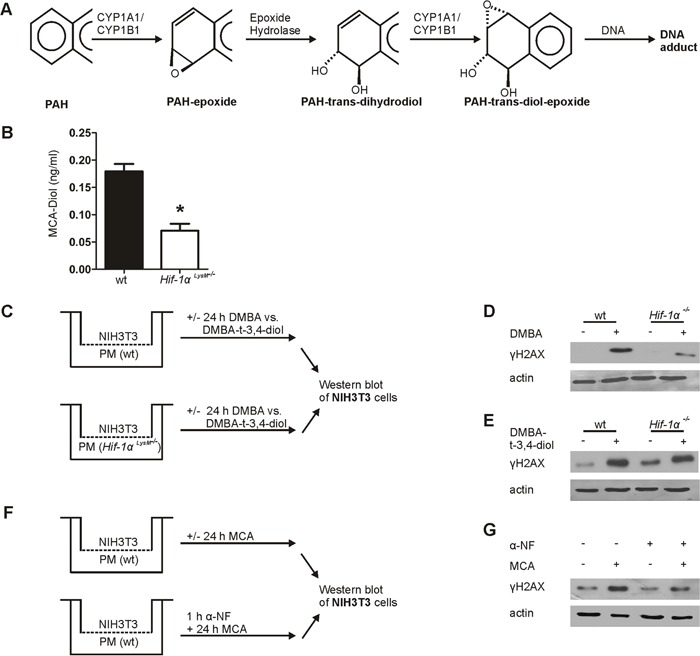
Figure 4. Metabolism of 3-methylcholanthrene (MCA) in peritoneal macrophages.

A. Schematic overview of the metabolic activation of polycyclic arylhydrocarbons (PAHs). B. Quantification of MCA-Diol in the supernatants of isolated peritoneal macrophages (PMs) from wt and Hif-1αLysM−/− mice stimulated with MCA for 8 h and quantified by LC-MS/MS. Bars are the mean ± SEM of n = 4 (each n containing 3 mice/genotype). * P < 0.05 compared to wt. C. Schematic illustration of the coculture of NIH3T3 cells with PMs isolated from wt or Hif-1αLysM−/− mice stimulated with 7,12-dimethylbenz[a]anthracene (DMBA), DMBA-trans-3,4-dihydrodiol (DMBA-t-3,4-diol), or DMSO for 24 h. Representative Western blot of phosphorylated γH2AX in NIH3T3 cell stimulated with D. DMBA or E. DMBA-t-3,4-diol from 4 experiments is shown. Actin served as a loading control. F. Schematic illustration of the coculture of NIH3T3 and wt PMs, prestimulated for 1 h with or without 10 nM α-napthoflavone (α-NF), a CYP-inhibitor, following stimulation with MCA or without for 24 h. G. Representative Western blot of phosphorylated γH2AX in NIH3T3 cells cocultured with wt peritoneal macrophages from 5 experiments is shown. Actin served as a loading control.
