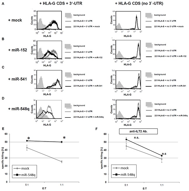
Figure 5. Impact of miR-mediated down regulation of HLA-G expression on the cytotoxicity of NK cells in vitro.

A–D. 721.221 cells expressing HLA-G with its 3′-UTR (221/HLA-G + 3′UTR) or expressing HLA-G without the 3′-UTR (221/HLA-G no 3′-UTR) were transduced with the respective mock vector (Figure 5A), the expression vector for miR-152 (positive control; Figure 5B), miR-541 (negative control; Figure 5C) and miR-548q (Figure 5D). The transfectants were analyzed for their HLA-G expression by flow cytometry. The experiments were performed three times. One representative experiment is shown as a histogram. E. 35S methionine-labelled 721.221/HLA-G + 3′-UTR cells were transduced with an empty vector (mock) or a miR-548q expression vector and then incubated for 5h with ILT2-expressing NK cells. Shown is the relative average killing ± standard derivation of three independent experiments. The HLA-G down regulation upon miR-548q expression caused a statistically significant (*p < 0.05) increase of NK cell mediated cytotoxicity when compared to the mock control, which can even be enhanced at a higher E:T ratio. F. From PBMCs freshly isolated NK cells were pretreated with an anti-ILT2 antibody. Due to this blockage of this main HLA-G receptor upon the NK cells no difference in the NK cell mediated cytotoxicity could be observed for the miR-548q overexpressing (HLA-G low) transfectants and the respective mock transfectants (HLA-G high). An increased E:T ratio also increased the cell lysis in both transfectants.
