eLife logoLink to eLife
. 2016 Aug 18;5:e16352. doi: 10.7554/eLife.16352

A hyperspectral method to assay the microphysiological fates of nanomaterials in histological samples

Elliott D SoRelle 1,2,3,4,, Orly Liba 1,2,4,5,, Jos L Campbell 1,6,, Roopa Dalal 7, Cristina L Zavaleta 1,6, Adam de la Zerda 1,2,3,4,5,*
Editor: Gaudenz Danuser8
PMCID: PMC5042654  PMID: 27536877

Abstract

Nanoparticles are used extensively as biomedical imaging probes and potential therapeutic agents. As new particles are developed and tested in vivo, it is critical to characterize their biodistribution profiles. We demonstrate a new method that uses adaptive algorithms for the analysis of hyperspectral dark-field images to study the interactions between tissues and administered nanoparticles. This non-destructive technique quantitatively identifies particles in ex vivo tissue sections and enables detailed observations of accumulation patterns arising from organ-specific clearance mechanisms, particle size, and the molecular specificity of nanoparticle surface coatings. Unlike nanoparticle uptake studies with electron microscopy, this method is tractable for imaging large fields of view. Adaptive hyperspectral image analysis achieves excellent detection sensitivity and specificity and is capable of identifying single nanoparticles. Using this method, we collected the first data on the sub-organ distribution of several types of gold nanoparticles in mice and observed localization patterns in tumors.

DOI: http://dx.doi.org/10.7554/eLife.16352.001

Research Organism: Mouse

eLife digest

Metallic elements like gold and silver can be made into particles that are one thousand times smaller than the width of a human hair. Researchers can create these “nanoparticles” in different sizes and shapes that exhibit unique properties. For example, gold can be made into rod-shaped particles that interact with infrared light. Other nanoparticles can be loaded with drug molecules and designed to bind to cancer cells. As a result, nanoparticles have been explored for use in a variety of biomedical imaging and therapy applications. However, we must fully understand how the nanoparticles bind to the cancer cells and how the body tolerates these nanoparticles before they can be used in humans.

Experiments that explore where nanoparticles accumulate in the body are typically called biodistribution studies. However, current techniques for studying biodistribution cannot simultaneously measure the uptake of particles into organs and reveal the fine structures inside the organs that interact with the particles.

SoRelle, Liba et al. aimed to address this problem by developing a new biodistribution technique called HSM-AD (short for hyperspectral microscopy with adaptive detection). This new technique combines a relatively recent method called hyperspectral dark-field microscopy, which can identify nanoparticles from their unique optical signatures, with versatile computer algorithms to detect nanoparticles.

HSM-AD is more sensitive than previously developed biodistribution techniques, and SoRelle, Liba et al. used it to produce highly detailed maps of nanoparticle uptake patterns in the organs of mice. These maps provide new insights into how cells and tissues in the body handle different nanoparticles. Moreover, HSM-AD was able to distinguish nanoparticles with unique shapes by their distinct optical signatures. Further experiments show that HSM-AD can reveal interactions between human tumor cells and nanoparticles specifically designed to target those cells.

HSM-AD will be a useful resource for researchers studying the effect of nanoparticles on the human body. Future studies will use this technique to explore which nanoparticles have the potential to be developed for medical uses.

DOI: http://dx.doi.org/10.7554/eLife.16352.002

Introduction

Nanoparticles (NPs) can be fashioned in precise shapes and sizes from a wide variety of materials. This synthetic versatility makes NPs excellent tools for wide-ranging biomedical applications including in vivo imaging (Jokerst et al., 2012; Durr et al., 2007), drug delivery (Hauck et al., 2008), photothermal therapy (Huang et al., 2006; von Maltzahn et al., 2009), and gene transfection (Huang et al., 2009). In particular, metal and metal oxide NPs made from gold, silver, iron, and titanium are commonly used in biomedicine owing to their unique electromagnetic properties (Giustini et al., 2011; Husain et al., 2015; Lee and El-Sayed, 2006). Once administered to a living subject, these NPs may exhibit vastly different pharmacokinetics and uptake profiles that are contingent on NP shape, size, surface coating, and other factors (Owens III and Peppas, 2006; Chen et al., 2009; Zhang et al., 2009; He et al., 2010; Decuzzi et al., 2010). These differences manifest not only at the scale of whole organs but also at the cellular level (Giustini et al., 2011; Yang et al., 2014; Sadauskas et al., 2009). Ideally, biodistribution studies should address various scales – from whole animal to tissue and cellular interactions – in order to understand a given NP’s in vivo behavior.

Current studies commonly employ inductively-coupled plasma (ICP) techniques (Niidome et al., 2006) or electron microscopy (EM) (Giustini et al., 2011) to interrogate metallic NP biodistribution. However, each of these techniques has notable disadvantages. ICP can be coupled to mass spectrometry (MS) or atomic/optical emission spectrometry (AES/OES) to quantify the presence of a metallic species in tissues of interest with high sensitivity (~10 parts per billion); incidentally, detection of large metal NPs with ICP relies upon dissolving samples in strong acids. The need to dissolve NP-containing samples has severe drawbacks with respect to characterizing particle uptake including the complete loss of spatial insights such as NP distribution patterns within the given tissue. Moreover, the sample preparation itself can impede detection sensitivity, especially for small tissue samples and tissues with intrinsically low NP uptake, which must be diluted in acid. Conversely, EM studies provide exquisite high-resolution images of NP uptake by individual cells. Unfortunately, EM requires cumbersome sample preparation and acquires qualitative data over fields of view that are too small to be tractable for whole organ studies. Fluorescence (Zhang et al., 2009; He et al., 2010; Poon et al., 2015) and radioactivity (Kreyling et al., 2015; Collingridge et al., 2003) detection can also be used to assess NP biodistribution, however these techniques typically require the addition of a labeling moiety to the NP prior to in vivo use. Aside from the potential that labels may detach or even alter NP pharmacokinetic properties, whole-organ studies with these techniques can be impeded by poor spatial resolution.

Hyperspectral dark-field microscopy (HSM) is a technique that obtains scattered light spectra from a sample on a per-pixel basis (Roth et al., 2015). HSM is capable of identifying individual nanoparticles in pure solutions and cell culture by their intrinsic scattering spectra without the addition of a labeling molecule (Fairbairn, 2013; Fairbairn et al., 2013; Patskovsky et al., 2015). This approach may be ideal for detecting metallic nanoparticles with unique visible and near-infrared (NIR) spectral signatures. Unlike current methods that characterize NP biodistribution, HSM simultaneously achieves diffraction-limited spatial resolution and excellent detection sensitivity without destroying the sample. HSM has been used to study NP uptake in cell culture (Yang et al., 2014; Fairbairn, 2013; Fairbairn et al., 2013; Patskovsky et al., 2015) and the induction of toxic effects in tissue (Husain et al., 2015), but its use for characterizing NP biodistribution has not yet been demonstrated due to several outstanding constraints. The primary limitation that has prevented HSM from being used in evaluating the biodistribution of NPs in tissue is the inability to accurately distinguish NPs from the background of tissue scattering. To abate this limitation, we use a modified dark-field microscope that uses oblique sample illumination to enable 150-fold brightness enhancement and ~15-fold better signal to noise ratio (SNR) than standard dark-field optics (Badireddy et al., 2012; Zhang et al., 2015). Another challenge with HSM detection stems from the reality that individual NPs within a given sample do not exhibit the exact same spectrum. Furthermore, the NP uptake within tissues inevitably results in a combination of the NP spectrum with tissue scattering, which can be spectrally diverse. Current approaches such as spectral angle mapping (Roth et al., 2015; Kruse, 1993; De Carvalho and Meneses, 1999; Luc, 2005; Roth et al., 2015) (originally developed for non-biological applications) and manual delineation (Husain et al., 2015; Roth et al., 2015) cannot adapt to these conditions and may yield high false positive and false negative detection rates. It has been observed that no HSM method to date has demonstrated robust capabilities for quantifying false positive rates or other diagnostic measures (Roth et al., 2015). Thus, HSM methods must be customized to address spectral mixing and diffraction effects as well as detection sensitivity and specificity if they are to be successfully used for microscopic analyses of complex biological samples.

Here, we demonstrate Hyperspectral Microscopy with Adaptive Detection (HSM-AD), the first HSM method based on adaptive clustering, as a viable alternative to current techniques for assessing whole-organ biodistribution and cellular uptake of NPs. In this study, we collected tissues of interest from mice that were injected with large gold nanorods (LGNRs) (SoRelle et al., 2015), gold nanoshells (Nanoshells), and silica-coated gold nanospheres (GNS@SiO2), and we developed pre-processing and adaptive algorithms to identify NPs that accumulated in tissue sections based on their spectral signatures. The implementation of an adaptive classification algorithm for spectral classification extended HSM’s single NP detection capabilities to tissue samples with negligible false-positive detection. HSM-AD was sufficiently robust for detecting NPs in images of different organ tissues and images acquired using variable illumination conditions. This approach may be preferable to conventional biodistribution assays for studies that simultaneously require quantification of relative NP uptake in various clearance organs and wide-field high-resolution images with histological detail.

Results

NP injection, tissue preparation, microscopy, and HSM-AD

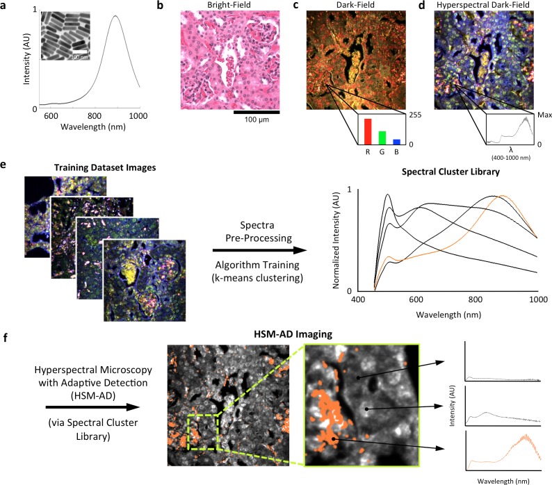
LGNRs (~100 × 30 nm) exhibiting a near infrared plasmonic peak (Figure 1a) were synthesized, biofunctionalized, and administered to healthy and tumor-bearing nude (Foxn1nu/nu) mice as previously reported (SoRelle et al., 2015; Liba et al., 2016). Mice were euthanized 24 hr post-injection, and various tissues were resected and fixed in 10% formalin. Fixed tissues were sectioned into 5 µm thick slices, mounted on glass slides, and stained with Hematoxylin and Eosin (H&E) as per standard histological preparation (Figure 1b). H&E-stained sections were imaged at 40x or 100x magnification in conventional dark-field and hyperspectral microscopy modes (CytoViva) (Figure 1—figure supplement 1). Conventional dark-field images (Figure 1c) were used to guide anatomical feature identification. All spectral data and quantitative comparisons presented in this report were derived from the analysis of hyperspectral images.

Figure 1. Overview of nanoparticle biodistribution analysis with HSM-AD.

(a) Large gold nanorods (LGNRs, ~100 × 30 nm) exhibiting near infrared plasmon resonance were synthesized, functionalized, and intravenously injected into live nude mice. (b,c) 24 hr post-injection, the animals were euthanized and tissues were resected and prepared as normal histological sections for characterization with bright-field (b) and dark-field microscopy (c) neither of which was able to visualize the distribution of the LGNRs. (d) The same section was then imaged with hyperspectral microscopy, which showed clear signs of LGNRs accumulation (denoted by red hues) in various areas of the tissue and exhibited spectral peaks matching the LGNR plasmon resonance. (e) We then trained an adaptive clustering algorithm for spectral identification of LGNRs with hyperspectral images from injected mice. The algorithm identified several characteristic spectra representing the tissue and the H&E staining, as well as one unique spectrum representing the LGNRs (depicted in orange), altogether representing a library of 5 spectra. Once a spectral cluster library is produced from the training dataset, images of unknown tissue samples can by analyzed for the presence of LGNRs via automated classification. (f) The resulting HSM-AD images depict the location of all points within the sample that exhibit the LGNR spectrum (orange for LGNRs, grayscale for tissue).

DOI: http://dx.doi.org/10.7554/eLife.16352.003

Figure 1.

Figure 1—figure supplement 1. Diagram of the CytoViva microscope used for dark-field and hyperspectral image acquisition.
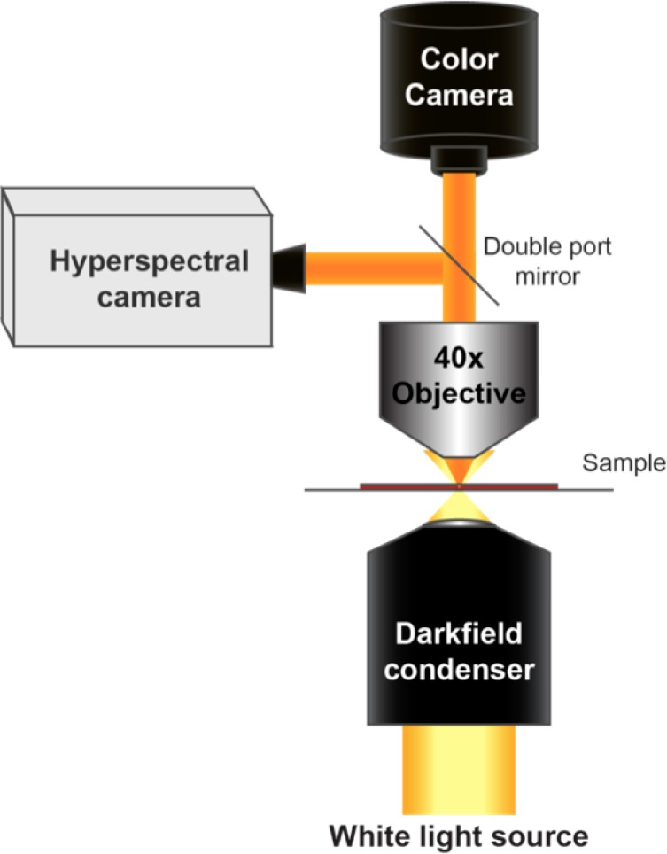
Figure 1—figure supplement 1.

Figure 1—figure supplement 2. Image segmentation, including method for dynamic threshold determination.

Figure 1—figure supplement 2.

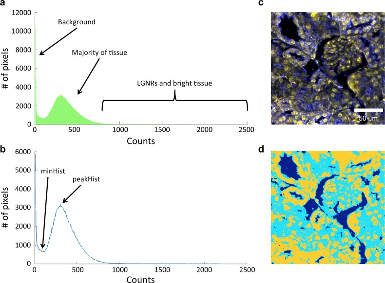
(a) A histogram of the peak intensities of each pixel in an image can be roughly divided into background (noise), tissue scattering, and LGNR and bright tissue scattering. (b) Detection of minHist and peakHist, as described in Methods. (c,d) A characteristic hyperspectral image (c) and its corresponding segmentation map (d) showing background (blue), tissue (cyan), and potential LGNRs and bright tissue (yellow).
Figure 1—figure supplement 3. Detailed flowchart of steps used in HSM-AD algorithm.

Figure 1—figure supplement 3.

Figure 1—figure supplement 4. Typical cluster results for pixel classification in an image of tissues with injected LGNRs.

Figure 1—figure supplement 4.

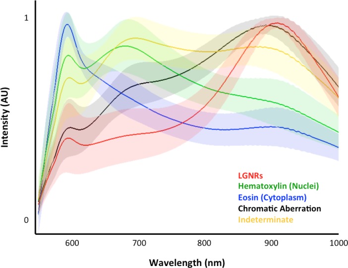
For a given image (>250,000 pixels), each pixel is binned into one of the five spectral clusters. This plot depicts the means (solid lines) and standard deviations (shaded areas) of all classified pixel spectra. Although the adaptive clustering algorithm is agnostic with respect to defining the spectral clusters (with the exception of chromatic aberration, which is user-defined), the learned clusters can be readily correlated to the major scattering components present in each sample, i.e., hematoxylin-stained nuclei (green), eosin-stained cytoplasm (blue), and LGNRs (red).

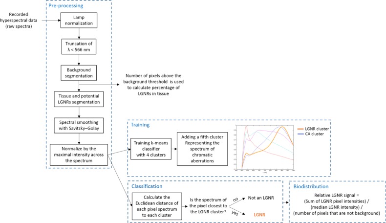
A hyperspectral camera with a detection range of 400–1000 nm was used to image scattered light from each sample in transmission mode. In the resulting images (Figure 1d), each pixel contains the spectral profile of the sample at the corresponding spatial position and can be used to detect LGNRs with near diffraction-limited resolution (1 μm). While standard dark-field images did not reveal notable differences between uninjected and injected samples, the hyperspectral images, in which three bands of the spectrum (800.0 nm, 700.6 nm, and 526.2 nm) were respectively color-coded as red, green, and blue, indicated that species with strong near infrared scattering (putative LGNRs, depicted in orange) were present in injected tissues but not observable in control tissues. We created HSM-AD, a method that combines pre-processing and adaptive classification algorithms to automatically detect and quantify LGNRs in hyperspectral images. The pre-processing stage is described in detail in the methods section (Figure 1—figure supplements 2,3). One of the first stages of processing includes vignette correction and a determination of whether each pixel in the image belongs to one of three categories—background, tissue, or potential LGNR—based on its average intensity across the measured spectrum. Only high-intensity pixels that belong to the potential LGNR group are classified by the adaptive algorithm (Figure 1—figure supplement 2). For the training of the adaptive algorithm, pre-processed images of tissue samples from LGNR injected mice were input into a standard k-means clustering algorithm (Bishop, 1995). Four initial clusters were identified using this scheme and were used to produce a spectral cluster library (Figure 1e). Owing to the unique spectral profile of LGNR scattering, the particle spectrum was automatically recognized as a separate cluster by the k-means algorithm. Next, a fifth cluster was manually added to the spectral cluster library to account for edge artifacts caused by chromatic aberrations that were frequently falsely detected as LGNRs (Figure 1—figure supplement 3). The five spectra in this library were used as cluster centers for automatic detection of LGNRs in tissue sections using a nearest centroid (or nearest-neighbor) classifier. In this scheme, pixels that exhibited a spectrum that was closest (in a Euclidean sense) to the LGNR cluster center were identified as containing LGNRs (denoted as LGNR+). The rest of the pixels were classified as not containing LGNRs (denoted as LGNR-). Initial validation of the algorithm shows that regions of the image that were detected as LGNR+ indeed exhibited the characteristic plasmonic peak at around 900 nm while pixels identified as LGNR- did not (Figure 1f). The mean and standard deviation spectra of pixels classified into each cluster for a representative image also indicate the high fidelity of the algorithm (Figure 1—figure supplement 4).

Characterization of sensitivity and specificity

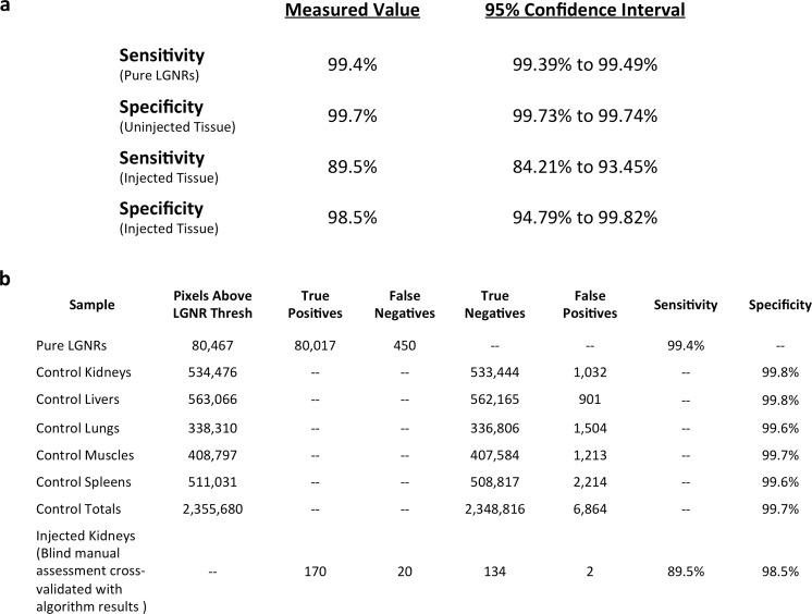
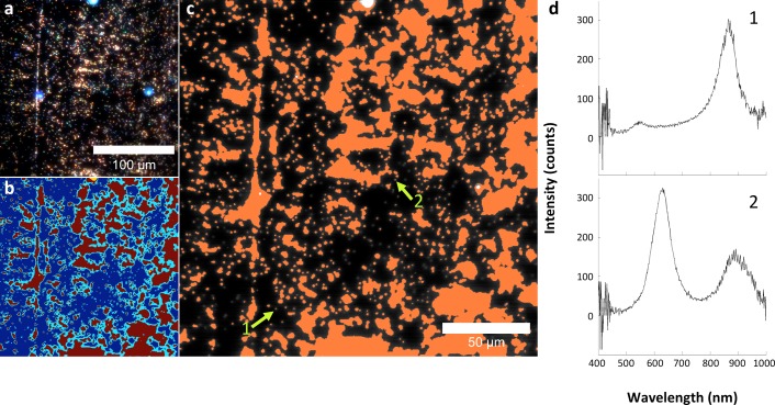
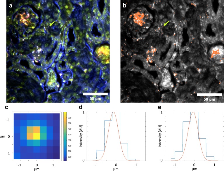
We characterized the sensitivity and specificity of HSM-AD by three methods. First, we measured the false positive rate in uninjected tissue samples to obtain a specificity of 99.7% (Figure 2—figure supplements 14). The false positives, which also have a spectral peak near 900 nm, usually appear near the edges of the tissue section. We attribute this red-shift of the spectrum to chromatic aberrations, ostensibly due to the spectral dependence of the diffraction diameter (Lipson and Lipson, 2010). Next, we measured the false negatives in an image of LGNRs in mounting media (CytoSeal 60, Electron Microscopy Sciences) on a glass slide and obtained a detection sensitivity of 99.4%. We attributed the false negatives to LGNRs with hybridized surface plasmon resonances (Funston et al., 2009), which resulted in spectral scattering that was different from the distinct plasmonic resonance of single LGNRs (Figure 2—figure supplements 5,6). Because all training and test samples were mounted using the same media, spectral shifts due to local refractive environments did not contribute to false detection (Figure 2—figure supplement 7). Independently we also calculated specificity and sensitivity by analyzing LGNR-injected tissue samples (see Methods). We obtained a sensitivity of 89.5% and a specificity of 98.5% using this approach. The high sensitivity of the automated algorithm is further evident from its ability to detect single LGNRs, both on a glass slide and in injected tissue samples (Figure 2—figure supplements 5,8).

NP biodistribution study

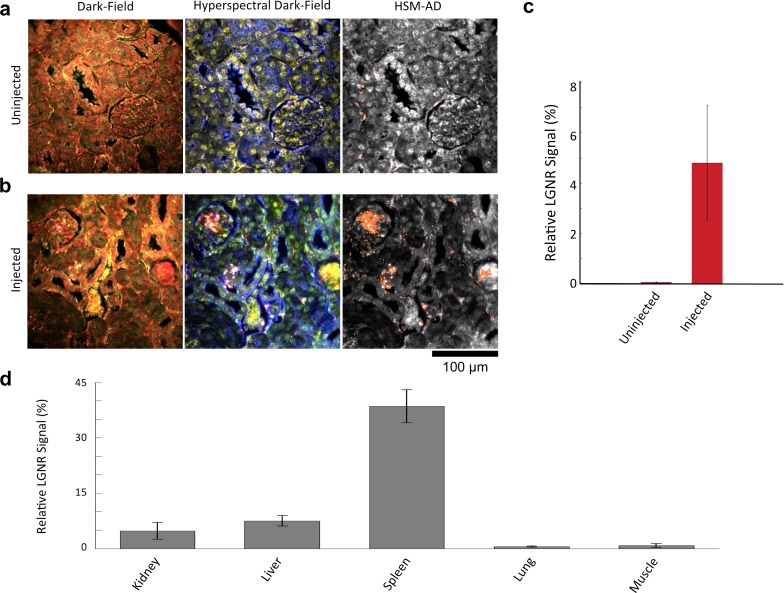
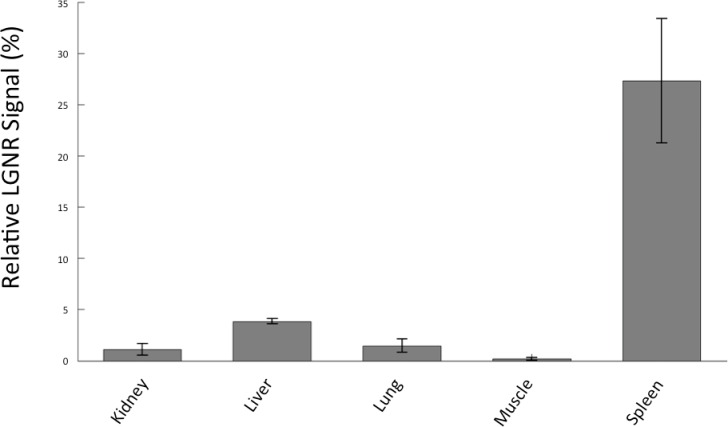
We demonstrated HSM-AD as a potential biodistribution technique by analyzing various tissues resected from mice (Figure 2). For quantitative measurements of LGNR uptake, we analyzed kidney tissue from uninjected (Figure 2a, Figure 2—figure supplements 2a,3a,4a) and injected (Figure 2b, Figure 2—figure supplements 9a,10a,11a) mice. Our analysis found a relative LGNR signal of 4.8% ± 2.3% in injected mouse kidney tissue. By comparison, a relative LGNR signal of 0.08% ± 0.01% was measured from uninjected samples, indicative of the method’s high specificity (Figure 2c, Figure 2—figure supplement 1). Similar low false positive rates were measured in other organ tissues (Figure 2—figure supplements 2b–e,3b–e,4b–e). In addition to the kidney, HSM-AD was used to analyze LGNR uptake in liver, lung, muscle, and spleen sections to demonstrate an alternative to common biodistribution techniques. While a conventional biodistribution study of LGNRs has not yet been reported, HSM-AD analysis indicated that LGNRs exhibited a similar uptake profile (mostly in the liver and spleen) as commonly-used smaller gold nanorods (Zhang et al., 2009; Niidome et al., 2006). The greatest relative LGNR signal (38.5% ± 4.5%) was observed in the spleen. LGNRs were also concentrated in the liver (7.5% ± 1.5%). Particle uptake was minimal in lung tissue (0.5% ± 0.1%) and muscle tissue (0.8% ± 0.5%) sections (Figure 2d, Figure 2—figure supplements 911). Tissue sections without H&E staining were also analyzed and yielded results similar to those obtained for H&E stained sections (Figure 2—figure supplements 1214).

Figure 2. Sensitivity and specificity validation of HSM-AD.

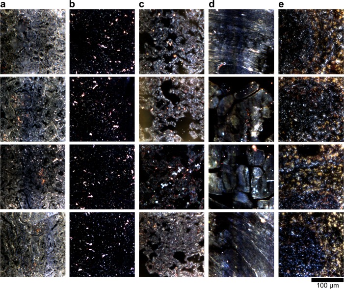
(a,b) Hematoxylin and Eosin (H&E) stained tissue samples (kidney) from uninjected (a) and injected (b) mice were imaged using dark-field and hyperspectral microscopy at 40x magnification and analyzed with HSM-AD to measure LGNR detection specificity and sensitivity. Conventional dark-field images highlight features including nuclei (salmon-pink), cytoplasm (green-brown), and erythrocytes (yellow-orange) within the tissues, but they reveal little information regarding the presence or absence of LGNRs. By comparison, putative LGNRs can be roughly identified as red-orange pixels in false-colored hyperspectral images, while nuclei and cytoplasm appear in green and indigo, respectively. (c) HSM-AD analysis of hyperspectral images demonstrates the absence of LGNRs in uninjected tissues and LGNR presence in injected samples (two-tailed Student’s t-test, p=0.054). Quantification of the relative LGNR signal from n = 4 tissue slices (representing a total of 1.04 million pixels) indicates that the false positive rate for LGNR detection (determined from uninjected tissues) is minimal. A detection specificity of 99.7% was determined from uninjected tissue sections, and a detection sensitivity of 99.4% was measured from samples of pure LGNRs analyzed using HSM-AD (Figure 2—figure supplement 1). (d) HSM-AD analysis of whole tissue sections (n = 4 for each tissue type) reveals quantitative differences in bulk LGNR uptake among various organs, in a manner analogous to conventional biodistribution methods. Quantitative data are presented as mean ± standard error of the mean (s.e.m.).

DOI: http://dx.doi.org/10.7554/eLife.16352.008

Figure 2—source data 1. Data used for diagnostic and 95% CIs.
DOI: 10.7554/eLife.16352.009
Figure 2—source data 2. Data for whole organ uptake quantification.
DOI: 10.7554/eLife.16352.010

Figure 2.

Figure 2—figure supplement 1. Measured sensitivity and specificity values for HSM-AD method.

Figure 2—figure supplement 1.

(a) Sensitivity values were calculated from pure LGNR samples in CytoSeal on a microscope slide and from blinded manual identification of LGNR spectra from injected sections cross-referenced with algorithm results. Specificity values were calculated directly from uninjected (LGNR-) tissue samples and from blinded manual identification of non-LGNR spectra cross-referenced with algorithm results. 95% confidence intervals were calculated for each sensitivity and specificity measurement. (b) Summary of raw data (pixel counts) for each measurement reported in (a).
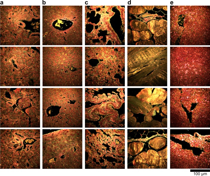
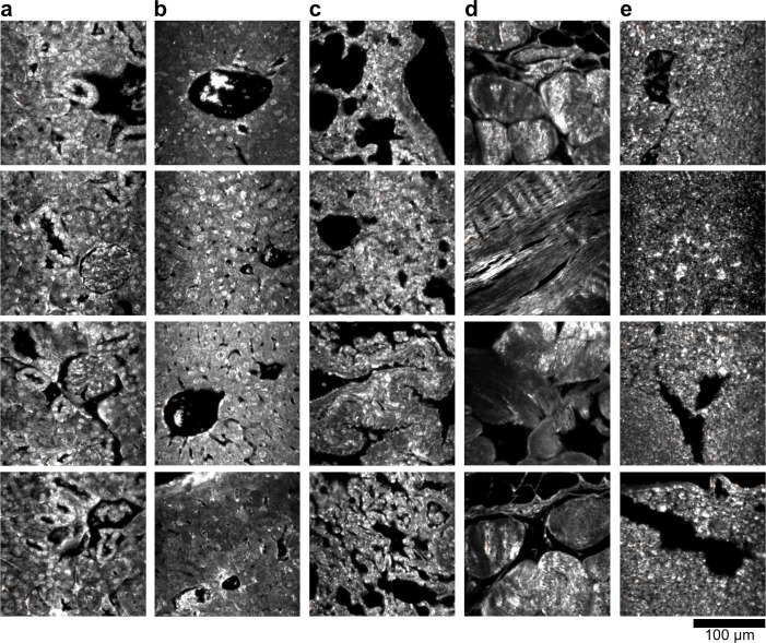
Figure 2—figure supplement 2. Dark-field images of additional uninjected H&E-stained tissue sections.

Figure 2—figure supplement 2.

(a) kidney, (b) liver, (c) lung, (d) muscle, and (e) spleen.
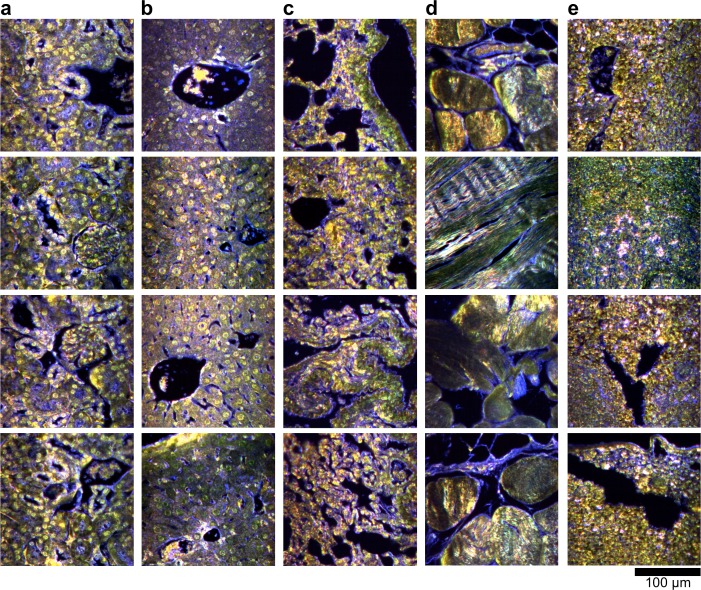
Figure 2—figure supplement 3. Hyperspectral images of additional uninjected H&E-stained tissue sections.

Figure 2—figure supplement 3.

(a) kidney, (b) liver, (c) lung, (d) muscle, and (e) spleen.
Figure 2—figure supplement 4. HSM-AD detection of additional uninjected H&E-stained tissue sections.

Figure 2—figure supplement 4.

(a) kidney, (b) liver, (c) lung, (d) muscle, and (e) spleen. Pixels identified as LGNR+ are denoted in orange. These analyzed samples were used for calculations of detection specificity.
Figure 2—figure supplement 5. Analysis of LGNRs on a glass slide.

Figure 2—figure supplement 5.

(a) Hyperspectral image of LGNRs embedded in CytoSeal on a glass slide. (b) a segmentation map showing background in blue & cyan, high-intensity LGNR- pixels in yellow, and LGNR+ pixels in red. (c) HSM-AD detection of the LGNR sample demonstrated high detection sensitivity. (d) Inspection of individual LGNR+ and LGNR- pixels validates the detection efficacy of HSM-AD. The split-peak spectrum of pixel 2, which is not identified as LGNR+, is possibly indicative of LGNR surface plasmon resonance hybridization.
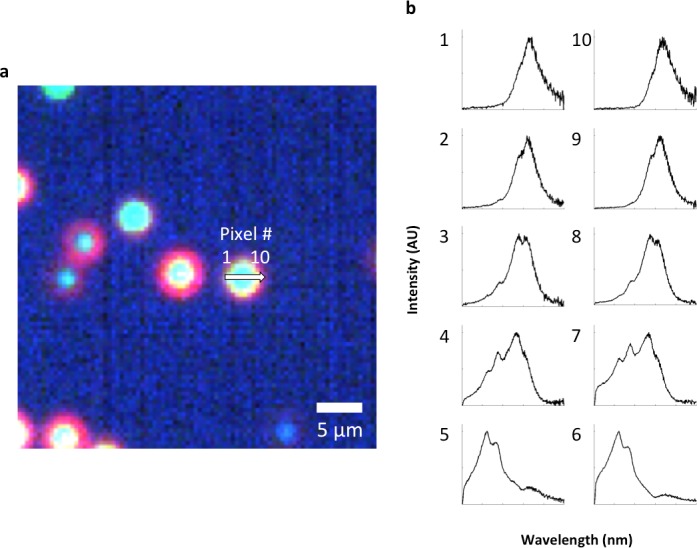
Figure 2—figure supplement 6. Spectral hybridization in partially aggregated LGNRs.

Figure 2—figure supplement 6.

(a) A sample of as-synthesized LGNRs (without additional surface functionalization) was centrifuged and resuspended to produce a sample with partial aggregation and imaged in water with the hyperspectral dark-field microscope. (b) Spectra from various distances from the aggregate center. Spectra from pixels near the edges of particle aggregates displayed scattering peaks similar to disperse LGNRs, while pixels closer to the centers of aggregates exhibited both blue-shifting and multiple spectral peaks.
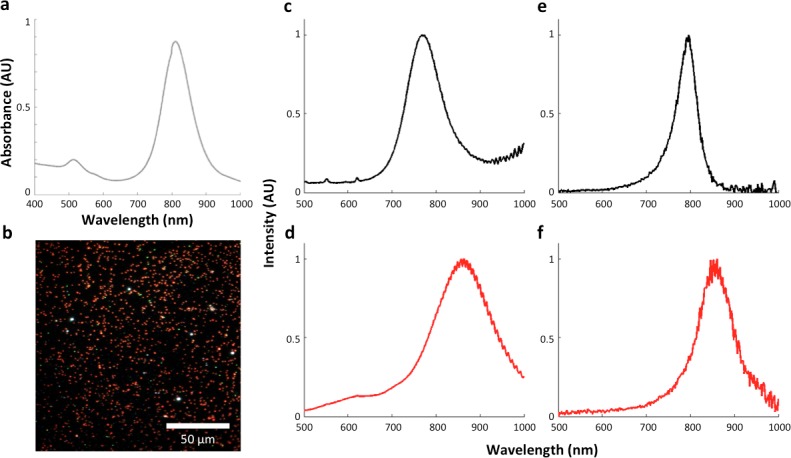
Figure 2—figure supplement 7. The influence of the local refractive index on the observed LGNR spectral peak.

Figure 2—figure supplement 7.

(a) LGNRs exhibited a spectral peak of ~800 nm when measured as a suspension in pure water with visible/near-infrared spectrometry. (b) Dark-field image of LGNRs in water. (c) The average spectrum of LGNRs in water has a peak that is slightly blue-shifted compared to the peak observed by spectrometry (a). (d) The average spectrum of LGNRs in CytoSeal (n = 1.5, matched to microscope immersion oil) produced a red-shift of ~80 nm in the average LGNR spectrum. (e) The spectrum of a single LGNR in water shows a similar peak to the average spectrum of LGNRs in water (c). (f) The spectrum of a single LGNR in CytoSeal shows a similar peak to the average spectrum of LGNRs in CytoSeal (d).
Figure 2—figure supplement 8. Evidence for single particle detection sensitivity with HSM-AD.

Figure 2—figure supplement 8.

(a, b) Hyperspectral dark-field image (a) and HSM-AD detection (b) of a tissue section which includes presumed single LGNRs, such as the point indicated by the green arrow. (c) A 2D plot of the scattering intensity around the LGNR intensity peak (~900 nm) of pixels in the vicinity of the LGNR+ pixel shown in (b). (d, e) 1D plots of normalized pixel intensity as a function of distance from the center pixel (blue traces) and theoretical intensity profiles of a point scatterer, calculated from a Gaussian point spread function (red traces). The measured intensity plots correlate well with the theoretical intensities in both vertical (d) and horizontal (e) directions. Along with the retention of the LGNR spectral peak, this result suggests that the identified location likely contains a single LGNR. If more than one LGNR were present in the same area of one pixel, the spectrum would possibly change due to plasmonic hybridization and the pixel would not be detected as LGNR+.
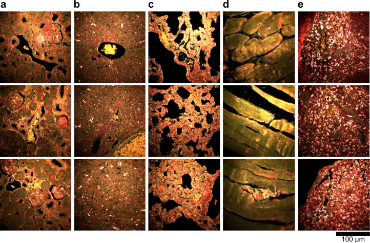
Figure 2—figure supplement 9. Dark-field images of additional LGNR-injected H&E-stained sections.

Figure 2—figure supplement 9.

(a) kidney, (b) liver, (c) lung, (d) muscle, and (e) spleen.
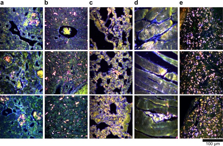
Figure 2—figure supplement 10. Hyperspectral images of additional LGNR-injected H&E-stained sections.

Figure 2—figure supplement 10.

(a) kidney, (b) liver, (c) lung, (d) muscle, and (e) spleen.
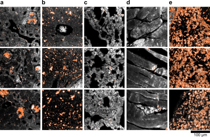
Figure 2—figure supplement 11. HSM-AD detection of additional LGNR-injected H&E-stained sections.

Figure 2—figure supplement 11.

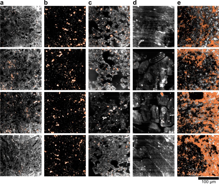
(a) kidney, (b) liver, (c) lung, (d) muscle, and (e) spleen. LGNR+ pixels are depicted in orange. Along with those in the main figures, these analyzed samples were used for calculations of detection sensitivity and specificity as well as whole-organ LGNR uptake.
Figure 2—figure supplement 12. Quantitative whole-organ biodistribution measured with HSM-AD on unstained tissue sections.

Figure 2—figure supplement 12.

All values are presented as the average relative LGNR signal (%) ± standard error of the mean (s.e.m) measured over four fields of view per organ. The results from unstained tissue sections are comparable to the results obtained for H&E stained sections.
Figure 2—figure supplement 13. Dark-field images of LGNR-injected unstained tissue sections.

Figure 2—figure supplement 13.

(a) kidney, (b) liver, (c) lung, (d) muscle, and (e) spleen.
Figure 2—figure supplement 14. HSM-AD detection of LGNR-injected unstained tissue sections.

Figure 2—figure supplement 14.

(a) kidney, (b) liver, (c) lung, (d) muscle, and (e) spleen. LGNR+ pixels are depicted in orange. These samples were used to calculate the values of LGNR uptake in unstained sections.
Figure 2—figure supplement 15. Spectral Angle Mapper (SAM) detection of LGNR+ kidney tissue.

Figure 2—figure supplement 15.

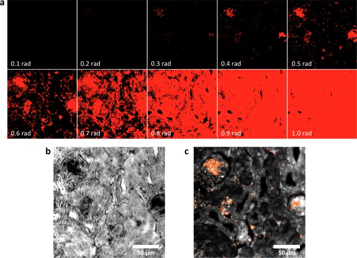
(a) Results of SAM classification of LGNR+ pixels (red masks) with various user-defined angular tolerance values (in radians, bottom-left of each panel). Selection of low angular tolerance results in poor detection sensitivity, while high tolerance leads to poor detection specificity. Along with other parameters, angular tolerance must be user-defined for each hyperspectral image. SAM classification was performed as described in reference 30. (b) Guide image corresponding to the SAM-classified masks in (a). (c) HSM-AD analysis of the same hyperspectral image. Diagnostic evaluation of this and related images yielded the sensitivity and specificity values reported in Figure 2—figure supplement 1. As noted in reference 21, such values are not readily extracted using SAM and related methods.

Sub-organ localization of LGNRs

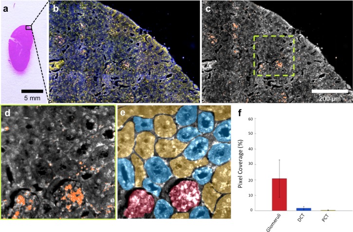
HSM-AD imaging of histological sections (Figure 3a) enabled sensitive LGNR detection with sub-cellular resolution over large fields of view (Figure 3b–c), which afforded more detailed characterizations of NP uptake than those achieved by typical biodistribution methods. We used these advantages to investigate and quantify the sub-organ distribution of LGNRs. This analysis revealed well-defined patterns of LGNR uptake that appeared to be largely influenced by factors including particle size, innate immunological function, and waste-filtering anatomical structures.

Figure 3. HSM-AD is capable of wide-field characterization of sub-organ distribution patterns of injected nanoparticles.

Figure 3.

(a–c) Millimeter-scale fields of view of histological sections of kidney. Photographed (a), acquired with near diffraction-limited resolution with a hyperspectral dark-field camera (b), and analyzed by HSM-AD (c) to reveal variable nanoparticle uptake within the fine anatomical structures. (d–f) As in conventional histology, micro-anatomical features of the kidney including glomeruli, Bowman’s spaces, proximal convoluted tubule (PCT), and distal convoluted tubule (DCT) networks can be clearly identified in HSM-AD images (d). The ability to distinguish such histological details enables region of interest (ROI) analysis to quantify sub-organ accumulation of LGNRs (e,f). Quantification of the relative LGNR signal in glomeruli (red ROI), PCT (yellow ROI), and DCT (blue ROI) regions (e) revealed that the vast majority (~13-fold greater than in either tubule network) of renal LGNR uptake is localized within glomeruli (f). This is likely due to the size-dependent inability of LGNRs to traverse the ultrafiltration barrier formed by endothelial cells within glomerular capillaries. All quantitative data are represented as mean ± s.e.m. for each ROI type, as calculated from 4 unique fields of view acquired at 40x magnification.

DOI: http://dx.doi.org/10.7554/eLife.16352.026

Figure 3—source data 1. Data for kidney sub-organ ROIs.
DOI: 10.7554/eLife.16352.027

The kidneys are responsible for filtering small, low molecular weight waste products from the bloodstream and diverting those products to the bladder for elimination (Rouiller, 2014). Waste-laden blood flows into capillary-dense structures called glomeruli within the kidney. Blood plasma containing small species including ions, biomolecules, cell fragments, and (in some cases) nanoparticles can extravasate from glomerular capillaries and traverse Bowman’s space before being collected into an extensive network of efferent tubes called Proximal Convoluted Tubules (PCT) and, further downstream, Distal Convoluted Tubules (DCT), which ultimately traffic waste from the kidney out to the bladder. Using our method, we observed that the vast majority of LGNRs within the kidney were concentrated in glomeruli and were virtually absent in the PCT and DCT, which are functionally downstream (Figure 3d–f). Glomerular uptake was 13-fold greater than uptake within the convoluted tubule network. These results can be explained by comparing the size of an individual LGNR with the narrow width of cellular junctions and the architecture of endothelial cells that form glomerular capillaries (Satchell and Braet, 2009). For reference, the sub-organ segmentation maps of all tissue images analyzed for quantification have been provided (Figure 4—figure supplement 1).

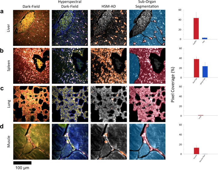
Along with the kidneys, the liver is instrumental in clearing waste from circulation. Partially because the size cutoff for hepatic filtration is notably larger than that of renal ultrafiltration (Longmire et al., 2008), we observed 1.6-fold greater LGNR uptake within the liver tissue compared to the kidney. Interestingly, the hepatic distribution of LGNRs also appeared to be non-uniform. Hepatocytes, which constitute the majority of liver tissue by mass, exhibited mild LGNR signal (2.9% ± 1.1%). By contrast, 15-fold more LGNR signal (43.5% ± 12.0%) in the liver was localized in a manner consistent with the shape, size, and number of Kupffer cells (Figure 4a, Figure 2—figure supplements 9b,10b,11b). We attribute this localization to the phagocytic function of Kupffer cells (Owens III and Peppas, 2006; Longmire et al., 2008). Thus, the variable localization of nanoparticles within the liver appears to be largely derived from the organ’s innate immunological functions. It is interesting to note that aggregation within Kupffer cells likely caused spectral hybridization of some LGNRs, an observation that is consistent with previous studies of cellular uptake of gold NPs (Chen, 2014). This spectral shifting, which was most prevalent at the centers of LGNR aggregates, caused a portion of LGNRs to remain undetected by HSM-AD. While these aggregates were undetected by algorithmic means, their manual identification as LGNRs was evident from the lack of similar morphological features in uninjected liver tissue.

Figure 4. HSM-AD reveals characteristic patterns of nanoparticle microbiodistribution contingent upon tissue function.

(a) LGNR accumulation in hepatic tissue occurs mostly in concentrated foci located within liver sinusoids. Along with the size, shape, and frequency of these foci, this pattern strongly suggests that these particles have been phagocytosed by Kupffer cells, the resident macrophages of the liver (red ROI). While there is mild uptake of LGNRs within liver hepatocytes (blue ROI), HSM-AD sub-organ quantification indicates that uptake by Kupffer cells is roughly 15-fold higher than hepatocytic accumulation. (b) The pattern of LGNR uptake in the spleen is also consistent with the physiological functions of various splenic tissues. A greater relative LGNR signal was observed in regions of splenic red pulp (red ROI), which is responsible for blood filtration, than in the white pulp follicles (blue ROI) that house B and T lymphocytes. (c,d) While LGNRs were prevalent within the liver and spleen tissues, HSM-AD results indicated minimal particle accumulation within the lung (c) or muscle (d) tissue samples (each < 1% relative LGNR signal for whole-tissue quantification). Interestingly, HSM-AD analysis demonstrated that the vast majority of LGNRs in muscle tissue sections were localized in blood vessels (red ROI) rather than within the muscle fiber tissue itself (blue ROI). Quantitative data are represented as mean ± s.e.m. as described previously.

DOI: http://dx.doi.org/10.7554/eLife.16352.028

Figure 4—source data 1. Data for liver, spleen, lung, and muscle sub-organ ROIs.
DOI: 10.7554/eLife.16352.029

Figure 4.

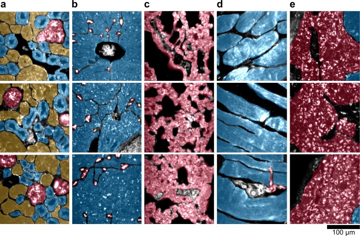
Figure 4—figure supplement 1. Sub-organ region of interest (ROI) segmentation for additional tissue sections used for quantitative results presented in Figures 3 and 4 of the main text.

Figure 4—figure supplement 1.

The ROI color schemes for each sub-organ feature are identical to those listed in the legends of the relevant figures in the main text. (a) kidney, (b) liver, (c) lung, (d) muscle, and (e) spleen.
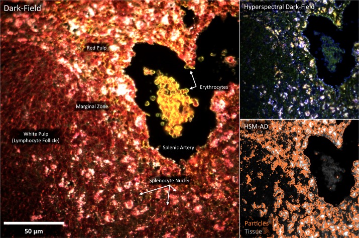
Figure 4—figure supplement 2. Detail of Figure 4b: spleen tissue histology correlated with LGNR uptake.

Figure 4—figure supplement 2.

Using HSM-AD, it is possible to cross-reference nanoparticle uptake patterns and tissue microstructures with sub-cellular resolution.

The spleen comprises several unique cell types arranged into tissues with diverse biological functions including blood filtration, innate immunity, and lymphocyte activation (Mebius and Kraal, 2005). The spleen is largely composed of red pulp, white pulp, and the boundary between these two tissues, commonly referred to as the marginal zone. Consistent with each tissue’s biological function, we observed 1.7-fold greater relative LGNR pixel coverage in splenic red pulp than white pulp (Figure 4b, Figure 2—figure supplements 9e,10e,11e). A similar result has been previously reported for carbon-based nanomaterials (Chen et al., 2015). We did not definitively identify marginal zone tissue, but the radial distribution of LGNRs around white pulp follicles indicated that a significant portion of white pulp uptake may in fact be within the marginal zone (Figure 4—figure supplement 2).

Despite its dense network of alveolar capillaries, lung tissue exhibited minimal LGNR accumulation relative to the organs described above (Figure 4c, Figure 2—figure supplement 9c,10c,11c). This finding was consistent with existing biodistribution data for smaller particles, which can be explained by the lungs’ major functions of gas exchange to and from the blood rather than biomolecule or particle filtration and clearance.

While whole-organ analysis indicated the presence of LGNRs in muscle tissue, HSM-AD revealed that muscle tissue itself (which consists largely of myocytes and dense networks of extracellular collagen) was virtually devoid of LGNRs (Figure 4d, Figure 2—figure supplement 9d,10d,11d). Rather, the apparently high LGNR presence was traced to blood vessels found in between muscle fiber bundles. As with the accumulation patterns described for other organs within this study, this distinction would not have been possible through conventional biodistribution methods.

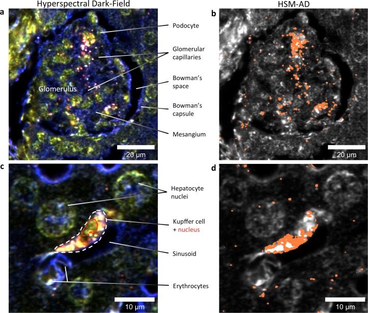
HSM-AD images acquired at higher objective magnification (100x) offered further insights into the cellular nature of LGNR uptake within the kidney and liver tissue (Figure 5). Within the kidney, LGNRs were observed mostly within or in close proximity to glomerular capillaries (Figure 5a,b). HSM-AD also revealed patterns of LGNR uptake within individual Kupffer cells resident in liver sinusoids (Figure 5c,d). LGNR signal was detected within the Kupffer cell cytoplasm, but not within the region of the cell nucleus. This pattern is consistent with the phagocytic function of Kupffer cells in clearing particulate matter from circulation. Interestingly, several bright regions within the Kupffer cell were not detected as LGNRs. We expect that these regions resulted from spectral hybridization of LGNRs, possibly due to aggregation induced by lysosomal acidification following particle phagocytosis.

Figure 5. HSM-AD reveals the sub-cellular localization of intravenously administered nanoparticles with histological precision.

Figure 5.

(a,b) Hyperspectral (a) and HSM-AD (b) images of a renal glomerulus acquired at 100x magnification. A majority of LGNRS are found within or in close proximity to glomerular capillaries. Trace levels of LGNRs are observed in the kidney tissue outside of Bowman's capsule. (c,d) Zoomed views of Hyperspectral (c) and HSM-AD (d) images of liver tissue acquired at 100x magnification. Several erythrocytes and a Kupffer cell (dashed white line) can be observed residing within a liver sinusoidal vessel. Within the Kupffer cell, the nucleus (dashed red line) can be distinguished. HSM-AD analysis indicated the prevalence of LGNRs within the Kupffer cell relative to surrounding hepatocytes. The minimal LGNR signal was detected in the region identified as the nucleus, consistent with cytoplasmic LGNR localization. Several bright regions within the cell were not identified as LGNRs; these regions likely result from particle aggregation within acidic lysosomes following uptake by the Kupffer cell.

DOI: http://dx.doi.org/10.7554/eLife.16352.032

HSM-AD detection and spectral unmixing of Nanoshells

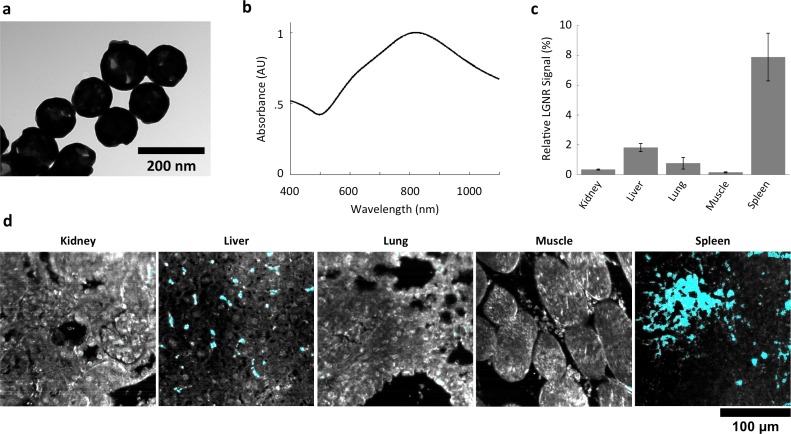
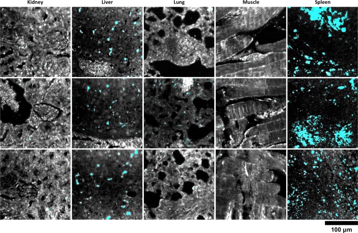
We also injected mice with Nanoshells, which are morphologically distinct from LGNRs (Figure 6a). While Nanoshells and LGNRs both exhibit near-infrared plasmonic peaks, the Nanoshell spectrum is substantially broader than the LGNR spectrum (Figure 6b). A spectral cluster library was developed for H&E-stained Nanoshell+ tissues and was then used to quantify Nanoshell uptake as described for LGNRs (Figure 6c). While Nanoshells and LGNR displayed related uptake patterns, several differences including negligible Nanoshell uptake in kidney tissue and Nanoshell concentration within the splenic white pulp were observed (Figure 6d, Figure 6—figure supplements 1,2, Nanoshell+ pixels are shown in cyan).

Figure 6. Characterization of gold nanoshell uptake after intravenous administration.

(a,b) Nanoshells (119 nm silica core with 14 nm-thick gold coating) exhibit distinct particle morphology and composition (a) that (like LGNRs) yield a near-infrared (~800 nm) spectral peak (b). However, the Nanoshell spectrum is markedly broader than the resonance observed for LGNRs. (c,d) HSM-AD revealed that Nanoshell uptake displays inter-organ distribution patterns somewhat similar to those observed for LGNRs, with maximal accumulation in the spleen (c). Values are represented as mean ± s.e.m. from four FOVs per tissue. However, there are notable distinctions including minimal Nanoshell uptake within kidney tissue and concentration of Nanoshells within splenic white pulp (d) (Nanoshell+ pixels are depicted in cyan).

DOI: http://dx.doi.org/10.7554/eLife.16352.033

Figure 6—source data 1. Data for Nanoshell uptake in organs.
DOI: 10.7554/eLife.16352.034

Figure 6.

Figure 6—figure supplement 1. Additional HSM-AD images of Nanoshell uptake used for quantification.

Figure 6—figure supplement 1.

Quantitative data from these FOVs were used to produce the bar graph in Figure 6c.
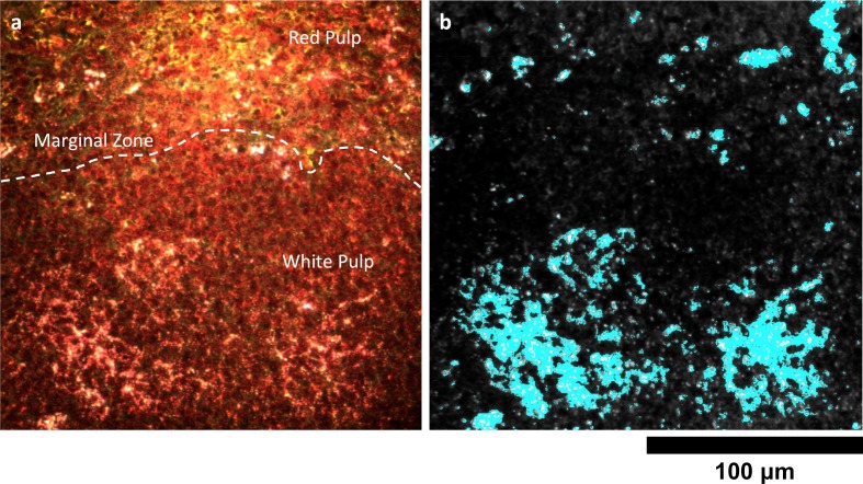
Figure 6—figure supplement 2. Detail of Nanoshell uptake in spleen tissue.

Figure 6—figure supplement 2.

(a) Conventional dark-field image of H&E-stained spleen tissue. The dashed white line approximately demarcates the marginal zone separating the red and white pulp. (b) HSM-AD indicates that, unlike LGNRs, Nanoshells are localized within white pulp follicles 24 hr after intravenous injection.
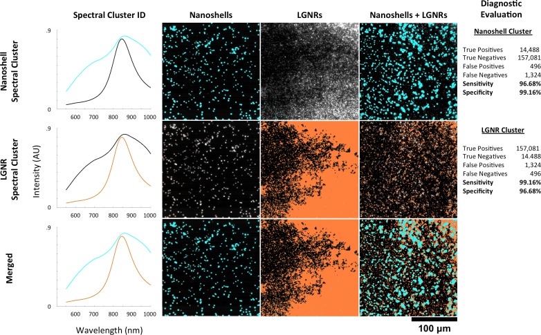
Figure 6—figure supplement 3. HSM-AD spectral unmixing of samples containing gold nanoshells and LGNRs.

Figure 6—figure supplement 3.

HSM-AD was trained on a hyperspectral image of a sample containing a mixture of Nanoshells and LGNRs. Using a target of two clusters, this training yielded one spectral cluster corresponding to the spectrum of the Nanoshells and another spectral cluster corresponding to LGNRs (far-left column). These clusters were then used to map images of Nanoshells + LGNRs, Nanoshells only, and LGNRs only. The presence of Nanoshells and LGNRs are marked using cyan and orange masks, respectively. HSM-AD classification using the Nanoshell cluster (top row) for all three sample types achieved 96.68% sensitivity and 99.16% specificity. HSM-AD classification using the LGNR cluster (middle row) for the samples achieved 99.16% specificity and 96.68% specificity. The bottom row depicts the merge of these two cluster maps. As a note, the reciprocal nature of the sensitivity and specificity values for the two different particle types results from the fact that, for pure particle solutions, the false positives for one particle type are false negatives for the other particle type and vice versa. The same reciprocal nature holds for true positives and true negatives as well. This relationship can be seen in the raw pixel counts used for diagnostic evaluation (far-right column).

HSM-AD was separately trained on a sample consisting of a mixture of pure Nanoshells and pure LGNRs. The spectral clusters identified during this training corresponded well to the spectra of each particle type and enabled high-specificity and high-sensitivity identification in samples of Nanoshells + LGNRs, Nanoshells-only, and LGNRs-only (Figure 6—figure supplement 3). These results demonstrate that HSM-AD can spectrally resolve plasmonic particles despite similarities in composition, although this capability was not tested in ex vivo tissues.

GNS@SiO2 detection in tissue

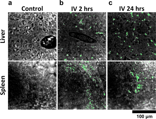
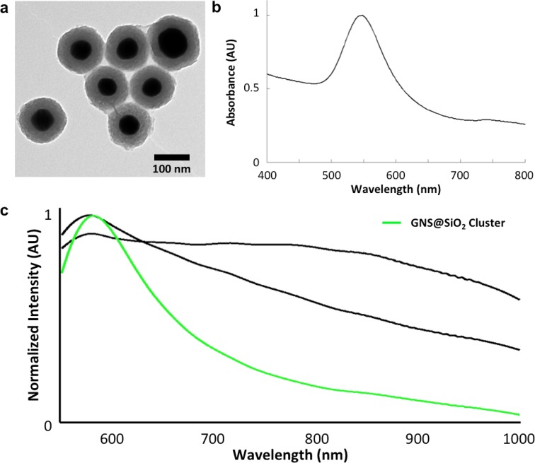
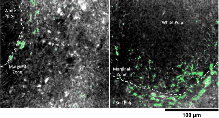
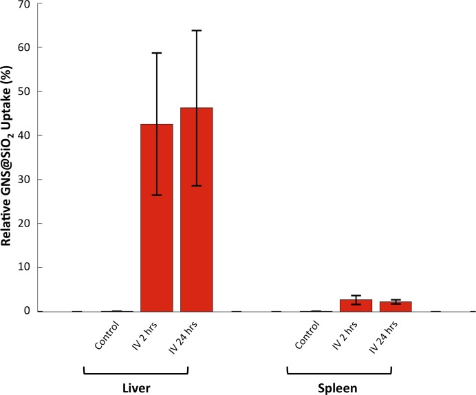
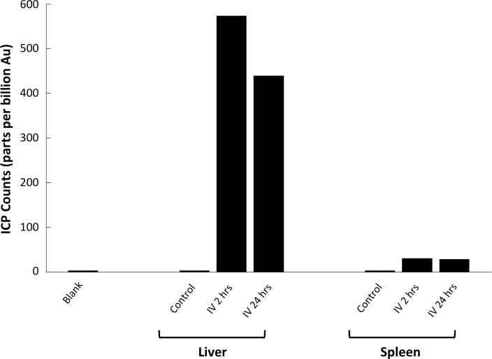
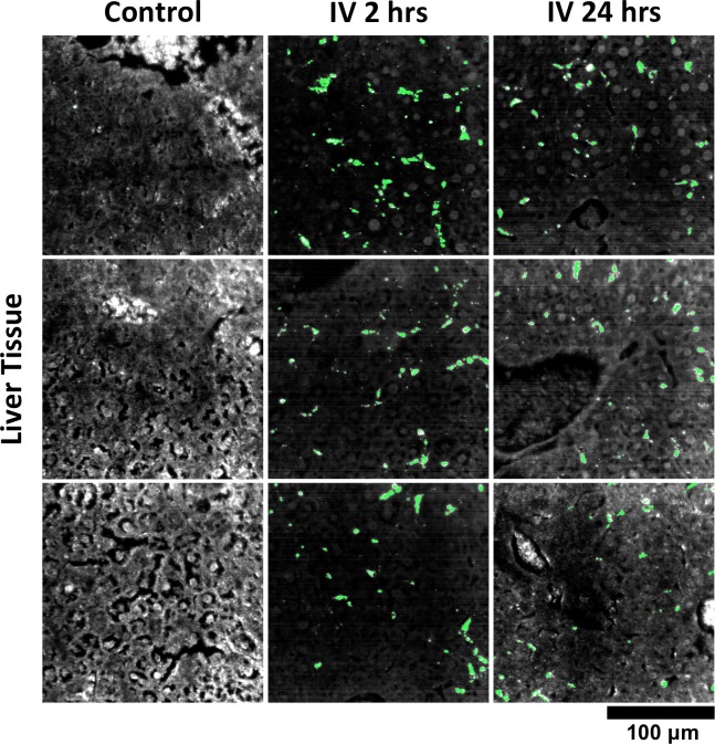
We used HSM-AD to characterize the tissue uptake of a third particle type, GNS@SiO2 (Figure 7, GNS@SiO2+ pixels are shown in green). Notably, GNS@SiO2 are distinct from LGNRs and Nanoshells in terms of shape, size, composition, particle surface, and plasmonic resonance (Figure 7—figure supplement 1a,b). Because GNS@SiO2 exhibit a visible regime plasmonic peak (~550 nm), HSM-AD analysis was performed on unstained tissue sections. First, a spectral cluster library was developed for GNS@SiO2 classification as described for LGNRs (Figure 7—figure supplement 1c). Control tissues classified with this library displayed negligible false positives (Figure 7a). GNS@SiO2 uptake in the liver and spleen was observed at 2 and 24 hr post-IV injection (Figure 7b,c). Interestingly, GNS@SiO2 uptake appeared to be even more localized to Kupffer cells than LGNR accumulation in the liver. Furthermore, GNS@SiO2 in the spleen are consistently found in the marginal zone, and presence within the red pulp and white pulp is minimal (Figure 7—figure supplement 2). Quantitative results from HSM-AD correlate well with those obtained using ICP-MS (Figure 7—figure supplements 3,4), although it should be noted that HSM-AD measurements are more relative rather than absolute with respect to the amount of gold present in each tissue. As for LGNR quantification, four FOVs per sample were analyzed (Figure 7—figure supplements 5,6).

Figure 7. HSM-AD analysis of GNS@SiO2.

(a) Classification of unstained control tissues yields negligible false-positive detection for GNS@SiO2 (green), which exhibit a peak plasmonic resonance of ~550 nm. (b,c) Intravenously-administered GNS@SiO2 accumulate in the Kupffer cells of the liver and the marginal zone of the spleen within 2 hr (b) and persist up to 24 hr (c).

DOI: http://dx.doi.org/10.7554/eLife.16352.038

Figure 7—source data 1. Data for GNS@SiO2 uptake in organs.
DOI: 10.7554/eLife.16352.039

Figure 7.

Figure 7—figure supplement 1. Structural and spectral characterization of GNS@SiO2.

Figure 7—figure supplement 1.

(a) TEM of GNS@SiO2. (b) Vis-NIR absorbance spectrum of GNS@SiO2 in water (SPR ~550 nm). (c) Spectral library clusters identified from training on images of unstained tissues (in CytoSeal) resected from mice injected with GNS@SiO2. The target number of clusters was set to three rather than five (used for LGNR detection) due to the absence of H&E staining. Cluster 2 (green) corresponds to the GNS@SiO2 and exhibits a red-shifted plasmonic peak relative to particles in water, as expected due to the difference in refractive environment.
Figure 7—figure supplement 2. Detail of GNS@SiO2 in spleen.

Figure 7—figure supplement 2.

HSM-AD reveals that GNS@SiO2 accumulate mostly in the marginal zone tissue between red and white pulp. The approximate boundaries between red pulp and white pulp follicles are marked by dashed white lines.
Figure 7—figure supplement 3. HSM-AD quantification of GNS@SiO2 uptake in liver and spleen tissue.

Figure 7—figure supplement 3.

Relative GNS@SiO2 uptake was measured using the positive ratio approach described in the methods section. All values represent the mean GNS@SiO2 uptake from 4 FOVs for each injection/tissue combination. Error bars represent standard error of the mean (s.e.m.).
Figure 7—figure supplement 4. Inductively-Coupled Plasma Mass Spectrometry (ICP-MS) quantification of GNS@SiO2 uptake in liver and spleen tissue.

Figure 7—figure supplement 4.

Quantitative measurements of atomic gold present in tissue samples prepared through microwave digestion. Counts are given in parts per billion (ppb) of Au.
Figure 7—figure supplement 5. Additional HSM-AD images of GNS@SiO2 uptake in liver tissue.

Figure 7—figure supplement 5.

Quantitative data from these FOVs were used to produce the bar graph in Figure 7—figure supplement 3.
Figure 7—figure supplement 6. Additional HSM-AD images of GNS@SiO2 uptake in spleen tissue.

Figure 7—figure supplement 6.

Quantitative data from these FOVs were used to produce the bar graph in Figure 7—figure supplement 3.

Tumor uptake of targeted and untargeted NPs

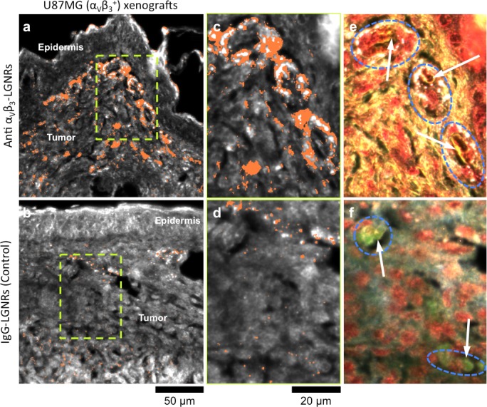
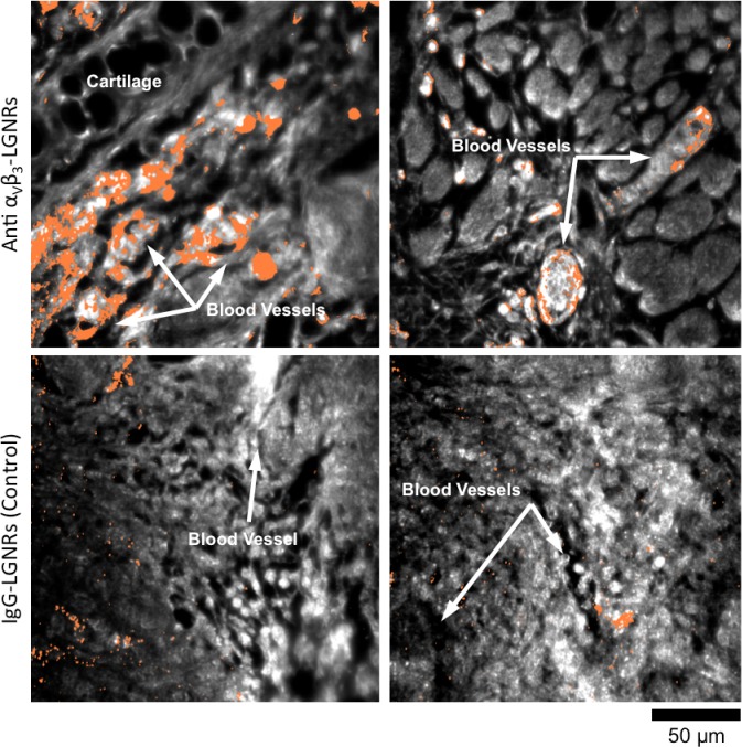
One hallmark of tumor growth is angiogenesis, the stimulated development of new blood vessels to provide nutrients to rapidly dividing cancer cells. This newly-formed vasculature is composed of endothelial cells that express high levels of cell adhesion receptors including αVβ3 integrin (Avraamides et al., 2008). Thus, αVβ3 is commonly used as a target biomolecule for tumor imaging (Sipkins et al., 1998). Such studies have demonstrated that NPs targeted to αVβ3 exhibit greater accumulation in tumors in vivo than NPs coated with non-specific antibodies or small molecules. We hypothesized that the presence or absence of specific molecular targeting moieties would influence tissue-NP interactions beyond simply the degree of accumulation in target tissues. To test this, we used HSM-AD to observe the spatial patterns of targeted and non-targeted LGNR uptake within U87MG (human glioblastoma cells, αVβ3+) tumor xenografts. We observed 7.4-fold greater relative LGNR signal of anti-αVβ3 LGNRs than isotype LGNRs in tumor tissue (Figure 8a–d). However, the most striking differences were in the localization patterns of each LGNR type. Anti-αVβ3 LGNRs were present in high density around the edges of small blood vessels within the tumor while isotype LGNRs showed no such association (Figure 8c–f, Figure 8—figure supplement 1). The prevalence of anti-αVβ3 LGNRs around the edges of tumor capillaries is highly consistent with the expression pattern of αVβ3 in angiogenic vessels. Moreover, isotype LGNRs found outside of the vasculature were notably dispersed compared to extravascular anti-αVβ3 LGNRs, which often appeared in small clusters. While NPs are known to accumulate in tumors regardless of molecular specificity due to leaky vasculature, these results indicated that the enhanced extravascular accumulation of anti-αVβ3 LGNRs may have originated from specific binding of αVβ3 integrins present on the U87MG cells themselves.

Figure 8. Active molecular functionalization affects nanoparticle uptake quantitatively and spatially within target tissues.

(a,b) HSM-AD images of sub-dermal U87MG tumor xenografts from mice injected with LGNRs display distinct accumulation patterns depending on the molecular specificity of the LGNR surface coating. Anti-αVβ3 LGNRs exhibit 7.5-fold greater accumulation in tumor tissue (a) than spectrally-identical LGNRs with non-specific IgG antibody coating (b) (n = 4 FOVs for Anti-αVβ3 LGNRs, n = 5 FOVs for IgG-LGNRs,two-tailed Student’s t-test, p=0.0041). The greater uptake of anti-αVβ3 LGNRs may result in part from specific LGNR binding to αVβ3 integrin, which is over-expressed by U87MG cells. (c–f) Validation of HSM-AD images with dark-field images of slightly higher spatial resolution further indicates that a substantial portion of anti-αVβ3 LGNRs are located along the edges of small capillaries within the tumor tissue (c,e) while no such association is observed for IgG-LGNRs (d,f). This observation is consistent with the nature of angiogenic tumor vasculature, which is also characterized by high expression levels of αVβ3 integrin in the vascular endothelium. Individual erythrocytes within angiogenic capillaries are denoted by white arrows, and capillary edges are approximately outlined by dashed blue ovals (e,f). Discrete regions of anti-αVβ3 LGNRs were also observed outside of the tumor vasculature, presumably due to either (1) specific binding to αVβ3integrin expressed by U87MG cells and/or (2) non-specific accumulation via the enhanced permeability and retention (EPR) effect characteristic of tumors. The absence of IgG-LGNR extravascular accumulation suggests the former of these mechanisms as the predominant source of anti-αVβ3 LGNR uptake in tumor tissue.

DOI: http://dx.doi.org/10.7554/eLife.16352.046

Figure 8—source data 1. Data for tumor uptake of targeted and untargeted LGNRs.
DOI: 10.7554/eLife.16352.047

Figure 8.

Figure 8—figure supplement 1. HSM-AD images of additional tumor tissue sections resected after targeted and untargeted LGNR injections.

Figure 8—figure supplement 1.

LGNR uptake in U87MG tumors is consistently greater and more localized to blood vessel endothelial cells (αVβ3+) when particles are conjugated with anti-αVβ3 antibodies (top row) rather than nonspecific IgG antibodies (bottom row).

Discussion

The necessity of sample digestion with strong acids for ICP quantification effectively reduces an entire organ (a remarkably rich dataset by any measure) down to a single number representative of bulk NP accumulation. While the quantification offered by ICP is certainly valuable, it provides minimal insight into the patterns and mechanisms of NP uptake within individual cells or tissues. Unlike ICP methods, HSM-AD provides additional dimensions of anatomical detail at optical resolution to facilitate better understanding of the biology behind quantitative measurements of NP uptake.

The primary solution for dealing with the limitations of ICP has been to use EM, which provides excellent spatial resolution (at the nanometer scale) and particle sensitivity (down to individual nanoparticles). However, EM can only scan minimal fields of view—a typical transmission EM (TEM) image for studying NP uptake covers ~1 × 1 µm. For comparison, TEM scanning of the same area depicted in Figure 3c would require ~460,000 TEM images, which is infeasible for single tissue studies and virtually unrealistic for multiple-organ studies. The necessity of thin samples (~10 nm) for TEM imaging compared to samples analyzed using HSM-AD (~1 µm optical focus) would further multiply the number of TEM scans (>46 million) required for equivalent volumetric imaging. Other biodistribution techniques based on radioactivity (Kreyling et al., 2015; Collingridge et al., 2003), photoacoustic (Poon et al., 2015), and fluorescence (He et al., 2010) detection have been used previously as alternatives to ICP and TEM. By comparison, HSM-AD offers roughly 100-fold higher spatial resolution (~1 µm vs ~100 µm) than current fluorescence and photoacoustic biodistribution methods. Fluorescence-based methods may also suffer from high false positive detection arising from tissue autofluorescence, as has been observed for renal capsule tissue (Poon et al., 2015). While HSM-AD was excellently suited for exploring the sub-organ localization of NPs, it has been observed that radiolabeling approaches may be poorly-equipped for accurately determining particle distribution within organs (Kreyling et al., 2015). Moreover, single-particle detection sensitivity was not demonstrated by any of these alternatives to ICP and TEM.

Biodistribution methods based on imaging mass spectrometry were recently demonstrated to enable sub-organ quantification of carbon nanomaterials (Chen et al., 2015). This impressive approach can obtain images of full mouse tissue sections (cm in scale), but the limited spatial resolution (50 µm) precludes the study of NP uptake within individual cells. Because detection relies upon particle fragmentation and ionization, it is unclear whether imaging mass spectrometry can achieve single particle sensitivity—calculations based upon the reported data indicate that ~10 (von Maltzahn et al., 2009) particles per pixel are required for detection. However, the cited spatial resolution negates many of the potential advantages of single-particle sensitivity such as direct observation of NP uptake by cells through endocytosis or adhesion to the cell membrane. More generally, while mass spectrometry provides an approach to biodistribution studies of certain materials, its use for identifying gold NPs has been constrained to NPs smaller than 10 nm and typically requires the inclusion of 'mass barcode' molecules as capping agents (such as alkanethiols) on the NP surface (Zhu et al., 2008; Harkness et al., 2010). Incidentally, gold NPs have previously been demonstrated as assisting matrices to improve mass spectrometry detection of biomolecules (Su and Tseng, 2007; Huang and Chang, 2007). Notably, many metallic NPs are compositionally similar yet spectrally distinct (for example, gold nanospheres, nanorods, nanoshells, etc.), which may confound results in mass spectrometry-based analysis of samples containing more than one NP species. We have demonstrated that HSM-AD can successfully identify gold nanoparticles with different spectra, shape, size, and composition in tissues. Furthermore, Nanoshells and LGNRs were discernible from each other in particle mixtures. While not directly tested in this work, HSM-AD may thus be capable of distinguishing such NPs from each other in tissues, enabling biodistribution studies of multiplexed NPs. Thus, we expect that HSM-AD will extend the advantages of high sensitivity and resolution to the analysis of a variety of metallic NPs with unique spectral properties.

As reported in previous studies (Chen, 2014; Okamoto et al., 2000), we observed that the scattering spectrum from gold NPs is heavily influenced by the local refractive index (n). Spectral shifts of ~80 nm were evident between preparations of LGNRs in water (n = 1) relative to the same particles prepared in CytoSeal (n = 1.5) (Figure 2—figure supplement 7), and similar shifts were observed for Nanoshells and GNS@SiO2. Thus, in order to generate reliable spectral cluster libraries, it is critical to train the adaptive algorithm using images prepared in similar fashion to the samples being studied. The orientation of anisotropic NPs within a sample has also been shown to influence the observed spectrum (Biswas et al., 2012). Because LGNRs are anisotropic, variation in particle orientation may affect detection sensitivity in our case, but this effect appears to be negligible in light of empirically measured sensitivity values. Because it relies on spectral identification of NPs, HSM-AD cannot detect NPs that have shifted their spectrum markedly, such as concentrated NP aggregation within cells. Such spectral hybridization is most prominent in Kupffer cells within the liver, which likely results in an artificially low measure of LGNR uptake in that organ. However, the absence of these bright aggregates in hyperspectral images of uninjected control tissues confirms their identity as LGNRs. Because of plasmon hybridization, LGNR aggregates produce shifted spectra that can resemble the scattering from H&E-stained tissue, which can impede automated detection. Thus, future efforts to detect such aggregates should rely on the analysis of unstained tissues.

We tested alternate machine learning approaches including support vector machine (SVM) and logistic regression for nanoparticle detection. We found that unsupervised k-means outperformed these other methods when trained on images of tissues containing nanoparticles (Liba and Shaviv, 2014). Further advantages of unsupervised k-means include its ease of use and no need to pre-label samples for analysis. While HSM-AD based on k-means clustering provides a robust general platform for nanoparticle detection, it is conceivable that certain studies may benefit from learning methods tailored to address specific applications.

HSM-AD imaging simultaneously achieves excellent sensitivity and specificity for detecting NPs in tissues with sub-cellular resolution. In addition to improved diagnostic capabilities, the automated and adaptive features of HSM-AD enable standardized high-throughput analysis previously absent from biomedical HSM studies (Roth et al., 2015). Unlike Spectral Angle Mapping (the current gold standard for HSM image analysis), HSM-AD does not require the manual steps typically needed to create target spectral libraries, define particle intensity and size thresholds, filter false positives from libraries, and calibrate angular tolerance for accurate classification on an individual image basis (Figure 2—figure supplement 15) (Roth et al., 2015). Along with an ability to image millimeter-scale fields of view on reasonable timescales (<30 min) and simple sample preparation, these properties make HSM-AD a favorable alternative to existing methods for characterizing NP biodistribution. Beyond biodistribution, this work demonstrates that HSM-AD can be used for post-injection validation of NP localization in target tissues as a function of surface modifications. Because HSM-AD is non-destructive, samples can be further analyzed by a variety of conventional microscopy techniques including immunohistochemistry to provide additional molecular detail. Collectively, the results presented herein indicate that HSM-AD provides a new approach for studying interactions of cells and whole tissues with spectrally unique NPs commonly used in biomedical imaging and therapeutic studies.

Materials and methods

LGNR preparation

LGNRs were synthesized using methods adapted from Ye et al (Ye et al., 2013). LGNRs were characterized using Transmission Electron Microscopy (TEM), visible/near-infrared spectrometry, and dark-field hyperspectral microscopy. As-synthesized LGNRs were prepared for biological use by removing excess CTAB from solution and coating the particles with poly(sodium 4-styrenesulfonate) (PSS, MW 70 kDa) as previously reported (SoRelle et al., 2015). PSS-coated LGNRs were then conjugated with IgG isotype antibody (clone eB149/10H5, eBioscience) for use in sub-organ biodistribution experiments. We also prepared LGNRs targeted to αVβ3 integrin (a cell-surface receptor that is overexpressed in angiogenic vasculature within tumors) by conjugating LGNRs with anti-αVβ3 antibody (clone 23C6, eBioscience).

Animal experiments and sample preparation

Healthy female nude (Foxn1nu/nu) mice (6–8 weeks old, Charles River Labs) were anesthetized with 2% isoflurane by inhalation and intravenously injected with 250 µL of IgG isotype-coated LGNRs at optical density (OD) 470. In separate experiments, mice bearing U87MG tumors in the right ear pinna were injected with either IgG isotype-coated LGNRs or anti-αVβ3-coated LGNRs. Additional details of these experimental protocols can be found in the literature (SoRelle et al., 2015; Liba et al., 2016). In nanoshell experiments, nude mice were injected with 200 µL of OD 50 (2.5 mg/mL) nanoshells composed of 119 nm silica cores and 14 nm-thick gold shells with PEG coating (Nano Composix, San Diego, CA). In GNS@SiO2 experiments, healthy female Balb/C mice were anesthetized as described previously and injected intravenously with 150 µL of 0.8 nM GNS@SiO2 particles composed of 60 nm gold cores and 30 nm-thick SiO2 shells (Oxonica, Mountain View, CA). For all experiments, mice were euthanized 24 hr (or 2 hr, for GNS@SiO2) post-injection, and tissues including kidney, liver, lung, spleen, thigh muscle, and (when applicable) tumor were immediately resected and preserved in 10% formalin. Tissues were also resected from uninjected mice for control imaging experiments. These tissues were subsequently embedded in paraffin and sectioned into 5 µm thick samples. Sections were prepared with and without H&E stains and mounted on microscope slides using CytoSeal 60 (Electron Microscopy Sciences) as the mounting medium. All animal experiments were performed in compliance with IACUC guidelines and with the Stanford University Animal Studies Committee’s Guidelines for the Care and Use of Research Animals. Experimental protocols (APLAC #s 27499 and 29179) were approved by Stanford University’s Animal Studies Committee.

Imaging system

All tissue samples were imaged with a modified dark-field microscopy setup as shown in Figure 1—figure supplement 1. Light from a broadband halogen lamp was coupled via an optical fiber into a custom dark-field condenser (CytoViva, Auburn, AL), which produced a light cone for sample illumination. Light scattered from the sample was collected using either a 40x magnification dark-field air objective lens (Olympus UPlanFLN 40x, 0.75 NA) or a 100x magnification oil immersion objective lens (Olympus UPlanFLN 100x, 1.3 NA) and directed to one of two cameras depending on detection mode. Conventional dark-field and hyperspectral images were collected for all samples in this study. Conventional dark-field images were collected with a Dagexcel-M cooled camera (Dage-MTI, Michigan City, IN). Hyperspectral images were collected with a hyperspectral camera (iXon3, Andor, Belfast, UK). Each image has 509 × 512 pixels. With a 40x lens, the sampling resolution is 410 × 408 nm, which produces a 209 × 209 µm field of view. With a 100x lens, the sampling resolution is 163 × 160 nm. Only Figure 5 shows images acquired at 100x magnification. The spectrum from each pixel was acquired with 361 uniform samples at wavelengths ranging from 400 nm to 1000 nm. The acquired raw spectra of each pixel were lamp-normalized using the Cytoviva software package (ENVI 4.8) and exported after normalization.

Data processing and automatic biodistribution detection

Processing of the hyperspectral images was done with Matlab (Mathworks, Natick, MA). Hyperspectral images were created by color-coding the spectrum by integrating over three bands. The band centers were 800.0 nm, 700.6 nm and 526.2 nm for the red, green and blue channels, respectively. The integration was weighted by a Gaussian window with a width of 80 spectrum samples. Each channel was scaled separately for optimal viewing.

Automatic detection of NPs required preprocessing the spectra prior to training and classification. First, due to noise at lower wavelengths, the spectra were truncated to disregard values below a cutoff of 566 nm. As part of HSM-AD, we initially segmented the image into background, tissue, or potential NPs based on each pixel’s average intensity across its spectrum. This segmentation allowed a more accurate calculation of the biodistribution by measuring the number of pixels that correspond to tissue inside the field of view. The segmentation of potential NPs helped to avoid classifying low intensity edges that may be falsely detected as NPs due to chromatic aberrations. Before measuring the intensity of each pixel, we also applied a correction for vignetting. Vignetting is a common artifact in photography and microscopy in which image brightness is reduced at the periphery compared to the image center. We assumed a natural illumination fall-off that follows the 'cosine fourth' law, in which the light fall-off is proportional to the fourth power of the cosine of the angle at which the light impinges on the sensor. We measured the radial falloff of several images and found that it can be approximated by cos4(θ), in which θ=tan1(R/d), where R is the calculated distance from the center of the image and d was found by fitting to be 2 mm. In order to correct the vignetting, we divided the intensities of each field of view by cos4(θ). Next, we calculated the segmentation thresholds adaptively for each image (to account for different exposure times and variations in tissue scattering). The thresholds were obtained by analyzing the histogram of pixel intensities of each image (Figure 1—figure supplement 2). The histogram was calculated with 510 bins and then re-sampled (using interpolation) every 5 intensity units. Pixels with the lowest intensities were segmented as background. The threshold for segmenting the background was calculated as the first minimum of the histogram (minHist) multiplied by a user-defined parameter (which is slightly larger than 1) to allow fine tuning of the background threshold. Next, we assumed that pixels representing tissue without NPs have a relatively consistent intensity lower than that of NPs and therefore correspond to a peak in the histogram. The threshold for differentiating between pixels that correspond to tissue and those that can be potential NPs can be determined by the peak of the histogram (peakHist) multiplied by a pre-defined parameter (larger than 1) which allows tuning of the threshold. This intensity-based segmentation was confirmed to be effective by comparing results over 20 separate fields of view from all analyzed tissue types. Pixels segmented as potential NPs were preprocessed by smoothing their spectra using a Savitzky-Golay algorithm (Orfanidis, 1995) implemented by a Matlab’s built-in function. Next, we normalized the spectrum of each pixel by the maximal intensity across its spectrum. Training and classification were performed only on pixels which were segmented as potential NPs.

Training of the k-means algorithm (Bishop, 1995) was initially done with 3, 4, and 5 clusters on 6 images of injected tissue sections, from which ~500,000 pixels were binned into the potential LGNR group. The 4 clusters found by the k-means algorithm matched the expected spectra of the injected and stained tissue. One of the spectra automatically matched the spectrum of LGNRs, owing to their distinct spectrum compared to tissue and staining dyes, two of the spectra represent the Hematoxylin and Eosin stains, and the fourth is an intermediate cluster representing the sum of both stains (see Figure 1—figure supplement 4). Testing the algorithm on uninjected tissue samples showed false detections of LGNRs near the edges of the tissue. We attributed these false positives to spectral red-shifting caused by chromatic aberrations due to the larger point spread function of longer wavelengths compared to the smaller point spread function of shorter wavelengths. To minimize the number of these false positives, we manually added a cluster representing the spectrum of the chromatic aberrations by averaging the spectra of falsely detected pixels from uninjected samples. Indeed, this cluster showed a spectral peak near the resonance of the LGNRs, albeit much broader. The initial clusters found by k-means with the added cluster representing chromatic aberrations were used for classification of pixels as LGNR+ or LGNR- with a nearest centroid (or nearest neighbor) classifier, based on the Euclidean distance to the cluster centers. We then measured the sensitivity and specificity and also qualitatively assessed the results on injected tissue section using the algorithm with different numbers of clusters. We chose to use the cluster library which includes 5 clusters (4 obtained through k-means and the 5th added manually) because it produced the best results and clusters that were more consistent with the actual spectra present in the samples. Several other machine learning algorithms were also explored for this purpose, including support vector machine (SVM) and logistic regression, but k-means yielded better results (Liba and Shaviv, 2014). The classification results are presented as detection maps in which the average intensity at each pixel is displayed in grayscale and the pixels that are LGNR+ are shown in orange. Similar training and classification steps were performed for samples containing GNS@SiO2 (3 clusters used) and Nanoshells (5 clusters for images of ex vivo tissues, 2 clusters for Nanoshells + LGNRs).

We characterized the sensitivity and specificity of HSM-AD by three methods. First, we measured the false positive and true negative rates in uninjected tissue samples to obtain the specificity. Note that false positives and true negative rates can be measured reliably from tissue-only samples due to the absence of LGNRs. Next, we measured the false negatives and true positives in an image of pure LGNRs in mounting media on a glass slide and obtained the detection sensitivity. Note that false negatives and true positives can be measured reliably from LGNR-only samples due to the absence of tissue and scattering media other than the particles themselves. In order to calculate the sensitivity and specificity for detecting LGNRs in tissue samples, we created a 'ground truth' data set by randomly choosing > 200 pixels, half of which were detected by the algorithm to contain LGNRs. We then manually (and in a manner blind to the results from the algorithmic classification) determined whether pixels were LGNR+ or LGNR- based purely on observing their raw spectra and looking for the unique plasmonic peak of LGNRs. Low-intensity pixels that were considered as the background or tissue were not considered in this calculation. By comparing the ground truth to the results of the automated algorithm we obtained the number of true positives, true negatives, false positives and false negatives, and used them to calculate additional measures of sensitivity and specificity. Confidence intervals for these measurements, presented in Figure 2—figure supplement 1, were calculated using the 'log method' (Altman et al., 2000) with the aid of a statistical calculator (MedCalc Software, 2016).

The biodistribution in each field of view was measured as the relative LGNR signal in an image. This measurement takes into account the amount of tissue versus background in a frame and also the signal of the detected LGNRs. For this calculation, we refer to LGNR signal as the average intensity around the plasmonic peak of the LGNRs (833–988 nm). The relative LGNR signal is the sum of the LGNR signal over LGNR+ pixels divided by the number of tissue pixels (i.e., the number of all pixels minus the number of background pixels) and divided by the median LGNR signal over the LGNR+ pixels in the field of view. For whole-organ analysis, we measured the relative LGNR signal in four fields of view for each organ (taken from the same mouse) and calculated the mean and standard deviation for each organ. For the sub-organ calculation, the measurement of relative LGNR signal yielded results with substantial variability due to a small number of pixels and high variability of intensity within several regions of interest. Therefore, a simpler pixel ratio (termed pixel coverage) was calculated by dividing the number of LGNR+ pixels by the number of tissue pixels for each region of interest (ROI). ROI maps are presented for each field of view in Figure 4 as well as for the additional images used for quantification (Figure 4—figure supplement 1). The same calculations also apply for quantification of other particle types.

Evidence of single particle detection

In order to determine whether HSM-AD is able to detect single LGNRs, we compared the theoretical point spread function of the microscope’s 40x lens with the shape of the increased intensity caused by an isolated LGNR+ pixel. The diffraction-limited point spread function of the microscope can be approximated by a Gaussian with a standard deviation of 0.25 μm (based on a numerical aperture of 0.75 and wavelength of 910 nm) (Lipson and Lipson, 2010), which may increase in the case of defocusing. The cross section of intensity in the wavelengths around the plasmonic peak of the LGNRs (833–988 nm) showed a close resemblance to the theoretical spot size (Figure 2—figure supplement 8). This analysis supports the capability to detect single LGNRs in tissue samples.

Acknowledgements

This work was funded in part by grants from the Claire Giannini Fund, the United States Air Force (FA9550-15-1-0007), the National Institutes of Health (NIH DP50D012179), the National Science Foundation (NSF 1438340), the Damon Runyon Cancer Research Center (DFS# 06-13), the Susan G Komen Breast Cancer Foundation (SAC15-00003), the Mary Kay Foundation (017-14), the Donald E and Delia B Baxter Foundation, the Skippy Frank Foundation, a seed grant from the Center for Cancer Nanotechnology Excellence and Translation (CCNE-T U54CA151459), and a Stanford Bio-X Interdisciplinary Initiative Seed Grant. AdlZ is a Pew-Stewart Scholar for Cancer Research supported by The Pew Charitable Trusts and The Alexander and Margaret Stewart Trust. EDS wishes to acknowledge funding from the Stanford Biophysics Program training grant (T32 GM-08294). OL is grateful for a Stanford Bowes Bio-X Graduate Fellowship. CLZ is supported by the National Cancer Institute of the National Institutes of Health under award numbers K22 CA160834 and R21 CA184608. JLC acknowledges funding from the Victorian government of Australia in the form of a Victorian Postdoctoral Research Fellowship. We would like to thank Debasish Sen, Byron Cheatham, Jamie Uertz, and Michelle Rincon for technical assistance. We wish to thank Dor Shaviv for help with testing alternative detection methods.

Funding Statement

The funders had no role in study design, data collection and interpretation, or the decision to submit the work for publication.

Funding Information

This paper was supported by the following grants:

  • Stanford University Biophysics Program, T32 GM-08294 to Elliott D SoRelle.

  • Stanford University Bowes Bio-X Graduate Fellowship to Orly Liba.

  • Victorian Government of Australia Postdoctoral Research Fellowship to Cristina L Zavaleta.

  • National Cancer Institute K22 CA160834 to Cristina L Zavaleta.

  • National Cancer Institute R21 CA184608 to Adam de la Zerda.

  • Claire Giannini Fund to Adam de la Zerda.

  • U.S. Air Force FA9550-15-1-0007 to Adam de la Zerda.

  • National Institutes of Health NIH DP50D012179 to Adam de la Zerda.

  • Damon Runyon Cancer Research Foundation DFS# 06-13 to Adam de la Zerda.

  • Susan G. Komen Breast Cancer Foundation, SAC15-00003 to Adam de la Zerda.

  • Mary Kay Foundation 017-14 to Adam de la Zerda.

  • Donald E. and Delia B. Baxter Foundation to Adam de la Zerda.

  • Skippy Frank Foundation to Adam de la Zerda.

  • Center for Cancer Nanotechnology Excellence and Translation CCNE-T U54CA151459 to Adam de la Zerda.

  • Stanford Bio-X Interdisciplinary Initiative Seed Grant to Adam de la Zerda.

  • National Science Foundation NSF 1438340 to Adam de la Zerda.

Additional information

Competing interests

The authors declare that no competing interests exist.

Author contributions

EDS, Designed the study, Performed all experiments, Acquisition of data, Analyzed the data, Wrote the paper, Approved the final version of this work.

OL, Designed the study, Performed all experiments, Acquisition of data, Analyzed the data, Wrote the paper, Approved the final version of this work.

JLC, Performed experiments, Acquisition of data, Analysis and interpretation of data, Approved the final version of this work.

RD, Prepared all histological sections, Conception and design, Acquisition of data, Approved the final version of this work.

CLZ, Performed experiments, Acquisition of data, Analysis and interpretation of data, Approved the final version of this work.

AdlZ, Designed the study, Analysis and interpretation of data, Wrote the paper, Approved the final version of this work.

Ethics

Animal experimentation: All animal experiments in this study were performed in compliance with IACUC guidelines and with the Stanford University Animal Studies Committee's Guidelines for the Care and Use of Research Animals (APLAC Protocol #27499 and #29179).

Additional files

Source code 1. Contains all MATLAB code used for HSM-AD.

DOI: http://dx.doi.org/10.7554/eLife.16352.049

elife-16352-code1.zip (46.4KB, zip)
DOI: 10.7554/eLife.16352.049

References

  1. Altman DG, Machin D, Bryant TN, Gardner MJ. Statistics with Confidence. BMJ Books; 2000. [Google Scholar]
  2. Avraamides CJ, Garmy-Susini B, Varner JA. Integrins in angiogenesis and lymphangiogenesis. Nature Reviews Cancer. 2008;8:604–617. doi: 10.1038/nrc2353. [DOI] [PMC free article] [PubMed] [Google Scholar]
  3. Badireddy AR, Wiesner MR, Liu J. Detection, characterization, and abundance of engineered nanoparticles in complex waters by hyperspectral imagery with enhanced Darkfield microscopy. Environmental Science & Technology. 2012;46:10081–10088. doi: 10.1021/es204140s. [DOI] [PubMed] [Google Scholar]
  4. Bishop CM. Neural Networks for Pattern Recognition. Clarendon Press; 1995. [Google Scholar]
  5. Biswas S, Nepal D, Park K, Vaia RA. Orientation sensing with color using plasmonic gold nanorods and assemblies. Journal of Physical Chemistry Letters. 2012;3:2568–2574. doi: 10.1021/jz3009908. [DOI] [PubMed] [Google Scholar]
  6. Chen AL, Hu YS, Jackson MA, Lin AY, Young JK, Langsner RJ, Drezek RA. Quantifying spectral changes experienced by plasmonic nanoparticles in a cellular environment to inform biomedical nanoparticle design. Nanoscale Research Letters. 2014;9:454. doi: 10.1186/1556-276X-9-454. [DOI] [PMC free article] [PubMed] [Google Scholar]
  7. Chen S, Xiong C, Liu H, Wan Q, Hou J, He Q, Badu-Tawiah A, Nie Z. Mass spectrometry imaging reveals the sub-organ distribution of carbon nanomaterials. Nature Nanotechnology. 2015;10:176–182. doi: 10.1038/nnano.2014.282. [DOI] [PubMed] [Google Scholar]
  8. Chen YS, Hung YC, Liau I, Huang GS. Assessment of the In vivo toxicity of gold nanoparticles. Nanoscale Research Letters. 2009;4:858–864. doi: 10.1007/s11671-009-9334-6. [DOI] [PMC free article] [PubMed] [Google Scholar]
  9. Collingridge DR, Glaser M, Osman S, Barthel H, Hutchinson OC, Luthra SK, Brady F, Bouchier-Hayes L, Martin SJ, Workman P, Price P, Aboagye EO. In vitro selectivity, in vivo biodistribution and tumour uptake of annexin V radiolabelled with a positron emitting radioisotope. British Journal of Cancer. 2003;89:1327–1333. doi: 10.1038/sj.bjc.6601262. [DOI] [PMC free article] [PubMed] [Google Scholar]
  10. De Carvalho OA, Meneses PR. Summaries of the 9th JPL Airborne EarthScience Workshop. JPL Publication; 1999. Spectral correlation mapper (SCM): An improvement on the spectral angle mapper (SAM) p. 9. [Google Scholar]
  11. Decuzzi P, Godin B, Tanaka T, Lee SY, Chiappini C, Liu X, Ferrari M. Size and shape effects in the biodistribution of intravascularly injected particles. Journal of Controlled Release. 2010;141:320–327. doi: 10.1016/j.jconrel.2009.10.014. [DOI] [PubMed] [Google Scholar]
  12. Durr NJ, Larson T, Smith DK, Korgel BA, Sokolov K, Ben-Yakar A. Two-photon luminescence imaging of cancer cells using molecularly targeted gold nanorods. Nano Letters. 2007;7:941–945. doi: 10.1021/nl062962v. [DOI] [PMC free article] [PubMed] [Google Scholar]
  13. Fairbairn N, Christofidou A, Kanaras AG, Newman TA, Muskens OL. Hyperspectral darkfield microscopy of single hollow gold nanoparticles for biomedical applications. Physical Chemistry Chemical Physics. 2013;15:4163–4168. doi: 10.1039/C2CP43162A. [DOI] [PubMed] [Google Scholar]
  14. Fairbairn N, Fernandes R, Carter R, Elliot TJ, Kanaras AG, Muskens OL. Single-nanoparticle detection and spectroscopy in cells using a hyperspectral darkfield imaging technique. Proceedings of SPIE 8595; 2013. [DOI] [Google Scholar]
  15. Funston AM, Novo C, Davis TJ, Mulvaney P. Plasmon coupling of gold nanorods at short distances and in different geometries. Nano Letters. 2009;9:1651–1658. doi: 10.1021/nl900034v. [DOI] [PubMed] [Google Scholar]
  16. Giustini AJ, Ivkov R, Hoopes PJ. Magnetic nanoparticle biodistribution following intratumoral administration. Nanotechnology. 2011;22:e16352. doi: 10.1088/0957-4484/22/34/345101. [DOI] [PMC free article] [PubMed] [Google Scholar]
  17. Harkness KM, Cliffel DE, McLean JA. Characterization of thiolate-protected gold nanoparticles by mass spectrometry. The Analyst. 2010;135:868–874. doi: 10.1039/b922291j. [DOI] [PMC free article] [PubMed] [Google Scholar]
  18. Hauck TS, Jennings TL, Yatsenko T, Kumaradas JC, Chan WCW. Enhancing the toxicity of cancer chemotherapeutics with gold nanorod hyperthermia. Advanced Materials. 2008;20:3832–3838. doi: 10.1002/adma.200800921. [DOI] [Google Scholar]
  19. He C, Hu Y, Yin L, Tang C, Yin C. Effects of particle size and surface charge on cellular uptake and biodistribution of polymeric nanoparticles. Biomaterials. 2010;31:3657–3666. doi: 10.1016/j.biomaterials.2010.01.065. [DOI] [PubMed] [Google Scholar]
  20. Huang HC, Barua S, Kay DB, Rege K. Simultaneous enhancement of photothermal stability and gene delivery efficacy of gold nanorods using polyelectrolytes. ACS Nano. 2009;3:2941–2952. doi: 10.1021/nn900947a. [DOI] [PMC free article] [PubMed] [Google Scholar]
  21. Huang X, El-Sayed IH, Qian W, El-Sayed MA. Cancer cell imaging and photothermal therapy in the near-infrared region by using gold nanorods. Journal of the American Chemical Society. 2006;128:2115–2120. doi: 10.1021/ja057254a. [DOI] [PubMed] [Google Scholar]
  22. Huang YF, Chang HT. Analysis of adenosine triphosphate and glutathione through gold nanoparticles assisted laser desorption/ionization mass spectrometry. Analytical Chemistry. 2007;79:4852–4859. doi: 10.1021/ac070023x. [DOI] [PubMed] [Google Scholar]
  23. Husain M, Wu D, Saber AT, Decan N, Jacobsen NR, Williams A, Yauk CL, Wallin H, Vogel U, Halappanavar S. Intratracheally instilled titanium dioxide nanoparticles translocate to heart and liver and activate complement cascade in the heart of C57BL/6 mice. Nanotoxicology. 2015;9:1013–1022. doi: 10.3109/17435390.2014.996192. [DOI] [PMC free article] [PubMed] [Google Scholar]
  24. Jokerst JV, Cole AJ, Van de Sompel D, Gambhir SS. Gold nanorods for ovarian cancer detection with photoacoustic imaging and resection guidance via Raman imaging in living mice. ACS Nano. 2012;6:10366–10377. doi: 10.1021/nn304347g. [DOI] [PMC free article] [PubMed] [Google Scholar]
  25. Kreyling WG, Abdelmonem AM, Ali Z, Alves F, Geiser M, Haberl N, Hartmann R, Hirn S, de Aberasturi DJ, Kantner K, Khadem-Saba G, Montenegro JM, Rejman J, Rojo T, de Larramendi IR, Ufartes R, Wenk A, Parak WJ. In vivo integrity of polymer-coated gold nanoparticles. Nature Nanotechnology. 2015;10:619–623. doi: 10.1038/nnano.2015.111. [DOI] [PubMed] [Google Scholar]
  26. Kruse FA, Lefkoff AB, Boardman JW, Heidebrecht KB, Shapiro AT, Barloon PJ, Goetz AFH. The spectral image processing system (SIPS)—interactive visualization and analysis of imaging spectrometer data. Remote Sensing of Environment. 1993;44:145–163. doi: 10.1016/0034-4257(93)90013-N. [DOI] [Google Scholar]
  27. Lee KS, El-Sayed MA. Gold and silver nanoparticles in sensing and imaging: sensitivity of plasmon response to size, shape, and metal composition. Journal of Physical Chemistry B. 2006;110:19220–19225. doi: 10.1021/jp062536y. [DOI] [PubMed] [Google Scholar]
  28. Liba O, Shaviv D. Automatic detection of nanoparticles in tissue sections. CS229 Machine Learning.2014. [Google Scholar]
  29. Liba O, SoRelle ED, Sen D, de la Zerda A. Contrast-enhanced optical coherence tomography with picomolar sensitivity for functional in vivo imaging. Scientific Reports. 2016;6:23337. doi: 10.1038/srep23337. [DOI] [PMC free article] [PubMed] [Google Scholar]
  30. Lipson A, Lipson SG, Lipson H. Optical Physics. Cambridge: Cambridge University Press; 2010. [Google Scholar]
  31. Longmire M, Choyke PL, Kobayashi H. Clearance properties of nano-sized particles and molecules as imaging agents: considerations and caveats. Nanomedicine. 2008;3:703–717. doi: 10.2217/17435889.3.5.703. [DOI] [PMC free article] [PubMed] [Google Scholar]
  32. Luc B, Deronde B, Kempeneers P, Debruyn W, Provoost S. Optimized Spectral Angle Mapper classification of spatially heterogeneous dynamic dune vegetation, a case study along the Belgian coastline. The 9th International Symposium on Physical Measurements and Signatures in Remote Sensing; 2005. p.  2. [Google Scholar]
  33. Mebius RE, Kraal G. Structure and function of the spleen. Nature Reviews Immunology. 2005;5:606–616. doi: 10.1038/nri1669. [DOI] [PubMed] [Google Scholar]
  34. MedCalc Software https://www.medcalc.org/calc/diagnostic_test.php Diagnostic test evaluation. 2016
  35. Niidome T, Yamagata M, Okamoto Y, Akiyama Y, Takahashi H, Kawano T, Katayama Y, Niidome Y. PEG-modified gold nanorods with a stealth character for in vivo applications. Journal of Controlled Release. 2006;114:343–347. doi: 10.1016/j.jconrel.2006.06.017. [DOI] [PubMed] [Google Scholar]
  36. Okamoto T, Yamaguchi I, Kobayashi T. Local plasmon sensor with gold colloid monolayers deposited upon glass substrates. Optics Letters. 2000;25:372–374. doi: 10.1364/OL.25.000372. [DOI] [PubMed] [Google Scholar]
  37. Orfanidis SJ. Introdction to Signal Processing. Prentice-Hall; 1995. [Google Scholar]
  38. Owens DE, Peppas NA, Owens III D. Opsonization, biodistribution, and pharmacokinetics of polymeric nanoparticles. International Journal of Pharmaceutics. 2006;307:93–102. doi: 10.1016/j.ijpharm.2005.10.010. [DOI] [PubMed] [Google Scholar]
  39. Patskovsky S, Bergeron E, Meunier M. Hyperspectral darkfield microscopy of PEGylated gold nanoparticles targeting CD44-expressing cancer cells. Journal of Biophotonics. 2015;8:162–167. doi: 10.1002/jbio.201300165. [DOI] [PubMed] [Google Scholar]
  40. Poon W, Heinmiller A, Zhang X, Nadeau JL. Determination of biodistribution of ultrasmall, near-infrared emitting gold nanoparticles by photoacoustic and fluorescence imaging. Journal of Biomedical Optics. 2015;20:e16352. doi: 10.1117/1.JBO.20.6.066007. [DOI] [PubMed] [Google Scholar]
  41. Roth GA, Sosa Peña MP, Neu-Baker NM, Tahiliani S, Brenner SA. Identification of metal oxide nanoparticles in histological samples by enhanced darkfield microscopy and hyperspectral mapping. Journal of Visualized Experiments. 2015;106:e16352. doi: 10.3791/53317. [DOI] [PMC free article] [PubMed] [Google Scholar]
  42. Roth GA, Tahiliani S, Neu-Baker NM, Brenner SA. Hyperspectral microscopy as an analytical tool for nanomaterials. Wiley Interdisciplinary Reviews: Nanomedicine and Nanobiotechnology. 2015;7:565–579. doi: 10.1002/wnan.1330. [DOI] [PubMed] [Google Scholar]
  43. Rouiller C, Kidney T. Morphology, Biochemistry, Physiology. Academic Press; 2014. [Google Scholar]
  44. Sadauskas E, Danscher G, Stoltenberg M, Vogel U, Larsen A, Wallin H. Protracted elimination of gold nanoparticles from mouse liver. Nanomedicine. 2009;5:162–169. doi: 10.1016/j.nano.2008.11.002. [DOI] [PubMed] [Google Scholar]
  45. Satchell SC, Braet F. Glomerular endothelial cell fenestrations: an integral component of the glomerular filtration barrier. AJP Renal Physiology. 2009;296:F947–F956. doi: 10.1152/ajprenal.90601.2008. [DOI] [PMC free article] [PubMed] [Google Scholar]
  46. Sipkins DA, Cheresh DA, Kazemi MR, Nevin LM, Bednarski MD, Li KC. Detection of tumor angiogenesis in vivo by alphaVbeta3-targeted magnetic resonance imaging. Nature Medicine. 1998;4:623–626. doi: 10.1038/nm0598-623. [DOI] [PubMed] [Google Scholar]
  47. SoRelle ED, Liba O, Hussain Z, Gambhir M, de la Zerda A. Biofunctionalization of large gold nanorods realizes ultrahigh-sensitivity optical imaging agents. Langmuir. 2015;31:12339–12347. doi: 10.1021/acs.langmuir.5b02902. [DOI] [PMC free article] [PubMed] [Google Scholar]
  48. Su CL, Tseng WL. Gold nanoparticles as assisted matrix for determining neutral small carbohydrates through laser desorption/ionization time-of-flight mass spectrometry. Analytical Chemistry. 2007;79:1626–1633. doi: 10.1021/ac061747w. [DOI] [PubMed] [Google Scholar]
  49. von Maltzahn G, Park JH, Agrawal A, Bandaru NK, Das SK, Sailor MJ, Bhatia SN. Computationally guided photothermal tumor therapy using long-circulating gold nanorod antennas. Cancer Research. 2009;69:3892–3900. doi: 10.1158/0008-5472.CAN-08-4242. [DOI] [PMC free article] [PubMed] [Google Scholar]
  50. Yang C, Uertz J, Yohan D, Chithrani BD. Peptide modified gold nanoparticles for improved cellular uptake, nuclear transport, and intracellular retention. Nanoscale. 2014;6:12026–12033. doi: 10.1039/C4NR02535K. [DOI] [PubMed] [Google Scholar]
  51. Ye X, Zheng C, Chen J, Gao Y, Murray CB. Using binary surfactant mixtures to simultaneously improve the dimensional tunability and monodispersity in the seeded growth of gold nanorods. Nano Letters. 2013;13:765–771. doi: 10.1021/nl304478h. [DOI] [PubMed] [Google Scholar]
  52. Zhang G, Yang Z, Lu W, Zhang R, Huang Q, Tian M, Li L, Liang D, Li C. Influence of anchoring ligands and particle size on the colloidal stability and in vivo biodistribution of polyethylene glycol-coated gold nanoparticles in tumor-xenografted mice. Biomaterials. 2009;30:1928–1936. doi: 10.1016/j.biomaterials.2008.12.038. [DOI] [PMC free article] [PubMed] [Google Scholar]
  53. Zhang P, Park S, Kang SH. Microchip electrophoresis with enhanced dark-field illumination detection for fast separation of native single super-paramagnetic danoparticles.  Bulletin of the Korean Chemical Society. 2015;36:1172–1177. doi: 10.1002/bkcs.10219. [DOI] [Google Scholar]
  54. Zhu ZJ, Ghosh PS, Miranda OR, Vachet RW, Rotello VM. Multiplexed screening of cellular uptake of gold nanoparticles using laser desorption/ionization mass spectrometry. Journal of the American Chemical Society. 2008;130:14139–14143. doi: 10.1021/ja805392f. [DOI] [PMC free article] [PubMed] [Google Scholar]
eLife. 2016 Aug 18;5:e16352. doi: 10.7554/eLife.16352.050

Decision letter

Editor: Gaudenz Danuser1

In the interests of transparency, eLife includes the editorial decision letter and accompanying author responses. A lightly edited version of the letter sent to the authors after peer review is shown, indicating the most substantive concerns; minor comments are not usually included.

[Editors’ note: this article was originally rejected after discussions between the reviewers, but the authors were invited to resubmit after an appeal against the decision.]
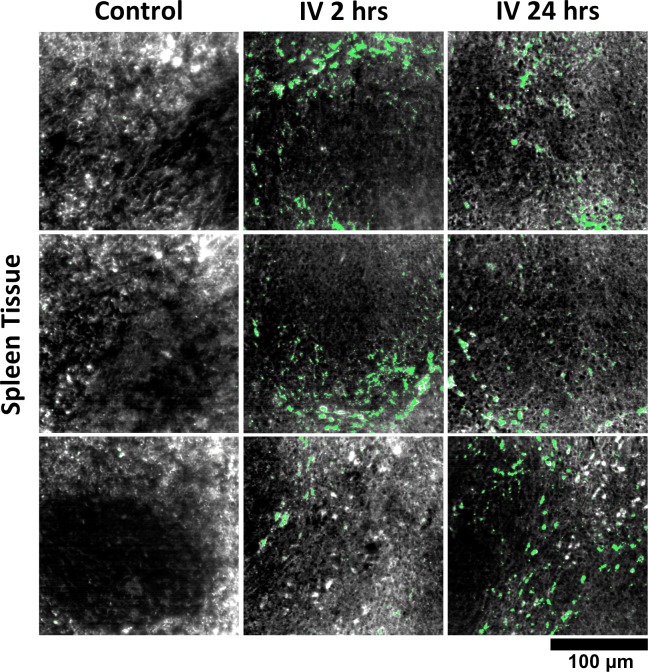
Thank you for submitting your work entitled "A hyperspectral method to assay the microphysiological fates of nanomaterials with single-particle sensitivity" for consideration by eLife. Your article has been reviewed by three peer reviewers, and the evaluation has been overseen by Gaudenz Danuser as the Reviewing Editor and Sean Morrison as the Senior Editor.

Our decision has been reached after consultation between the reviewers. Based on these discussions and the individual reviews below, we regret to inform you that your work will not be considered further for publication in eLife.

As you will see from the full reviews below, all reviewers praise the quality of the study and its relevance and timeliness. However, all three reviewers raised from different angles the same concern: How new and versatile is the technique. Reviewer #1 provides a number of papers that your approach should be carefully compared to – potentially with additional validation experiments. Similarly, Reviewer #3, an expert in machine learning, is not convinced that the approaches taken are all state-of-the-art. Some cross-validation with other methods would help easing this concern for a future reader of a publication. Reviewer #2 raises the most critical point: although the claim of generality is made, the data presented relies on a single type of nano-particles. In the discussion among the reviewers following the initial evaluation it became clear that manuscript could only be considered if data from multiple nano-particles were shown. We suppose that acquisition of such complementary data will exceed the typical time eLife grants for a revision. Therefore, we have decided at this point to reject the manuscript. That said, there is significant merit in the combination of approaches, leading to a significant result. Thus, with a more thorough comparison of your approaches to others and a demonstration of applicability to other nano-particles we would consider a new submission of this work and make our best effort to send the manuscript back to the same editor and reviewers.

Reviewer #1:

In their work the authors propose a technique for quantifying nanoparticles distribution in histological samples. Hyperspectral imaging is based on the collection of images containing spectral information across a large (relatively speaking) range of the electromagnetic spectrum. Typical applications are found for military, geoscience, or environmentally studies. Here for each pixel within an image for example the signal across the visible or infrared spectrum is collected and hyperspectral data cubes are built and analyzed. Because different objects possess different optical properties, knowing a priori specific signatures allow classifying the information present within a scene.

In recent years this technique has found quite some success within the biomedical imaging field. Because metallic nanoparticles scatter light quite strongly at specific "resonant" wavelengths, the combination of darkfield microscopy and an hyperspectral approach has made it possible to successfully detect nanoparticles at high imaging speed and to separate them from the cellular background. Also the technique offers greater resolution when compared to other tools for studying biodistributions.

In the manuscript the authors report specifically on an imaging processing method to quantify nanoparticles distributions in tissue samples. The imaging setup is a standard one from CytoViva for "enhanced darkfield microscopy" equipped with a CCD camera for hyperspectral imaging. Previous work in the field is already present in my opinion. See for example a review paper from Brenner's group detailing recent works ("Hyperspectral microscopy as an analytical tool for nanomaterials"). Different groups have also presented several works demonstrating single-nanoparticle detection. See for example "Single-nanoparticle detection and spectroscopy in cells using a hyperspectral darkfield imaging technique" and others from Musken's group, and also another nice paper from Meunier group "Hyperspectral darkfield microscopy of PEGylated gold nanoparticles targeting CD44-expressing cancer cells" where 3D nanoparticles tracking is also demonstrated.

Because the authors concentrate on ex vivo tissue sections I found strange that very recent intriguing work from Brenner's group is not cited considering it is focusing basically on a very similar subject (i.e. "Identification of Metal Oxide Nanoparticles in Histological Samples by Enhanced Darkfield Microscopy and Hyperspectral Mapping" on JoVE 2015). It would have been nice to see a discussion and analysis of this work and in which respect the presented work differs from the cited one.

Having said that, I found the paper very interesting and very well written. There is a lot of work and data are very compelling. Also I think it could be of great interest. My only concern deals with the novelty of it (specifically see the paper mentioned above from JoVE).

Also, because the paper deals with the development of a machine learning algorithm I found myself a little bit in difficulty giving a judgement in this regard because I'm not a specialist of this particular field and I'm familiar with only the most common and basic approaches. From what I've seen a lot of sophisticated work has been done for hyperspectral classification in areas outside from the biological one (e.g. recently deep-learning based approaches for feature extraction for hyperspectral imaging have been implemented from different groups). Therefore, I honestly cannot judge in this regard and perhaps maybe someone with a specific imaging processing background in the field of hyperspectral classification (not necessarily limited to biomedical microscopy, but a more broad one) could perhaps provide more insights into it and comment on the novelty of the computational approach used here.

Reviewer #2:

Overall, this is an interesting paper that seems to have been executed with significant care. The figures are of high quality and the text is well written.

Strengths:

The authors use hyperspectral imaging to detect large gold nanorods (100 x 30 nm) in ex vivo tissues with single particle detection capabilities and single micron resolution.

They analyze histologically stained tissue slices from various organs, and the appropriate controls. From a technical standpoint, everything looks fine. I was expecting this technique to not work for aggregated NPs, especially in liver Kupfer cells, and they report exactly that, the honesty of which I appreciate.

Unlike many papers that provide descriptive multivariate models for their data, in this one, they use a training dataset to build the model, and then they use the model on unknown samples. This shows that the model has a high predictive power. I would be curious to see how well this method could distinguish between different types of plasmonic particles. Also, the particles used are ~30x100 nm2.

Weaknesses:

In my opinion the biggest weakness of this paper is that the technique is only demonstrated for one single nanoparticle type, and especially one that is not commonly used, i.e. large gold nanorods that are 100 x 30 nm in size.

However, in the Discussion the authors specifically claim that their technique is applicable to many nanoparticle shapes "(for example, gold nanospheres, nanorods, nanocages, etc.)" […]. "ABIDE is capable of distinguishing such NPs from each other by spectral differences, enabling biodistribution studies of multiplexed NPs." This is not only a bold claim, which importantly is not supported by any data.

The technique is only interesting enough for a broad audience if multiple different nanoparticle shapes such as spheres, cages, regular sized nanorods etc., can be analyzed with this method.

And my concern is that the technique will run into issues with discriminating some of the other shapes from cellular components, as can already be predicted when considering the curves in "Figure 1—figure supplement 4". The large gold nanorods have a plasmonic peak in the near infrared, which may be much easier to discriminate by the machine-learning algorithm than for shapes such as e.g. spheres.

The authors need to show convincingly that this technique works for the other nanoparticle shapes as they claim.

Reviewer #3:

The paper presents methods and results of combining mathematical modeling, data analysis, hyper spectral imaging, related to imaging of nano materials with applications in cancer, angiogenesis, and others. I find this area really interesting, and the potential impact large, although tissue and cell physiology are not my area of expertise. Though I find the ideas very stimulating, the main issue that I have is that the mathematical modeling/data analysis used here is not very clear, and may not be near top shelf work. Combined with certain choices of validation expanded below, my understanding is that the contributions to imaging methodology is not sufficiently novel, nor inspire a lot of confidence that results are as best as possible. See below for detailed comments.

Regarding modeling and data analysis, a few questions. The topic of discerning the contributions of different elements (in this case nano materials versus others such as Eosin, Hematoxylin, etc.) from spectral measurements is a well studied one. While I understand the intricacies of this imaging experiment are not the same as other more well studied spectral unmoving problems, I'm not convinced that the wealth of other methods for linear and nonlinear unmixing don't apply here. In the paper I did not find any discussion related to this. Classification methods of the type authors claim to have attempted (nearest neighbor, SVMs, etc.) could be applied on such unmixed data, and to me this would constitute a more standard way of doing things. As is, the methodology regarding data analysis coupled to imaging does not seem very novel, and it is not presented in a way that can be related to many other already proposed methods to other seemingly similar applications.

Regarding the data analysis for validation, a couple of things seemed unclear to me. For the detection of false positives and false negatives, were these evaluations performed separately? And if so, is the validation criterion computed by checking whether there is one single pixel indicating nonmaterial (in case of false positives) present? Similar comments for the reverse situation (false negatives). It would be necessary to know the specifics of these details better, and even better have the data (e.g. histograms and if pixel counts are used, how thresholds are utilized). Also, how is the user defined parameter (it seems a manually selected threshold is used to initially determine if a pixel is potentially LGNR) handled during validation? Is data used in the validation stage also used by the user when selecting this parameter? It is a little confusing when in the subsection “Data processing and automatic biodistribution detection” the authors comment "We then manually (and in a manner blind to algorithmic classification)…". How is this possible given that for a positive detection the pixel must be classified as potential LGNR first? Am I missing something, or is it possible that authors are mixing training and testing data?

A few more comments below:

The Results section reads more like a Methods section. If format is to be followed, I'd suggest only describing results in the Results section.

Results, second paragraph: K-means is most commonly referred as a clustering algorithm. Once clusters are identified, one can use this information to design a multitude of classification methods, but authors must specify which they used. Presumably the simplest would be the nearest neighbor cluster center. Are the cluster centers utilized as ground truth?

[Editors’ note: what now follows is the decision letter after the authors submitted for further consideration.]

Thank you for resubmitting your work entitled "A hyperspectral method to assay the microphysiological fates of nanomaterials with single-particle sensitivity" for further consideration at eLife. Overall, your revised article has been favorably evaluated by Sean Morrison (Senior editor), a Reviewing editor, and three reviewers.

The manuscript has been improved but there are some remaining issues that need to be addressed before acceptance. They mainly concern the tendency of 'over-selling' some of the approaches and accomplishments of the work. We especially encourage you to reconsider the use of an acronym for this technology. We are concerned about the use of acronyms as cheap eye-catchers. Moreover, a new expert in machine learning has been brought to the panel because previous Reviewer #3 was unavailable. Given the rapid pace of the machine learning field it seems inappropriate to call the application of a clustering method (and even a very classic one, which is taught in undergraduate computer science course) 'machine learning'. It is perfectly fine to use standard methods if they solve the task, but there is a discomfort among reviewers that machine learning is merely used as a catchy phrase in this case. This could fire back on your work. We thus encourage you to revise some of the language in your manuscript, including the Abstract, and to address the few other comments listed below.

Reviewer #1:

I've found that the authors’ comments are very appropriate and to the point even though in some cases I'm not too familiar with some of the points discussed. My major concern is always related to my original comment. Previous similar work from Brenner using a basically identical setup hardware is already present in the literature. Because the scientific contribution of the submitted paper consists in proposing an analysis procedure based on a machine learning algorithm with the intent of extending the work of Brenner and co-workers and others, it is critical to determine the novelty of the proposed algorithm. Overall I found the data provided by the authors very compelling and particularly interesting.

Reviewer #2:

The authors are now showing feasibility data from two other nanoparticle shapes, and have therefore satisfactorily addressed my previous main concern whether ABIDE may be versatile enough with regards to different nanoparticle shapes and sizes.

There are a few remaining issues.

General comment: The revised version does not include any tracked changes or other markings to indicate where changes were made, which made the review of the paper quite difficult. I am making this comment not because I want to review another version with track changes, but to make it clear that this limitation may have reduced my ability to catch all remaining or new issues.

Specific comments:

1) In the PowerPoint slides the authors provided in their rebuttal (for the reviewers only), it says "Example of new in vivo data". I find this misleading, as I could not find any data in the entire paper that was acquired "in vivo". A reviewer who does not carefully examine the manuscript may be misled by the rebuttal summary slides that this is in fact all acquired in vivo and not catch this discrepancy between the summary and the actual paper. I am not expecting the authors to provide true in vivo data, but would like to clarify what the authors meant by that.

2) The title of the paper is overstated with regards to claiming "single particle sensitivity" and this needs to be changed or else would be misleading. Figure 2—figure supplement 8 is the only figure that shows any data that would support that, and only in the very large gold nanorods (which by some definitions would not represent a nanoparticle). "Likely" as is stated in the figure legend is probably an honest assessment by the authors, but not enough to make such a major claim, and there is no evidence for this to work in the other two nanoparticles that are now included. I suggest replacing "…with single particle sensitivity" with "… in tissue sections" or "… in histological slices".

I would be willing to accept the manuscript pending these clarifications/changes if the other reviewers agree that their areas of expertise were addressed sufficiently as well.

Reviewer #4:

This revised manuscript describes an interesting and straightforward development of an approach for detecting nanoparticles in hyperspectral dark field images. It has been significantly improved based on comments in the initial reviews. The results presented demonstrate an impressive ability to detect and quantify these nanoparticles in tissue images. From an image processing/analysis/machine learning point of view, the approach is not novel or instructive (clustering spectra, especially with manual tuning, barely qualifies to be called machine learning, and does not seem to warrant a new acronym). Hence the significance of the manuscript must derive from the future importance of the method's application, something this reviewer is not qualified to judge.

eLife. 2016 Aug 18;5:e16352. doi: 10.7554/eLife.16352.051

Author response


[Editors’ note: the author responses to the first round of peer review follow.]

As you will see from the full reviews below, all reviewers praise the quality of the study and its relevance and timeliness. However, all three reviewers raised from different angles the same concern: How new and versatile is the technique. Reviewer #1 provides a number of papers that your approach should be carefully compared to – potentially with additional validation experiments. Similarly, Reviewer #3, an expert in machine learning, is not convinced that the approaches taken are all state-of-the-art. Some cross-validation with other methods would help easing this concern for a future reader of a publication. Reviewer #2 raises the most critical point: although the claim of generality is made, the data presented relies on a single type of nano-particles. In the discussion among the reviewers following the initial evaluation it became clear that manuscript could only be considered if data from multiple nano-particles were shown. We suppose that acquisition of such complementary data will exceed the typical time eLife grants for a revision. Therefore, we have decided at this point to reject the manuscript. That said, there is significant merit in the combination of approaches, leading to a significant result. Thus, with a more thorough comparison of your approaches to others and a demonstration of applicability to other nano-particles we would consider a new submission of this work and make our best effort to send the manuscript back to the same editor and reviewers.

We thank the editors and reviewers for their fair and constructive feedback on our manuscript. Below, we have provided detailed responses to the concerns raised by each reviewer. In summary, the following key revisions have been made to our manuscript:

a) New Nanoparticles Tested with Favorable Results:Extensive ex vivo data are now presented for two additional nanoparticle – Gold Nanoshells and silica-coated Gold Nanospheres (GNS@SiO2) – each of which exhibit distinct spectral properties, shapes, and compositions from the LGNRs we reported in the initial manuscript (please see Figures 6 and 7, as well as all related figure supplements). We also demonstrated the ability to spectrally multiplex Nanoshells and LGNRs using our method, despite their highly overlapping spectrum (see Figure 6—figure supplement 3). Specific details of each new particle type and experimental result are noted in the response to Reviewer #2’s comments.

b) Direct Comparison of ABIDE with Existing Hyperspectral Methods: We have updated the Introduction and Discussion sections of our manuscript with comparisons to the existing studies recommended by Reviewer #1. Extensive comparisons are provided in the response to Reviewer #1’s comments below. An additional experimental comparison with the cited spectral angle mapping (SAM) approach is also now included in the manuscript (Figure 2—figure supplement 15).

c) Cross-Validation and Comparison with Other Machine Learning Approaches: The results of k-means analysis were compared with those from other prevalent machine learning methods including SVM. A detailed report of results from these various methods is listed as Liba and Shaviv, 2014 in our manuscript, and our rationale for choosing k-means analysis is summarized within the response to Reviewer #3. Additional discussion of alternate machine learning approaches has been added to the manuscript.

Summary of new data:

Figure 2—figure supplement 1: now contains raw data for sensitivity/specificity (panel b);

Figure 2—figure supplement 15: new data comparing ABIDE to spectral angle mapping (SAM);

Figure 6 and all supplements: new data for ABIDE detection and spectral unmixing of gold nanoshells;

Figure 7 and all supplements: new data for ABIDE detection of gold nanospheres.

Reviewer #1:

In their work the authors propose a technique for quantifying nanoparticles distribution in histological samples. Hyperspectral imaging is based on the collection of images containing spectral information across a large (relatively speaking) range of the electromagnetic spectrum. Typical applications are found for military, geoscience, or environmentally studies. Here for each pixel within an image for example the signal across the visible or infrared spectrum is collected and hyperspectral data cubes are built and analyzed. Because different objects possess different optical properties, knowing a priori specific signatures allow classifying the information present within a scene.

In recent years this technique has found quite some success within the biomedical imaging field. Because metallic nanoparticles scatter light quite strongly at specific "resonant" wavelengths, the combination of darkfield microscopy and an hyperspectral approach has made it possible to successfully detect nanoparticles at high imaging speed and to separate them from the cellular background. Also the technique offers greater resolution when compared to other tools for studying biodistributions.

In the manuscript the authors report specifically on an imaging processing method to quantify nanoparticles distributions in tissue samples. The imaging setup is a standard one from CytoViva for "enhanced darkfield microscopy" equipped with a CCD camera for hyperspectral imaging.

We thank the reviewer for this concise overview of hyperspectral imaging and its applications for context. Similar descriptions can be found in the Introduction and Discussion sections of our manuscript.

Previous work in the field is already present in my opinion. See for example a review paper from Brenner's group detailing recent works ("Hyperspectral microscopy as an analytical tool for nanomaterials"). Different groups have also presented several works demonstrating single-nanoparticle detection. See for example "Single-nanoparticle detection and spectroscopy in cells using a hyperspectral darkfield imaging technique" and others from Musken's group, and also another nice paper from Meunier group "Hyperspectral darkfield microscopy of PEGylated gold nanoparticles targeting CD44-expressing cancer cells" where 3D nanoparticles tracking is also demonstrated.

We thank Reviewer #1 for bringing these references to our attention. We have added a comparison of these previous methods with the method described in our manuscript to the Discussion section. In general these existing works should be acknowledged (they are now cited in our revised manuscript), but we do not believe they compromise the novelty or utility of our current study. Specifically, our manuscript reports new capabilities for diagnostic evaluation of particle identification in HSM images, improvements for standardizing HSM analysis and reproducibility, validated single-particle detection in animal tissues, the first demonstration of HSM for systemic biodistribution studies, and the novel nanoparticle microbiodistribution data itself (now 3 particle types). Further details are provided below:

“Hyperspectral microscopy as an analytical tool for nanomaterials” (Brenner): This review article is listed in our original manuscript. We cited this review (and several other studies the review cites) in the Introduction section of our manuscript, as it provides very useful context for biological applications of hyperspectral imaging. Importantly, the review by Brenner also highlights the outstanding problems and limitations of hyperspectral imaging, several of which can be resolved by our method (summarized below). Please see the following limitations mentioned in the Brenner review article and how our methods can address these limits:

“More significantly is that while [Hyperspectral Imaging] enables the detection of nanomaterials, no study has precisely quantified the rate of false-positive identification or assessed its efficacy in the presence of stains used in histology or immunohistochemistry.” (Brenner, WIRE Nanomed Nanobiotechnol 2015)

We provide false-positive quantification measurements validated through several approaches, along with full diagnostic characterizations of detection sensitivity and specificity. Notably, these capabilities are demonstrated in stained histological samples. Sentences highlighting this existing problem have been added to the Introduction and Discussion.

“[Hyperspectral Imaging] is also sufficiently novel that there are few standardized methods to be applied, with the result that each lab may use differing source power, exposure time, or other parameters which can result in significant variance in spectra of similar materials analyzed under different conditions.” (Brenner, WIRE Nanomed Nanobiotechnol 2015)

Because of the adaptive nature of our algorithms with respect to a given image’s intensity histogram, the detrimental effects of variance in incident illumination power and exposure time can be minimized using our method. This provides potential benefits in terms of result reproducibility and the development of standardized methods, which is now emphasized in the manuscript.

"Single-nanoparticle detection and spectroscopy in cells using a hyperspectral darkfield imaging technique" (Muskens): Reviewer #1 is correct that this paper provides a nice demonstration of single particle detection with hyperspectral imaging – we have now added the paper by Muskens as a useful reference for validation of our own single particle detection capabilities (i.e., validation of empirical signals against theoretical optics predictions, etc.). We do wish to clarify that the cited Muskens paper demonstrates single particle detection in pure nanoparticle samples and, in the case of another Muskens paper (Fairbairn et al., Phys. Chem. Chem. Phys. 15(12) 2013), in cell culture. It should be noted that the ability to detect particles in cell culture does not guarantee that such detection will be possible in tissues (especially stained sections) due to the complex spectral scattering environment. By comparison, we use our method to demonstrate single-particle detection capabilities in such tissue sections. We see this as a distinction of our manuscript relative to existing work.

“Hyperspectral darkfield microscopy of PEGylated gold nanoparticles targeting CD44-expressing cancer cells” (Meunier): As Reviewer #1 notes, this paper also demonstrates single particle detection. As for the papers from Muskens’ group, we note that this work is limited to particle detection in cell culture rather than whole tissues following systemic nanoparticle administration. For completeness, we have now incorporated this reference into our manuscript’s Introduction section. We would like to clarify that the 3D particle tracking demonstrated in the paper from Meunier is achieved through image acquisition at multiple focal planes (z-axial scanning) in fixed samples followed by reconstruction – in principle, this approach could also be applied for 3D characterization of tissue sections using our methods.

A general note on the hyperspectral methods used by Muskens and Meunier: both groups implement a super-continuum tunable light source. While we expect that our methods can translate to hyperspectral imaging with laser-based sources, the success of spectral cluster identification may be influenced by the chosen spectral resolution (laser sampling density/sparsity). While not necessarily prohibitive, higher spectral sampling density with a tunable laser typically leads to longer scan times. Sufficiently high spectral resolution is important for the accurate classification of spectra into clusters. Also, as Meunier notes, the use of laser-based sources can introduce speckle that can corrupt image formation and quantification. Meunier also points out the following with respect to using laser-based hyperspectral imaging:

“It should be mentioned that additional experimental efforts are needed to obtain spectrally and spatially homogeneous imaging field comparable to the conventional white light illumination used in push-broom hyperspectral imaging systems (CytoViva, PARISS). Accurate spectral and spatial characterization of single and aggregated AuNPs is essential for the improvement of nanoplasmonic-based imaging, disease detection and treatment in complex biological environment” (Meunier, J. Biophotonics 8(1-2) 2015).

Thus, spectral stability and uniformity are further advantages of the system and methods we demonstrate relative to existing work in the field, although this is a technical point related more to optical setup and hardware rather than analytical methods.

Because the authors concentrate on ex vivo tissue sections I found strange that very recent intriguing work from Brenner's group is not cited considering it is focusing basically on a very similar subject (i.e. "Identification of Metal Oxide Nanoparticles in Histological Samples by Enhanced Darkfield Microscopy and Hyperspectral Mapping" on JoVE 2015). It would have been nice to see a discussion and analysis of this work and in which respect the presented work differs from the cited one.

Thank you to Reviewer #1 for bringing this work to our attention. Reviewer #1 is correct that the focus of the work by Brenner is related to our own study, and for this reason we have now added it as a reference (Roth et al., 2015) and addressed its relation to our work in the Discussion section as requested. For completeness, there are several advantages to our approach, which are described below:

Our method’s accuracy and practicality enable new applications of hyperspectral imaging: Brenner et al.describe (in excellent detail) the use of commercially-available ENVI software for analyzing hyperspectral images acquired using a CytoViva microscope (similar to the one we use in our study). As noted in the “Protocol” portion of the paper in JoVE, this analysis involves separate user-initiated steps for 1) appropriate selection of reference spectra for particles of interest, 2) removal of false-positive spectra, and 3) use of the reference spectra produced in 1) and 2) for spectral angle mapper (SAM) classification of images of interest. Within the SAM analysis steps themselves, users must manually set factors including intensity thresholds and target size parameters in order to obtain accurate (i.e., high-specificity, high-sensitivity) representations of the presence of a given nanomaterial in the sample. Following SAM classification, the authors note that mapped hyperspectral images are then analyzed by additional third party software (ImageJ) to obtain quantitative data on particle uptake/presence. Finally, the authors note that this quantitative data should be exported to other programs for statistical analysis. This process can then be repeated for additional images.

By comparison, the methods described in our manuscript achieve spectral identification, diagnostic validation, and quantification rapidly using an open-source code to process many images in a single run. Because the processing is automatic, we believe our method provides a means for higher throughput analysis, the importance of which should not be overlooked if the technique is to be used for biodistribution studies that require analysis of numerous images of various tissues.

Because it is adaptive, our method requires minimal user-defined inputs. In addition to manual definition of intensity thresholds (see below) or expected particle size (which may be highly variable depending on routes of uptake/accumulation in tissues and therefore cannot be assumed a priori), SAM classification requires users to define angular tolerance values that can heavily impact the resulting detection sensitivity and specificity (see Figure 2—figure supplement 15). This may introduce significant detection error that can be difficult to quantify.

The existing method reported by Brenner in JoVE produces spectral libraries through methods that are susceptible to manual biases and non-comprehensive sampling. Thus, libraries may not be faithfully reproduced by different users or for different samples. From the JoVE Protocol section “Creation of Reference Spectral Libraries”:

3.1.3 “…click onto pixels of interest on the datacube, particularly the brightest ones or those that can be confidently identified as representing the material of interest…take note particularly of their lowest and highest value and which wavelength corresponds to it.” The authors then note in section 3.1.4 that these user-observed values should be entered as parameters into the software’s “Particle Filter” function to identify pixels with spectra that will be added to the spectral library file used for subsequent image classification.

For images with more than 200,000 pixels, manual assessment of this type is likely inadequate to determine the relevant intensity and spectral ranges that will allow complete and accurate particle identification in subsequent steps. In other words, the results for any image analyzed with a spectral library defined in this way will only be as specific/sensitive as the initial user’s discretion allows. Accuracy and reproducibility among users and images (even for similar samples under different illumination) become significant concerns for this reason. Brenner’s group notes this specific issue in their review article from the same year as well.

Comparison of article scope and novelty: The cited work by Brenner successfully demonstrates the ability to identify nanomaterials of different composition following administration to samples of porcine skin. While this demonstration is relevant to our manuscript, we note the following novel aspects of our manuscript that are not within the scope of this or other previous hyperspectral imaging work:

The machine learning approach we implemented realizes several new improvements to existing issues in the field of hyperspectral imaging (as noted in the review by Brenner, see above). While the machine learning method is not novel per se, its application to improve the quality of biological hyperspectral image analysis is the first such demonstration to our knowledge.

While Brenner and several other groups referenced by our work have rightfully discussed the potential of hyperspectral imaging for performing studies of biodistribution, our manuscript provides the first empirical demonstration of hyperspectral imaging as a viable biodistribution technique. Thus in terms of potential clinical impact and future use, we believe our manuscript marks a significant advance for hyperspectral imaging as a biomedical resource.

In addition to demonstrating biodistribution capabilities, we provide novel data on the sub-organ localization of nanoparticles (LGNRs, Nanoshells, and GNS@SiO2) that have not been previously reported. The microbiodistribution data is itself a novel resource that can inform future uses (clinical or otherwise) of the studied nanoparticles.

We also report the first hyperspectral study of molecularly-targeted particle uptake in tissue (tumor xenografts from live animal models). [We note the previous demonstration of CD 44-targeted particle uptake in cell culture by Meunier et al.] While this data is itself unique, the greater takeaway is that ABIDE may be an ideal tool for future assessments of nanoparticles used for targeted imaging and therapy in animal models of disease.

The study by Brenner’s group in JoVE reports the detection of nanomaterials in tissue, but the authors do not assess single particle detection sensitivity in these tissues. Our manuscript provides one such demonstration of single-particle sensitivity in tissue.

The cited work does not demonstrate spectral unmixing capabilities to distinguish different particles types present in a single sample, which we have now demonstrated (see Figure 6—figure supplement 3).

Having said that, I found the paper very interesting and very well written. There is a lot of work and data are very compelling. Also I think it could be of great interest. My only concern deals with the novelty of it (specifically see the paper mentioned above from JoVE).

We thank Reviewer #1 for this very positive consideration of our work. We believe our work provides several notable novel advances relative to current work in the fields of biodistribution and hyperspectral imaging. For more details on these novel aspects, please refer to previous addresses of existing literature.

Also, because the paper deals with the development of a machine learning algorithm I found myself a little bit in difficulty giving a judgement in this regard because I'm not a specialist of this particular field and I'm familiar with only the most common and basic approaches. From what I've seen a lot of sophisticated work has been done for hyperspectral classification in areas outside from the biological one (e.g. recently deep-learning based approaches for feature extraction for hyperspectral imaging have been implemented from different groups). Therefore, I honestly cannot judge in this regard and perhaps maybe someone with a specific imaging processing background in the field of hyperspectral classification (not necessarily limited to biomedical microscopy, but a more broad one) could perhaps provide more insights into it and comment on the novelty of the computational approach used here.

We thank Reviewer #1 for the positive review of our work and for the suggestion of several useful references that we have added to our manuscript. A more detailed discussion of the machine learning methods tested for our study is addressed in the response to Reviewer #3’s comments.

Reviewer #2:

Overall, this is an interesting paper that seems to have been executed with significant care. The figures are of high quality and the text is well written.

Strengths:

The authors use hyperspectral imaging to detect large gold nanorods (100 x 30 nm) in ex vivo tissues with single particle detection capabilities and single micron resolution.

They analyze histologically stained tissue slices from various organs, and the appropriate controls. From a technical standpoint, everything looks fine. I was expecting this technique to not work for aggregated NPs, especially in liver Kupfer cells, and they report exactly that, the honesty of which I appreciate.

We thank the reviewer. Anecdotally, Reviewer #2 may find it interesting that ABIDE detection of silica-coated gold nanoshells (GNS@SiO2) in Kupffer cells did not seem to be impeded by spectral hybridization. We suspect this may be due in part to the steric effects of the silica shell around the gold core, however it may also be attributed to smaller spectral shifts relative to the original peak upon aggregation. (See liver images in Figure 7 and Figure 7—figure supplement 5).

Unlike many papers that provide descriptive multivariate models for their data, in this one, they use a training dataset to build the model, and then they use the model on unknown samples. This shows that the model has a high predictive power.

Thank you to Reviewer #2 – we appreciate these compliments on our work.

I would be curious to see how well this method could distinguish between different types of plasmonic particles. Also, the particles used are ~30x100 nm2.

As part of the revisions to our work, we have included new data detailing the detection of two additional plasmonic particles: gold nanoshells (Nanoshells) and silica-coated gold nanospheres (GNS@SiO2). These particles exhibit very different spectra from the LGNRs originally tested. In particular, the GNS@SiO2 have ~550nm SPR vs ~850nm SPR for LGNRs. The size, shape, and material composition/surface coating of both Nanoshells and GNS@SiO2 are also distinct from LGNRs. Results for these two new particle types are found in Figures 6 and 7. Please also refer to new results for spectral unmixing of gold Nanoshells and LGNRs (Figure 6—figure supplement 3). We hope this (and other) new data helps answer Reviewer #2’s questions regarding the ability to distinguish between plasmonic particles.

Weaknesses:

In my opinion the biggest weakness of this paper is that the technique is only demonstrated for one single nanoparticle type, and especially one that is not commonly used, i.e. large gold nanorods that are 100 x 30 nm in size.

Please see the response above and refer to Figures 6, 7, and related figure supplements. While Reviewer #2 is correct that LGNRs are not widely used, we note that one feature of our manuscript is the first report of biodistribution data for these particles. While not in the scope of the current work, we hope that the use of LGNRs will become more widespread based on their advantages over regular GNRs in several respects, which we have recently reported.

However, in the Discussion the authors specifically claim that their technique is applicable to many nanoparticle shapes "(for example, gold nanospheres, nanorods, nanocages, etc.)" […]. "ABIDE is capable of distinguishing such NPs from each other by spectral differences, enabling biodistribution studies of multiplexed NPs." This is not only a bold claim, which importantly is not supported by any data.

Thank you to Reviewer #2 for this important point. Because we do not explicitly test gold nanocages or ex vivo spectral unmixing capabilities, we have revised these statements accordingly to reflect the particle types tested herein and the specific experiments we have performed. They now reflect our demonstrated abilities to characterize multiple particle types in vivo and to spectrally resolve different particle types within mixtures (although not yet demonstrated in tissue sections). Please refer to Figures 6, 7, and their related figure supplements in the revised manuscript. Notably, the Nanoshells and LGNRs used in spectral unmixing tests exhibited similar near-infrared plasmonic peaks, yet they can be resolved from each other, likely as a result of differences in spectral width.

The technique is only interesting enough for a broad audience if multiple different nanoparticle shapes such as spheres, cages, regular sized nanorods etc., can be analyzed with this method.

We agree with Reviewer #2 that the ability to identify a number of nanoparticles is of key importance to a general audience. To address this, we have added extensive ex vivo analysis of gold nanoshells and spherical gold nanoparticles administered intravenously to mice. The particles we used are commercially-available Nanoshells (Nano Composix, San Diego, CA) and GNS@SiO2 (Oxonica, Mountain View, CA) that have been used in numerous applications to date (several are listed below). Please note that the GNS@SiO2 used in our experiments are currently under evaluation for potential clinical applications.

Examples of prior work using GNS@SiO2:

“Noninvasive molecular imaging of small living subjects using Raman spectroscopy” (Keren et al., PNAS 2008);

“A brain tumor molecular imaging strategy using a new triple-modality MRI-photoacoustic-Raman nanoparticle” (Kircher et al., Nat. Med. 2012);

“Affibody-functionalized gold-silica nanoparticles for Raman molecular imaging of the epidermal growth factor receptor” (Jokerst et al., Small 2011);

“Preclinical evaluation of Raman nanoparticle biodistribution for their potential use in clinical endoscopy imaging” (Zavaleta et al., Small 2011).

And my concern is that the technique will run into issues with discriminating some of the other shapes from cellular components, as can already be predicted when considering the curves in "Figure 1—figure supplement 4". The large gold nanorods have a plasmonic peak in the near infrared, which may be much easier to discriminate by the machine-learning algorithm than for shapes such as e.g. spheres.

The authors need to show convincingly that this technique works for the other nanoparticle shapes as they claim.

Reviewer #2 is correct that some particles may exhibit similar spectra to cellular components, a point that we acknowledged in our Discussion section. One way to circumvent this problem is to perform analysis on unstained tissue sections (in which case, two of the curves (for H&E stains) shown in Figure 1—figure supplement 4 would not be present). In fact, we found that it was important to analyze unstained tissue sections when detecting GNS@SiO2. While this requires consideration of sample preparation methods, we found that GNS@SiO2 detection and characterization in unstained tissues was as successful as our original detection of LGNRs, despite the ~300 nm difference in spectral peaks. Thus, scattering from (unstained) cellular components does not appear to preclude the detection of commonly-used nanoparticles that lack near infrared spectral signatures. We believe this new data provides a demonstration of the broader utility of our reported methods. Reviewer #2 is correct that spectra in the near-infrared will be easier to identify in stained sections. However, our results for GNS@SiO2 indicate that particles with visible spectrum signatures can also be studied with our methods, given the aforementioned preparation of unstained sections.

Reviewer #3:

The paper presents methods and results of combining mathematical modeling, data analysis, hyper spectral imaging, related to imaging of nano materials with applications in cancer, angiogenesis, and others. I find this area really interesting, and the potential impact large, although tissue and cell physiology are not my area of expertise. Though I find the ideas very stimulating, the main issue that I have is that the mathematical modeling/data analysis used here is not very clear, and may not be near top shelf work. Combined with certain choices of validation expanded below, my understanding is that the contributions to imaging methodology is not sufficiently novel, nor inspire a lot of confidence that results are as best as possible. See below for detailed comments.

Thank you to Reviewer #3 for the positive comments on our work. We hope that the revisions made to our manuscript and the responses below provide an adequate address of the reviewer’s concerns. As a general point, we acknowledge that the novelty of our work does not lie exclusively within a major advance in the field of machine learning. Rather, we believe our work’s novelty and (perhaps more importantly) utility resides in the combination of: 1) using machine learning to improve the state of biomedical hyperspectral imaging, 2) the specific biological insights and new data reported for nanoparticle microbiodistribution, and 3) the general biomedical capabilities developed for future applications (i.e., high-resolution biodistribution, studies of molecular targeting for therapy and imaging agent evaluation, etc.).

Furthermore, it is important to note that k-means clustering and nearest neighbor classification are only two parts of the full algorithm, which also includes defining an adaptive signal intensity threshold and pre-processing of the data. We believe that the full approach is novel and it is what makes ABIDE invariant to image acquisition parameters and applicable in many cases.

For more detail regarding novelty, please refer to the responses to Reviewer #1. Regarding whether our results are the best possible for machine learning methods, please refer to the detailed responses below.

Regarding modeling and data analysis, a few questions. The topic of discerning the contributions of different elements (in this case nano materials versus others such as Eosin, Hematoxylin, etc.) from spectral measurements is a well studied one. While I understand the intricacies of this imaging experiment are not the same as other more well studied spectral unmoving problems, I'm not convinced that the wealth of other methods for linear and nonlinear unmixing don't apply here. In the paper I did not find any discussion related to this. Classification methods of the type authors claim to have attempted (nearest neighbor, SVMs, etc.) could be applied on such unmixed data, and to me this would constitute a more standard way of doing things. As is, the methodology regarding data analysis coupled to imaging does not seem very novel, and it is not presented in a way that can be related to many other already proposed methods to other seemingly similar applications.

As part of the effort to detect the LGNRs in stained tissue sections, we explored various machine learning approaches. We actually started with supervised learning approaches, such as SVM and logistic regression, but then we found that k-means, when trained on slides of stained tissue that was injected with LGNRs, performed much better in detecting the LGNRs. This is because of the un-supervised nature of k-means, which automatically found the correct target LGNR spectrum, which turned out to be slightly different in tissue sections compared to the spectra of LGNRs dispersed on a glass slide. These results are explained in a report, which we referenced in our manuscript (Liba and Shaviv, 2014 in the revised manuscript), and have also been uploaded along with the revision.

An additional advantage of k-means as an unsupervised learning method is that it does not require pre-labeling the samples, which may be tedious. Rather, the researchers run the algorithm and choose the cluster that best fits their target spectrum (as determined through independent measurements such as Vis-NIR spectrometry).

We did not add the information regarding the different machine learning approaches in order to help the reader focus on the biomedical application, as we assumed the majority of readers will come from the application field and not from the computational side. If the reviewer believes that this information will help the readers, we are happy to include the comparison of machine learning methods as a supplementary document.

Furthermore, we believe that the ease of use of k-means and its popularity will encourage researchers to apply ABIDE for their research, while more complicated methods, such as deep-learning or unmixing, would make this work out of reach for researches who are not working in this field (k-means is more readily applied). We are hopeful that researchers who are familiar with or would like to try other methods, would be able to easily do so in post processing.

It may very well be that k-means is not the optimal algorithm to use for all applications, but we found that it is a great place to start in terms of result quality and diagnostic evaluation (particularly with respect to current methods for hyperspectral analysis, such as spectral angle mapping, described in the response to Reviewer #1). As in many machine-learning projects, it may be that the best algorithm would be tailored to the specific problem. For this demonstration, we sought to provide a generalizable method that will be broadly applicable and accessible for researchers working with various samples, particle types, and experimental designs.

As a note to the reviewer, we have also attempted linear unmixing methods and independent component analysis (ICA), however these did not produce qualitatively better results than k-means and therefore we did not perform a rigorous analysis on them. We believe they did not work well because the LGNR spectral component is not sufficiently independent from the tissue spectral components, and especially spectra that result from chromatic aberrations (this is discussed in detail within our manuscript’s results and methods sections). However, we are continuing to explore the potential use of linear and nonlinear unmixing for hyperspectral image analysis.

Regarding the data analysis for validation, a couple of things seemed unclear to me. For the detection of false positives and false negatives, were these evaluations performed separately? And if so, is the validation criterion computed by checking whether there is one single pixel indicating nonmaterial (in case of false positives) present? Similar comments for the reverse situation (false negatives). It would be necessary to know the specifics of these details better, and even better have the data (e.g. histograms and if pixel counts are used, how thresholds are utilized). Also, how is the user defined parameter (it seems a manually selected threshold is used to initially determine if a pixel is potentially LGNR) handled during validation? Is data used in the validation stage also used by the user when selecting this parameter? It is a little confusing when in the subsection “Data processing and automatic biodistribution detection” the authors comment "We then manually (and in a manner blind to algorithmic classification)…". How is this possible given that for a positive detection the pixel must be classified as potential LGNR first? Am I missing something, or is it possible that authors are mixing training and testing data?

First we would like to reassure that the training data sets were completely separate from the testing data sets. Training (meaning, calculating the clusters with k-means) was performed on images of LGNR injected tissue while testing was performed on different images and is detailed below.

The calculation of false positives and false negatives was done in two separate stages:

False negatives and true positives were calculated on images which include pure LGNRs only. The false negatives are the number of pixels (above the algorithm-defined threshold) that do not classify as LGNRs. Please see Figure 2—figure supplement 5 for an example of ABIDE detection in a pure LGNR sample, as well as the methods section describing this calculation.

False positives and true negatives were calculated on images, which include tissue only (i.e., no LGNRs present). The false positives are the number of pixels (above the algorithm-defined threshold) that classify as LGNRs. Please refer to Figure 2—figure supplement 4 for qualitative visualization of typical false positive detection. Also, Figure 1—figure supplement 2A, B shows an example histogram for a tissue-only sample with annotations depicting the threshold values (minHist = noise threshold and peakHist = particle threshold) determined by the algorithm for each image.

In addition to that, we performed another validation of the algorithm by testing it on slides of tissue that were injected with LGNRs (4 separate sections of kidney from LGNR-injected animals were used for this purpose). In that case, we observed and manually classified the spectra of >200 randomly-selected pixels (>50 pixels per each of the 4 FOVs) and compared our observation with the results of the algorithm (we were independent and blind to the results of the algorithm during this process, as was mentioned in line 411 of our initial submission). This analysis provided a measurement of FP, TP, FN and TN. This test was done on different images than the training data.

We note that the raw data for sensitivity and specificity measurements were provided as ancillary text documents in the original submission. Following the reviewers comment, we have included the numbers of measurements (pixel counts) in each category alongside the ratios previously provided. These raw data have been added as Figure 2—figure supplement 1B.

An adaptive threshold was calculated and used before the classification process. The adaptive threshold is a result of the algorithm described in the paper (section “Data processing and automatic biodistribution detection” and Figure 1—figure supplement 2), and has no user-defined parameters, other than “sanity parameters” ensuring the threshold is in a certain range. The algorithm, which determines the threshold, was qualitatively validated on over 20 images during its development. In the validation process we manually validated that the pixels that have enough intensity to be LGNRs fall above the LGNR threshold and that all tissue falls above the tissue threshold. Furthermore, we manually validated these “threshold maps” (such as the one in Figure 1—figure supplement 2D) for all the images used in the manuscript, and in general for our research.

To further clarify, only pixels above the image-adaptive LGNR threshold were considered for classification purposes. Only these pixels appear in the calculation of TP, FP, TN, FN.

A few more comments below:

The Results section reads more like a Methods section. If format is to be followed, I'd suggest only describing results in the Results section.

Thank you to Reviewer #3 for this feedback. We wanted to ensure that readers could readily follow the logic of key experiments and their results, and we felt that including some key aspects of our methodology was relevant to this aim. More detailed methods needed for reproducibility were avoided in the Results section and instead provided in the Methods section.

Results, second paragraph: K-means is most commonly referred as a clustering algorithm. Once clusters are identified, one can use this information to design a multitude of classification methods, but authors must specify which they used. Presumably the simplest would be the nearest neighbor cluster center. Are the cluster centers utilized as ground truth?

We thank the reviewer for this clarification. Indeed we used a nearest centroid (or nearest neighbor) classifier, based on the Euclidean distance to the cluster centers. We have now updated the manuscript to clarify this aspect of the algorithm.

We are not sure what the reviewer means by “ground truth” in this context. In the sense that each spectral cluster of interest can be cross-validated with spectral measurements from other techniques (i.e., Vis-NIR spectrometry, HSM of pure particles), the clusters do constitute a ground truth (as in the real, empirically assayed particle or dye spectra).

[Editors’ note: the author responses to the re-review follow.]

The manuscript has been improved but there are some remaining issues that need to be addressed before acceptance. They mainly concern the tendency of 'over-selling' some of the approaches and accomplishments of the work. We especially encourage you to reconsider the use of an acronym for this technology. We are concerned about the use of acronyms as cheap eye-catchers. Moreover, a new expert in machine learning has been brought to the panel because previous Reviewer #3 was unavailable. Given the rapid pace of the machine learning field it seems inappropriate to call the application of a clustering method (and even a very classic one, which is taught in undergraduate computer science course) 'machine learning'. It is perfectly fine to use standard methods if they solve the task, but there is a discomfort among reviewers that machine learning is merely used as a catchy phrase in this case. This could fire back on your work. We thus encourage you to revise some of the language in your manuscript, including the Abstract, and to address the few other comments listed below.

We would like to thank the editors and reviewers for their favorable consideration and constructive comments on our work. The newest version of our manuscript has addressed the outstanding concerns as described in the point-by-point response to reviewer comments provided below. In brief:

The title and Abstract have been revised to avoid over-selling.

The acronym “ABIDE” (Automated Biodistribution Detection) has been replaced with the abbreviation “HSM-AD” (Hyperspectral Microscopy with Adaptive Detection). This is a modification of the existing abbreviation “HSM.” We believe the abbreviation “HSM-AD” is more descriptive of our method and should relieve the concern about eye-catcher acronyms. Generally, we note that some form of abbreviation is necessary to aid readability.

The phrase “machine learning” has been replaced throughout the manuscript with more specific language that describes the relevant components of the algorithm. These changes should avoid misleading readers.

Reviewer #1:

I've found that the authors’ comments are very appropriate and to the point even though in some cases I'm not too familiar with some of the points discussed.

We thank Reviewer #1 for the positive consideration of our revised manuscript.

My major concern is always related to my original comment. Previous similar work from Brenner using a basically identical setup hardware is already present in the literature. Because the scientific contribution of the submitted paper consists in proposing an analysis procedure based on a machine learning algorithm with the intent of extending the work of Brenner and co-workers and others, it is critical to determine the novelty of the proposed algorithm.

We wish to emphasize the advances of our work with respect to previously published studies including the work by Brenner, as noted in extensive detail within our original point-by-point response document. Summarily, the novel aspects of the algorithm relative to existing work include:

a) The first use of hyperspectral imaging for a full systemic biodistribution study;

b) Single-particle detection sensitivity in tissues;

c) The demonstration of a fully adaptive (i.e., corrects for variable acquisition parameters and illumination) and automated (i.e., notably higher throughput image analysis) method for qualitative and quantitative assessments of biological samples using hyperspectral imaging;

d) This method can provide diagnostic values to aid image interpretation. Moreover, the detection method itself achieves high sensitivity and high specificity.

We also note the novel aspects of the experimental data obtained using the algorithm package:

a) Sub-organ localization patterns of three nanoparticle types with unique shapes, spectra, and sizes. Numerous insights are provided regarding the influence of size and surface coating on differences in both intra- and inter-organ biodistribution. For spherical gold particles, the hyperspectral imaging results correlated well with biodistribution data we obtained using ICP-MS;

b) The first use of hyperspectral microscopy to detect molecularly-targeted nanoparticle uptake in tumors.

The algorithm’s high-throughput and ease of use offer notable practical advances in hyperspectral analysis. We believe there is significant value in this sense because it enables a wide host of studies that rely on the analysis of large imaging datasets (for example, systemic biodistribution studies).

Overall I found the data provided by the authors very compelling and particularly interesting.

Again, thank you to Reviewer #1 for helpful and positive comments throughout the review process.

Reviewer #2:

The authors are now showing feasibility data from two other nanoparticle shapes, and have therefore satisfactorily addressed my previous main concern whether ABIDE may be versatile enough with regards to different nanoparticle shapes and sizes.

Thank you to Reviewer #2. We are pleased that these additions have addressed the reviewer’s original concerns.

There are a few remaining issues.

General comment: The revised version does not include any tracked changes or other markings to indicate where changes were made, which made the review of the paper quite difficult. I am making this comment not because I want to review another version with track changes, but to make it clear that this limitation may have reduced my ability to catch all remaining or new issues.

We thank the reviewer for the concern. Please note that we did in fact include two versions of the manuscript during the first round of revisions: one with tracked changes and one without. The tracked changes version was included as manuscript item #13, entitled: “Document with markup showing changes from original full submission to the revised manuscript.”

Specific comments:

1) In the PowerPoint slides the authors provided in their rebuttal (for the reviewers only), it says "Example of new in vivo data". I find this misleading, as I could not find any data in the entire paper that was acquired "in vivo". A reviewer who does not carefully examine the manuscript may be misled by the rebuttal summary slides that this is in fact all acquired in vivo and not catch this discrepancy between the summary and the actual paper. I am not expecting the authors to provide true in vivo data, but would like to clarify what the authors meant by that.

Thank you to Reviewer #2 for catching this error. Because we are analyzing tissue sections, all data are necessarily ex vivo. We have ensured that this is accurately reflected throughout the manuscript.

2) The title of the paper is overstated with regards to claiming "single particle sensitivity" and this needs to be changed or else would be misleading. Figure 2—figure supplement 8 is the only figure that shows any data that would support that, and only in the very large gold nanorods (which by some definitions would not represent a nanoparticle). "Likely" as is stated in the figure legend is probably an honest assessment by the authors, but not enough to make such a major claim, and there is no evidence for this to work in the other two nanoparticles that are now included. I suggest replacing "…with single particle sensitivity" with "… in tissue sections" or "… in histological slices".

We have revised the title pursuant to Reviewer #2’s request. The new title is: “A hyperspectral method to assay the microphysiological fates of nanomaterials in histological samples.”

I would be willing to accept the manuscript pending these clarifications/changes if the other reviewers agree that their areas of expertise were addressed sufficiently as well.

Thank you to Reviewer #2 for the helpful comments and positive consideration of our work throughout the review process.

Reviewer #4:

This revised manuscript describes an interesting and straightforward development of an approach for detecting nanoparticles in hyperspectral dark field images. It has been significantly improved based on comments in the initial reviews. The results presented demonstrate an impressive ability to detect and quantify these nanoparticles in tissue images.

We thank the reviewer for the positive consideration of our work.

From an image processing/analysis/machine learning point of view, the approach is not novel or instructive (clustering spectra, especially with manual tuning, barely qualifies to be called machine learning, and does not seem to warrant a new acronym).

We agree that the method we have used for clustering and classification is not novel and is indeed widely used. The novelty of our work lies in the application of these methods for the detection of particles and obtaining their biodistribution using hyperspectral imaging, which has not been demonstrated previously.

Please note that the described method, which we refer to as HSM-AD (previously “ABIDE”), includes two auxiliary algorithms in addition to clustering and classification: adaptive thresholding (Figure 1—figure supplement 2) and pre-processing of the hyperspectral signals (Figure 1—figure supplement 3). The combination of these methods make HSM-AD non-trivial and, most importantly, critical for the automated and generalizable detection of particles in tissue samples.

Following the reviewers comment, we have removed the use of the term “machine-learning” from the manuscript. Please refer to the document with tracked changes to see these modifications.

We believe that using an abbreviation for our method will improve readability. Therefore, we have changed the acronym “ABIDE” to “HSM-AD” (Hyperspectral Microscopy with Adaptive Detection) to avoid overly catchy terms. Please note that HSM is not a new acronym but rather an existing abbreviation.

Hence the significance of the manuscript must derive from the future importance of the method's application, something this reviewer is not qualified to judge.

We note that there are several significant aspects of the current manuscript (described in the response to Reviewer #1, the point-by-point response from the first round of revisions, and in the conclusion section of the main text). For example, we were able to characterize unique patterns of particle accumulation within liver, kidney, and spleen tissue that have not previously been reported.

Reviewer #4 correctly notes that the manuscript should be relevant for future applications. We believe that our method can be used in the future for rigorous preclinical assessments of nanoparticles intended for diagnostic or therapeutic uses. As a specific example, we are currently using our method to evaluate the uptake and clearance of a clinical nanoparticle candidate (GNS@SiO2) following intravenous versus oral administration routes. The method’s high sensitivity, specificity, and imaging resolution provide particle biodistribution data with greater detail than the data obtained from existing techniques like ICP (see Figure 7 and related supplements for intravenous data from these particles). For example, our method can be used to evaluate the extent of elimination of particles from a tissue of interest over time, which is critical for preclinical evaluations by the FDA and other regulatory agencies.

Associated Data

    This section collects any data citations, data availability statements, or supplementary materials included in this article.

    Supplementary Materials

    Figure 2—source data 1. Data used for diagnostic and 95% CIs.

    DOI: http://dx.doi.org/10.7554/eLife.16352.009

    DOI: 10.7554/eLife.16352.009
    Figure 2—source data 2. Data for whole organ uptake quantification.

    DOI: http://dx.doi.org/10.7554/eLife.16352.010

    DOI: 10.7554/eLife.16352.010
    Figure 3—source data 1. Data for kidney sub-organ ROIs.

    DOI: http://dx.doi.org/10.7554/eLife.16352.027

    DOI: 10.7554/eLife.16352.027
    Figure 4—source data 1. Data for liver, spleen, lung, and muscle sub-organ ROIs.

    DOI: http://dx.doi.org/10.7554/eLife.16352.029

    DOI: 10.7554/eLife.16352.029
    Figure 6—source data 1. Data for Nanoshell uptake in organs.

    DOI: http://dx.doi.org/10.7554/eLife.16352.034

    DOI: 10.7554/eLife.16352.034
    Figure 7—source data 1. Data for GNS@SiO2 uptake in organs.

    DOI: http://dx.doi.org/10.7554/eLife.16352.039

    DOI: 10.7554/eLife.16352.039
    Figure 8—source data 1. Data for tumor uptake of targeted and untargeted LGNRs.

    DOI: http://dx.doi.org/10.7554/eLife.16352.047

    DOI: 10.7554/eLife.16352.047
    Source code 1. Contains all MATLAB code used for HSM-AD.

    DOI: http://dx.doi.org/10.7554/eLife.16352.049

    elife-16352-code1.zip (46.4KB, zip)
    DOI: 10.7554/eLife.16352.049

    Articles from eLife are provided here courtesy of eLife Sciences Publications, Ltd

    RESOURCES