Abstract
Critical to the design and assessment of interventions for enteropathy and its developmental consequences in children living in impoverished conditions are non-invasive biomarkers that can detect intestinal damage and predict its effects on growth and development. We therefore assessed fecal, urinary and systemic biomarkers of enteropathy and growth predictors in 375 6–26 month-old children with varying degrees of malnutrition (stunting or wasting) in Northeast Brazil. 301 of these children returned for followup anthropometry after 2-6m. Biomarkers that correlated with stunting included plasma IgA anti-LPS and anti-FliC, zonulin (if >12m old), and intestinal FABP (I-FABP, suggesting prior barrier disruption); and with citrulline, tryptophan and with lower serum amyloid A (SAA) (suggesting impaired defenses). In contrast, subsequent growth was predicted in those with higher fecal MPO or A1AT and also by higher L/M, plasma LPS, I-FABP and SAA (showing intestinal barrier disruption and inflammation). Better growth was predicted in girls with higher plasma citrulline and in boys with higher plasma tryptophan. Interactions were also seen with fecal MPO and neopterin in predicting subsequent growth impairment.
Biomarkers clustered into markers of 1) functional intestinal barrier disruption and translocation, 2) structural intestinal barrier disruption and inflammation and 3) systemic inflammation. Principle components pathway analyses also showed that L/M with %L, I-FABP and MPO associate with impaired growth, while also (like MPO) associating with a systemic inflammation cluster of kynurenine, LBP, sCD14, SAA and K/T. Systemic evidence of LPS translocation associated with stunting, while markers of barrier disruption or repair (A1AT and Reg1 with low zonulin) associated with fecal MPO and neopterin.
We conclude that key noninvasive biomarkers of intestinal barrier disruption, LPS translocation and of intestinal and systemic inflammation can help elucidate how we recognize, understand, and assess effective interventions for enteropathy and its growth and developmental consequences in children in impoverished settings.
Introduction
Children living in impoverished areas around the world often develop stunted growth from 4 to 24 months of age, a concern that is heightened by potential lasting consequences of impaired cognitive development [1–5]. Increasingly sensitive detection methods are revealing that multiple and repeated intestinal infections are even more common (with and without overt diarrheal illnesses) in young children living in impoverished areas lacking in adequate sanitation or clean water [6, 7]. These heavy pathogen burdens may associate with growth faltering or cognitive impairment [8–14]. Undernutrition is also common and may further worsen the impact of enteric infections on growth and development, leading to a potential “vicious cycle” of malnutrition and enteric infections [15–17]. These potential relationships can be causally dissected in animal models, where certain enteric infections, such as Cryptosporidium or enteroaggregative Escherichia coli [18, 19] or even certain mixed Bacteroidetes and Proteobacteria [20] can further worsen the growth impairment and intestinal damage in undernourished states. In the latter case, a malnourishing diet resulted in growth failure and microbiome alterations, but without intestinal damage unless certain mixed microbiota were added. This problem of environmental enteropathogens inducing enteric dysfunction has been recognized for over half a century in tropical, developing areas and among Peace Corps volunteers [15, 21, 22]. More recently this clinical entity has been associated with the concept of disruption of gut barrier function, passage of microorganisms and/or their bioproducts from the intestinal lumen to the lamina propria, and mucosal inflammation, ultimately leading to damage of villous architecture and further impaired digestive and absorptive functions and barrier disruption, thereby generating a vicious cycle of impaired gut functions referred to as “Environmental Enteropathy” (EE) [23]. Numerous extensive studies have suggested that different assessments of intestinal barrier disruption, local and systemic inflammatory responses can associate with not only impaired growth, but also with poor household environments that likely predispose to enteric infections and EE [24–29]. Yet this hugely important problem for healthy child growth and development remains poorly understood [30]. Our objective therefore was to examine a wide range of potential fecal, urinary and plasma biomarkers to determine how they associate with each other and with malnutrition (defined by height or weight for age) and to determine which ones best predict impaired subsequent growth.
The need is great for biomarkers that can predict both EE onset and its longer term growth and developmental impact as well as to evaluate potential interventions designed to ameliorate these mechanisms and improve clinical outcome. We therefore examined potential biomarkers that might associate functional and structural “enteropathy” with malnutrition or with subsequent growth impairment in children enrolled in a study of malnutrition at a nutrition clinic serving several impoverished communities in and near Fortaleza, Ceará in Northeast Brazil. In particular, we assessed potential plasma, urine and fecal biomarkers of intestinal epithelial or barrier disruption, evidence of bacterial product translocation and intestinal and systemic inflammatory responses, and intestinal permeability.
Methods
Study location and population
The study was conducted at Promotion of Nutrition and Human Development (IPREDE) clinic located in Fortaleza, CE, Brazil. Details of the geographic location, population, demographics, environmental and socioeconomic status have been described elsewhere [31]; this outpatient clinic serves children from several impoverished communities in and near Fortaleza, often with moderate or severe malnutrition, having weight-for-age z-scores and height-for-age z-scores (WAZ or HAZ) ranging from -2 to -6. Standard nutrition education and supplementation was provided according to local standards based on World Health Organization guidelines [32]. The study started in Aug, 2010 and ended with follow up in Sep, 2013. Malnourished or “case” children were initially defined as having WAZ scores <-2 and, to the extent possible, age and gender matched ‘non-malnourished controls’ were defined as having a WAZ better than -1. However, others have correlated linear growth (or HAZ) with EE or other outcomes such as cognitive impairment [3]; hence we have examined both WAZ and HAZ in these analyses. Children were excluded if they had underlying recognized disease (other than malnutrition) or if they did not have a responsible parent or guardian ≥16 years old. After obtaining informed consent from the responsible parent or guardian, a total of 402 children aged 6–26 months (201 malnourished and 201 age and gender-matched non-malnourished) were enrolled between August 30, 2010 and July 12, 2013; 375 provided fecal and blood samples and completed a lactulose-mannitol absorption test, and were asked to return for followup in 2–6 months.
The MAL-ED case-control study protocol and consent forms were approved by the local institutional review board (IRB) at Universidade Federal do Ceará, the national IRB, Conselho Nacional de Ética em Pesquisa, Brasília, DF, Brazil, and the IRB at the University of Virginia, VA. The consent forms were reviewed and signed by the responsible parents or caregivers at the time of the screening process of the study protocol.
Anthropometry and biomarker assessments and statistical analyses
Detailed methods for anthropometric measurements, assessment of blood, stool, and urine biomarkers and statistical methods are provided in S1 File. As shown in Table 1, 375 children were enrolled and had initial anthropometry and were invited to provide a fecal specimen, have a L/M absorption test and to have blood obtained for testing potential biomarkers of intestinal barrier function, intestinal and systemic inflammation and injury repair. Shown in Table 2 are the 13 plasma, 4 fecal and urinary tests with at least 274 valid results obtained from within 1 month of enrollment. Markers of barrier function included urinary L/M absorption, fecal A1AT, Reg-1, and plasma LPS (acute levels, by neutralization luminescence [LUM] assay), IgG and IgA anti-LPS anti-FliC, zonulin, I-FABP and, with limited amounts of plasma available, claudin-15. Fecal MPO and neopterin were tested to assess intestinal inflammation, and plasma SAA, sCD14, LBP, citrulline, tryptophan and kynurenine were measured to assess systemic inflammatory responses and as potential predictors of intestinal injury repair. These 18 markers of intestinal barrier disruption and inflammation had adequate samples for assay in at least 274 (to 321) children at enrollment for study of associations with enrollment stunting (or wasting). Of several fecal biomarkers that have been used to assess intestinal inflammation, we selected myeloperoxidase (MPO) as our main test fecal marker of acute neutrophilic intestinal inflammation because of its ready availability, relatively less influence by age or breast-feeding and its potential use in murine models of enteropathy. In a separate analysis comparing fecal MPO, lactoferrin, calprotectin and lipocalin-2, we found that they correlate well with each other [33].
Table 1. Demographic information for study participants (Children and caregivers; n = 375 at study start, except where otherwise noted).
| Child | |
| Gender (n, % male) | 180/375 (48%) |
| Age Months (mean ± SD) | 14.3 ± 5.4 (n = 375) |
| Birth WAZ (mean ± SD) (by caregiver’s report) | -1.07 ± 1.7 (n = 365) |
| Breastfeeding (n, %Yes) | 210/375 (56%) |
| Diarrhea on Day of Visit (n, %Yes) | 10/375 (2.7%) |
| Caregiver (n = 375)* | |
| Age Years (mean ± SD) | 26.3 ± 6.5 n = 373 |
| Years Education | 7.9 ± 3.0 n = 373 |
| Age of 1st Pregnancy | 18.6 ± 4.2 n = 373 |
*Mother, n = 337;Grandmother, n = 27; Father, n = 9; other, n = 2.
Table 2. Frequencies of biomarker testing, including 13 tests on plasma, 4 on fecal and L/M absorption testing on urine as shown.
*Of 326 children with samples obtained within 1 month of study start.
| Plasma Biomarkers | # with Valid results* |
| SAA | 281 |
| LBP | 281 |
| sCD14 | 277 |
| I-FABP | 281 |
| kyn | 283 |
| try | 283 |
| AdjFlicIgA | 292 |
| AdjLPSIgG | 292 |
| AdjFlicIgG | 292 |
| AdjLPSIgA | 292 |
| citrulline | 283 |
| LPS_Nutri_Enz (LUM) | 289 |
| Zonulin | 288 |
| Fecal and Urine (L/M) Biomarkers | |
| MPO | 321 |
| REG1 | 315 |
| A1AT | 289 |
| Neo | 234 |
| L/M (and %L and %M) | 274 |
In addition, followup anthropometry at 2–6 months after initial sampling enabled us to assess these biomarkers as predictors of subsequent growth. We also assessed their associations with each other as well as with separately studied comparisons of fecal lactoferrin, calprotectin and lipocalin, and plasma hsCRP, and claudin-15. Repeated Measures MANOVA analyses were used to model mean growth from Study Start to follow-up anthropometric measurement obtained within 2 to 6 months later by children with low and high levels of each biomarker while controlling for child age and gender. Interaction effects of gender with biomarker were also assessed for each model. Similarly interaction effects of study start growth status (stunting present or not) of biomarkers on growth were assessed, although these results were not significant.
Missing data analysis (Pearson correlations between known characteristics of participants retained or failed to return for followup or were missing biomarker results because of sample limitations) showed that children with caregivers having fewer years of education were missing more of three biomarkers (A1AT, Reg1, MPO, r ≤ -0.18) compared to their more educated counterparts. Boys were disproportionately missing A1AT results (r = 0.15 p = 0.003), older children were more likely to be missing Neopterin (r = 0.12 p = 0.017) while nonbreastfeeding (r = -0.10, p = 0.37) and more wasted children (r = -0.12, p = 0.026) are missing more L/Ms. To account for bias in followup, multiple regression was used to impute missing data for all biomarkers with at least 274 (73% of 375) complete data. Principle Components Analyses (PCA) were then used to combine related combinations of 11 barrier function, 2 intestinal inflammation and then 7 systemic biomarkers. These related sets of biomarkers were then correlated among each other as well as to children’s growth status and growth trajectory initially using Partial Pearson correlations and ultimately using Multiple Regression. All analyses predicting anthropometric status and growth trajectories included controlling for child age and gender.
Results
Markers of malnutrition (individual biomarkers at enrollment study start, ss)
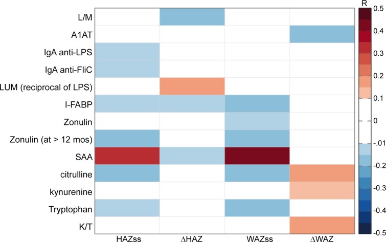
Several plasma biomarkers suggesting prior intestinal barrier disruption were significantly correlated with stunting or wasting, as assessed by HAZ or WAZ at study start, respectively, in children when they were first enrolled (ie at study start, ss). As shown in Fig 1 (and in S1 Table), these included plasma IgA anti-LPS antibody, zonulin (in all children for WAZ and, for HAZ in children >12 months old), and I-FABP. IgA anti-flagellin and anti-LPS antibodies (n = 292; r = 0.15, p = 0.011 and r = 0.14, p = 0.017) as well as intestinal fatty acid binding protein (I-FABP; n = 281; r = 0.14; p = 0.024) associate with worse stunting (i.e. lower HAZ SS scores) at the time of enrollment, suggesting greater prior exposure to LPS and release of an intestinal cytosolic protein, thus enterocyte damage, respectively. In addition, citrulline (as a marker of intestinal injury repair) and lower SAA (suggesting impaired defenses) correlated with stunting at enrollment.
Fig 1. Heat map showing all significant partial Pearson correlations of barrier and systemic biomarkers with HAZ or WAZ at enrollment (ie. study start, ss) or with changes in (Δ) HAZ or WAZ.
Significant correlations (at p<0.05; * = p<0.01) between biomarkers and growth, controlling for child age and gender. HAZ = height for age Z score; WAZ = weight for age z score; ss = study start; Δ = change in HAZ or WAZ at 2-6m followup; numbers range from 230 to 292 except for zonulin at age >12m where n = 172. Full r, p and df values are provided in S1 Table.
Predictors of subsequent growth (individual biomarkers with delta HAZ)
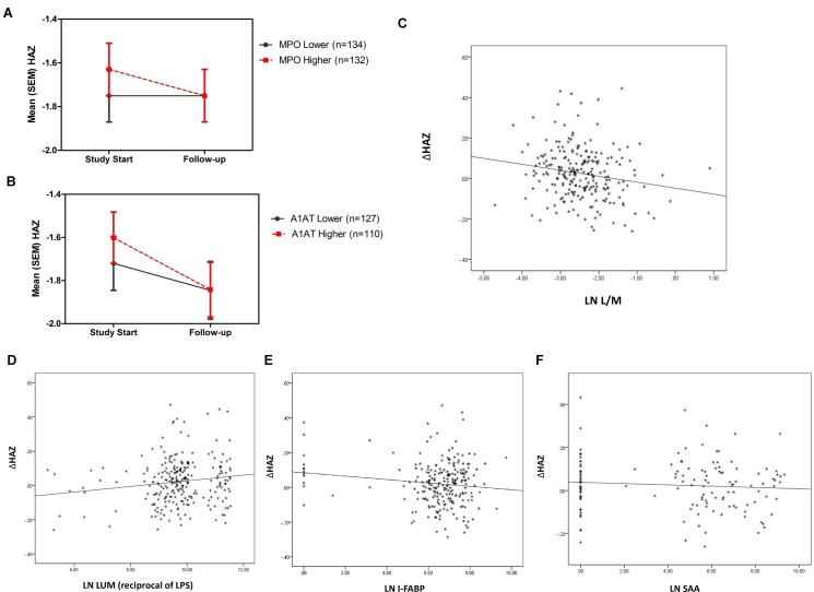
Several biomarkers of intestinal inflammation and barrier disruption (or “environmental enteropathy”) at the time of sampling were predictive of subsequent growth. Of importance, subsequent growth was significantly worse in those with higher fecal MPO or A1AT (Fig 2A and 2B), and growth impairment correlated with higher L/M, LPS, I-FABP and SAA (Fig 1 and Fig 2C–2F) in showing fecal and systemic markers of intestinal inflammation and barrier disruption or enteropathy).
Fig 2. Fecal MPO, Fecal alpha-1-antitrypsin (A1AT), and plasma LPS, FABP and SAA each predicts subsequent growth impairment.
a: For MPO, p = 0.028; n = 266 when correcting for age and gender, and independent of breastfeeding status (that showed no correlation in these 6-26m old children) and of age. b: For A1AT, n = 237; p = 0.042; and A1AT also correlates with “catchup WAZ” as well, p = 0.035 after correcting for age and gender. c: For urine L/M, higher values correlated (controlling for age and gender) with impaired growth (delta HAZ) (r = -0.173; p = 0.009; n = 230). d: For plasma LPS (ie lower LUM), higher values correlated with impaired growth (delta HAZ) (r = 0.151; p = 0.017; n = 251). e: For plasma FABP, higher values correlated with impaired growth (delta HAZ) (r = -0.134; p = 0.042; n = 231). f: For plasma SAA, higher values correlated with impaired growth (delta HAZ) (r = -0.132; p = 0.046; n = 231).
Claudin-15 (Cdn-15), even with relatively few (n = 35–42) matching samples, significantly correlates with lower L/M, A1AT, Reg-1 and lipocalin-2, all markers (when lower) of 'better' mucosal integrity/barrier function, and all consistent with the previously recognized associations of Cdn-15 with healthy absorptive surface function and barrier function in animal models [34] (see S2 Table).
Citrulliine predicts growth in girls and tryptophan in boys
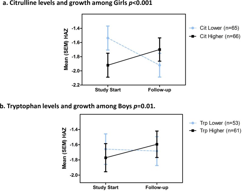
Shown in Fig 3 (Fig 3A and 3B) are the associations of plasma citrulline (esp. in girls) and tryptophan (in boys) with improved growth. The linear association of plasma citrulline levels with weight gain is significant for all 246 children (r = 0.15, p = 0.018). Bivariate analyses show this is predominantly accounted for by improved growth among the 131 girls (p<0.001). Conversely, the association of plasma tryptophan with better subsequent growth was only seen in the 114 boys (p = 0.010). Furthermore, higher kynurenine/tryptophan ratios correlate with fecal MPO and systemic hsCRP (and negatively with citrulline) (S1 Table).
Fig 3. Plasma citrulline (in girls) and tryptophan (in boys) predicts subsequent better growth.
A. citrulline (for which gender differs significantly) in girls (p<0.001; n = 131; median citrulline = 23.97 umol/L) and B. tryptophan in boys (p = 0.010; n = 114; median tryptophan = 66umol/L) predicts subsequent better growth.
Interaction of MPO and neopterin
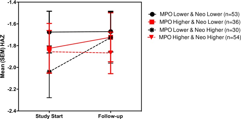
As shown in Fig 4 (Fig 4), Repeated Measures MANOVAs also shows an interaction effect in predicting growth: high MPO when combined with high neopterin associates with poorest growth. Thus, MPO combines with neopterin to show subsequent growth impairment; however, neopterin may also, in the absence of intestinal inflammation (ie low MPO), reflect “healthier” host cellular immune responses and thus correlate with better growth (again showing a significant interaction of MPO with neopterin).
Fig 4. Repeated measures MANOVAs show interactions in predicting growth between MPO and Neo: high MPO when combined with high neopterin associate with poorest growth.
Clustering of biomarkers with each other
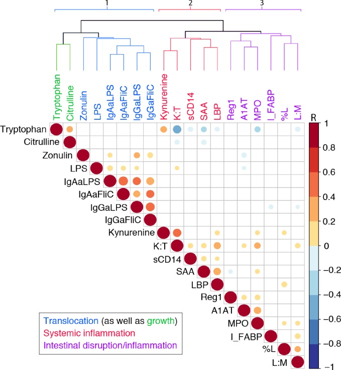
The biomarkers tested tend to cluster as markers of intestinal barrier function (including LPS translocation), intestinal inflammation, or systemic inflammation as shown in Fig 5 (Fig 5) and in S2 Table. Specifically, in Fig 5 a cluster dendrogram with Pearson correlations among those biomarkers with ≥274 values are three main groups: the first representing the translocation markers, LPS and IgA and IgG anti-LPS or FliC as well as zonulin and the 2 predictors of subsequent growth, tryptophan and citrulline. The second group shows predominantly systemic responses to disrupted barrier function and translocation, while the third group are the markers of specific intestinal barrier disruption or local intestinal inflammation. Thus groups 1 and 3 likely lead to the group 2 systemic responses in associating with each other as well as fitting our concept of the pathogenesis of enteropathy. Shown in Fig 5 for the ≥274 biomarkers available within 1 month of enrollment and, more extensively for all tests done in S2 Table, fecal MPO correlates with fecal A1AT, and Reg-1, as well as with urinary L/M ratios, % lactulose and even with kynurenine, K/T ratios and (negatively) with tryptophan. Fecal MPO also correlated significantly with neopterin, albeit in smaller numbers available for neopterin at study start (n = 233; but with n = 279, r = 0.238, p<0.001 for all samples tested shown in S2 Table). Published separately using a subset of 77 of these fecal samples for comparative simultaneous studies of fecal MPO, lactoferrin (LF), calprotectin (FC) and Lipocalin-2 (Lcn-2) [33], we found that the neutrophil markers, MPO, LF and FC correlated best, but also, albeit less tightly, with Lcn-2. The full sample shows that fecal Reg-1 also correlated with A1AT and the intestinal epithelial biomarker Lcn-2 as well as with the inflammation biomarkers, lactoferrin, MPO or neopterin (Fig 1). In addition, fecal MPO correlates with several plasma biomarkers suggesting systemic inflammatory responses, including hsCRP (again, in S2 Table, with smaller numbers, but still significant at p<0.005; r = 0.238; n = 142), as well as with SAA, kynurenine, K/T and negatively with tryptophan.
Fig 5. Cluster dendrogram with Pearson correlations among those biomarkers with ≥274 values showing three main groups: 1) translocation markers, LPS and IgA and IgG anti-LPS or FliC as well as zonulin and the 2 potential predictors of subsequent growth, tryptophan and citrulline; 2) predominantly systemic responses to disrupted barrier function and translocation; and 3) markers of specific intestinal barrier disruption or local intestinal inflammation.
As discussed, groups 1 and 3 may predispose to group 2 systemic responses in associating with each other as shown in the heat map as would fit our concept of the pathogenesis of enteropathy.
Pathway model, using clusters of biomarkers to predict malnutrition and growth
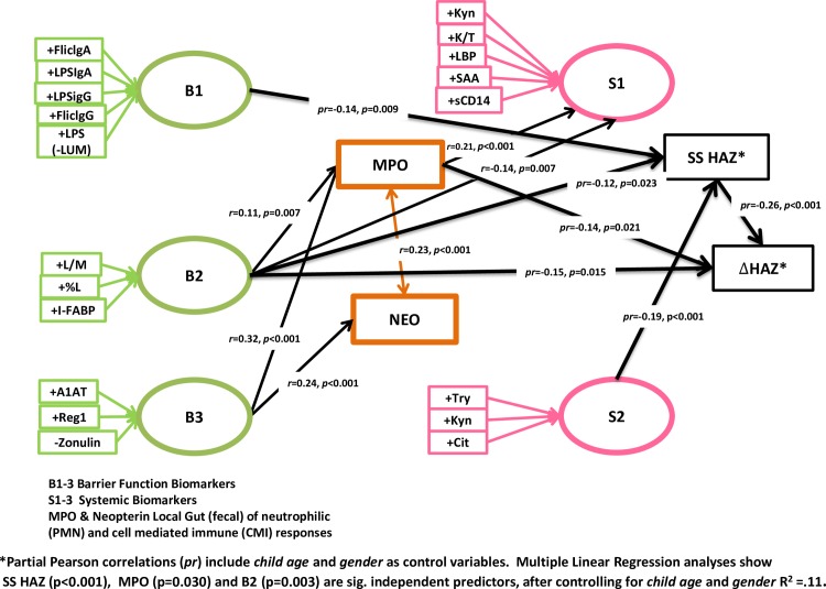
Shown in Fig 6 (Fig 6) are the results of principle components factor analyses to assess parsimonious linear pathways of grouped biomarkers as they relate to children’s growth status and subsequent growth. Available biomarker data with sufficient numbers for this principle component analysis that can be considered to reflect disruption of intestinal barrier function fall into 3 groups as shown, reflecting (B1) LPS ‘translocation’, (B2) absorptive function and epithelial cell damage (L/M and I-FABP), and (B3) further barrier disruption (ex Reg1 and A1AT) with tight junction modulation (zonulin). Most importantly, these factor analyses reveal associations of two pathways that associate with impaired subsequent growth: B2 (disrupted absorptive function and epithelial damage) and MPO (indicating intestinal inflammation); B2 additionally associates with MPO.
Fig 6. Path model, using Principle Components Analyses (Equamax rotation solution maximizing independence of groups) showing associations among 1) Barrier (green), 2) Local Gut (orange) and 3) Systemic (pink) sets of biomarkers, as well as their predictive utility regarding linear growth.
In addition, group B1 (markers of past and recent LPS translocation) associates with stunting at the time of enrollment (‘study start,’ or HAZss); and B2 barrier/absorptive disruption with the systemic cluster (S1: Kyn, K/T, LBP, sCD14, and SAA). Like B2 (barrier/absorptive disruption), the other cluster showing barrier disruption, B3, also associates separately with MPO, and with neopterin. Finally, stunting at enrollment highly significantly predicted ‘catch-up’ growth (ie stunted children indeed did show greater growth that has been termed ‘catch-up’ growth).
Multiple Linear Regression analysis showed HAZss (p<0.001), MPO (p = 0.041) and B2 (p = 0.021) are significant independent predictors of subsequent growth impairment (delta HAZ), after controlling for child age and gender (R2 = .11, p<0.001), or 11% of the growth impairment seen in these children.
Discussion
The need for simple, sensitive biomarkers of environmental enteropathy (EE) in readily available fecal, blood or urine specimens lies in the magnitude of its impact and the need to understand and develop effective interventions. One in every three children in developing areas become stunted in their critically formative first 2 years of life, likely related at least in part to increased exposure to environmental pathogens, often as they are weaned in settings of inadequate water and sanitation [35–37]. The potential developmental consequences of enteropathy with or without overt diarrhea or stunting can be devastating to the full physical and neurocognitive development in one third of the world’s children growing up in impoverished areas. Thus early recognition and intervention in those at greatest risk is of paramount importance. Our findings focus attention on intestinal barrier and absorptive function and inflammation that might reflect increased functional gut permeability (assessed by L/M permeability testing and by serum zonulin levels) and on translocation of microbial products like LPS which then may trigger local and systemic inflammatory responses leading to further structural impairment of gut barrier (A1AT) and, ultimately, stunting growth that characterize EE.
With respect to biomarkers that associate with already stunted children, we find increased systemic IgA antibody against the microbial markers, LPS or FliC (suggesting prior translocation of microbial products), as well as Intestinal-FABP (a marker of enterocyte death) and, in children over 12 months of age, plasma zonulin all suggesting prior intestinal barrier disruption. Similarly, the negative association of the LPS ‘cluster’ (B1) with HAZss is seen in the Principle Components (Partial Pearson correlation) analyses shown in Fig 6. Conversely, the association of reduced SAA in stunted children may reflect an impairment in their host defense responses. In addition, greater citrulline and tryptophan levels in stunted children suggest their involvement in the needed intestinal injury repair or ‘catch-up’ growth, that is clearly greater in these stunted children.
In contrast, biomarkers that help ‘predict’ subsequent growth show the importance of an initial disruption of the intestinal barrier, translocation of LPS and local intestinal and inflammatory signaling as well as the importance of key building blocks like citrulline and tryptophan (albeit with opposite sex associations). Children with higher fecal MPO or fecal A1AT grow less well, and L/M, LPS (reciprocal of LUM), I-FABP and SAA all correlate significantly with impaired subsequent growth. Similarly, the interaction of the 2 fecal markers, MPO and neopterin shows additional power to predict subsequent growth impairment.
The differential association of select biomarkers in girls versus boys likely reflect differences in body composition and basal metabolic rates in males vs females, which are present well before the onset of puberty [38–46]. In addition, boys in low and middle income countries (LMICs) are at greater risk of stunting than girls—especially boys of families with the lowest socioeconomic resources (http://bmcpediatr.biomedcentral.com/articles/10.1186/1471-2431-7-17). Hence, just as children's growth is monitored using sex-adjusted anthropometric indices, the power of biomarkers to predict growth must be considered in the context of sex differences in growth. Further work to understand the biological basis of sexual dimorphisms in growth under adverse conditions is urgently needed.
Indeed, as shown in the cluster dendrogram in Fig 5 (Fig 5), the biomarkers themselves tend to cluster into 3 main groups, reflecting intestinal translocation, intestinal mucosal barrier disruption and inflammation, and systemic inflammatory responses that likely contribute to the potential long-term growth, developmental and metabolic consequences that have been attributed to repeated intestinal infections and “environmental enteropathy” in early childhood [5]. Those that indicate early tight junction effects (zonulin) cluster with the indicators of past or recent LPS translocation and even the potential “catch-up growth” markers, tryptophan and citrulline, while those that indicate intestinal cell and structural barrier disruption (I-FABP, L/M, %L, A1AT and Reg1) cluster with the marker of intestinal inflammation, MPO. Finally, markers of systemic acute phase or proinflammatory responses (SAA, Kynurenine, K/T, sCD14 and LBP) cluster to indicate the systemic responses to intestinal barrier disruption and inflammation that we postulate effect the troubling lasting growth, developmental and metabolic consequences noted above. Somewhat similar clustering of intestinal and systemic biomarkers has also been seen in children in Bangladesh [47].
Furthermore, our multivariate pathway analyses show that barrier function, intestinal inflammation and systemic markers are linearly associationed with each other as well as either previous stunting (ie HAZss at study start) or with subsequent growth (delta HAZ). As observed with single biomarker analyses, this more parsimonious pathway analysis elucidates the direct association of L/M, %L and I-FABP (B2, barrier/absorptive disruption) with growth impairment as well as its indirect association through MPO.
The antibody as well as ‘acute’ LPS cluster was associated with stunting at the time of sampling (as was seen with the IgA antibodies in the individual partial Pearson correlations shown in Table 2; and LPS, as reciprocal LUM, was correlated with subsequent growth impairment as well). As lower LUM values reflect high LPS-neutralizing enzyme activity and thus higher recent systemic LPS exposure, they correlate with subsequent growth impairment (Fig 2D), and with higher IgA or IgG anti-FliC or anti-LPS (r = 0.300, p<0.001 for IgAαFliC; r = 0.211, p<0.001 for IgAαLPS; r = 0.199, p = 0.001 for IgGαLPS; to r = 0.146; p = 0.014 for IgGαFliC). Thus, while IgA or IgG antibody against LPS or FliC likely reflect prior translocation of enteric bacterial components, hence stunting at enrollment; the more current LPS exposure, reflected by the reciprocal luminescence assay for LPS neutralizing activity correlates better with subsequent growth, even though it also correlates with anti-LPS and FliC antibody that had associated with pre-enrollment stunting. Therefore, this LPS-neutralizing enzyme activity assay may provide a better predictor of subsequent growth, irrespective of past stunting. Furthermore, it is clear that having antibody against LPS does not protect against subsequent stunting that is associated with recent evidence of LPS exposure. Several extensive studies have assessed LPS using plasma IgG anti-endotoxin core antibody (EndoCAb) as well as different measures of endotoxin and some, but not others have found impressive associations with impaired intestinal barrier function and growth [25, 28, 48]. These differences may relate to batch or other differences in available assays and hence are the reason we have used different assessments in this work. Taken together, however, consistent with our findings in this study, numerous studies suggest that different assessments of intestinal barrier disruption, and of local and systemic inflammation can be associated with impaired growth.
The third ‘barrier’ cluster (B3) of increased A1AT, Reg-1 and lower zonulin correlated with increased fecal MPO and with increased neopterin. In addition, the tendency for stunted children to experience “catch-up growth” was clearly shown in the association of stunting at study start (HAZss) with better subsequent growth as shown in Fig 6. The host attempt to “catch up” when challenged by growth impairing conditions has also been shown in intestinal cells, where “catch-up” intestinal cell kinase (ICK) is stimulated both in vitro and in vivo by even brief protein deprivation [49].
Intestinal inflammation can also further disrupt barrier function in a vicious cycle. Several fecal biomarkers have emerged as helpful tools in the assessment of intestinal inflammation or immune stimulation. As noted in our methods section, and having studied others (lactoferrin, calprotectin and lipocalin-2 with MPO) [33], we selected MPO and neopterin. The marker of intestinal epithelial cell repair, Reg-1B correlates with Lcn-2 [50], as it does with the marker of more severe barrier disruption, A1AT [51] in these fecal samples.
Although a poorly sensitive assay of structural intestinal barrier disruption, the serum protein that is inherently resistant to digestive degradation, A1AT has offered another biomarker that has shown correlations with epithelial destruction in previous studies [52]. Like lactulose absorption in the L/M test, these markers of barrier disruption could well represent key initiating or, with A1AT, later events in the development and progression of EE. A recent report by Brown et al [20] suggests that dietary deficiencies alone are insufficient to trigger inflammatory or villus architectural disruption without certain microbial components.
Plasma citrulline, an amino acid predominantly synthesized in proximal intestinal epithelial cells and only minimally present in the diet, appears to reflect healthy epithelium that is strikingly reduced in short bowel syndrome, AIDS enteropathy and other villus atrophy syndromes [53]. Its association with better subsequent growth, albeit with initial stunting suggests that it may play a key role in ‘catch-up’ growth, which, when possible is definitely greater in stunted children.
The acute phase protein, SAA (serum amyloid A protein) can be triggered by selected microbiota (segmented filamentous bacteria, SFB) [54] and has been shown to induce epigenetic alterations via JMJD3 that removes the methylation from the H3K27 repressor of the promotors of IL-23, G-CSF and TREM-1, and thus drives proinflammatory cytokine production [55, 56]. Hence, its association with fecal MPO, plasma hsCRP, sCD-14, LBP, kynurenine and K/T ratios (albeit with better HAZ at study start, perhaps reflecting more robust host responses, while also predicting poorer subsequent growth as shown in Fig 2F, and negatively with tryptophan suggest its potential role as not only a biomarker, but a potential pathway involved in the development of environmental enteropathy.
REG1B, the product of regenerating gene1 and marker of intestinal cell damage has been previously associated with growth failure in children in Bangladesh [50]. It has also been recently shown to cluster with sCD14, A1AT, MPO, calprotectin, and neopterin in these children linking environmental enteropathy (EE) with oral vaccine failure [57].
Soluble CD14 is secreted by monocytes and macrophages in response to LPS, and hence joins LBP in reflecting systemic responses to LPS translocation [58]. The marker of enterocyte death, intestinal fatty acid binding protein (I-FABP) [59] joins biomarkers of structural barrier disruption, including L/M and %L as well as A1AT and Reg1 as an indicator of monocyte responses to LPS bioactivity, sCD14 has also been associated with increased IL6, CRP, SAA, D-dimer and increased mortality in HIV-infected individuals [60], as well as directly measured LPS and I-FABP, all presumably reflecting an enteropathy with increased microbial (or microbial product) translocation [61]. The association of zonulin with LBP, IgG anti-LPS and anti-FliC, low tryptophan and malnutrition (lower SS WAZ for all children and, if greater than 12 months old, also SS HAZ) is consistent with the involvement of this tight junction modulator early in development of EE [62], while the reduced zonulin with increased A1AT or neopterin may reflect compensatory zonulin pathway down-regulation secondary to epithelial disruption. Interestingly in the Principle Components analyses (Fig 5), reduced zonulin with increased A1AT and Reg1 associated with both increased MPO and neopterin.
To our knowledge, this is the first evaluation of urinary claudin-15 as a biomarker for healthy gut function. Previous studies by Thuijls and colleagues used Western blotting to measure significant elevations of urinary claudin-3 in patients with active IBD versus those in remission or healthy controls [63]. Here, we successfully used ELISA to quantify urinary claudin-15 and uncovered inverse correlations with urinary L/M, and fecal A1AT, lipocalin-2, and Reg-1. Claudin-15 is highly expressed throughout the intestine [64] in an age-dependent manner [65]. Interestingly, lactating rats upregulate small intestinal Cld-15 expression in association with villous hypertrophy and enhanced calcium absorption [66], whereas Cld-15 ko display megaintestine and sodium deficiency and glucose malabsorption in the small intestine [67]. Taken together, this suggests that increased urinary Cld-15, like urinary mannitol, is a positive biomarker of gut health.
Potential pathways involved may thus include enteric pathogens (especially bacteria and protozoa) that compound dietary insufficiencies to disrupt intestinal mucus and epithelial barrier function, thereby enabling translocation of microbial LPS that triggers both local intestinal as well as systemic inflammatory signaling. Intestinal inflammation can also further disrupt barrier function in a vicious cycle. Both pathogen exposure and inadequate diet are products of poverty, with its concomitant inadequate water, sanitation and food. Our principle components analyses of biomarkers reveal that correlated groups of biomarkers that can be said to reflect barrier disruption have significant correlations with subsequent growth (as does initial HAZ) as well as with fecal and systemic biomarkers and selected metabolites.
Our findings in this report thus support the hypothesis that the systemic effects of potential intestinal barrier disruption and local inflammation on growth are likely occurring through effects of systemic inflammation that have indirect effects through impairing the hepatic synthesis of such key growth mediators as IGFBP-3 and IGF-1, as suggested by DeBoer et al [in press]. However, additional key pathways may also include effects via citrulline or via tryptophan metabolism. These key metabolites may become less available for intestinal injury repair because of increased demands or because of altered host or microbial metabolism or signaling. For example, the activation of indoleamine 2,3-dioxygenase-1 (IDO-1) by microbial translocation or LPS has been shown to be triggered in dendritic cells and monocytes to skew CD4+ T-cell differentiation away from T-helper (Th17) to regulatory T cells (Tregs) [68]. We thus find that lower plasma tryptophan levels not surprisingly associate with biomarkers of barrier disruption and intestinal and systemic inflammation, including higher L/M, zonulin, fecal MPO and lactoferrin, and higher hsCRP, SAA, sCD14, kynurenine and K/T ratios; while lower tryptophan is associated with lower citrulline. Indeed, in metabonomic studies of all metabolites in the urines of these children, we have recently found that tryptophan is likely depleted via both gut microbial metabolism (through the indole pathway to measureable increases in urinary 3-indole sulfate) as well as via the endogenous IDO/kynurenine pathway (as indicated by the increased nicotinic acid, N-methyl nicotinamide and 2-PY metabolites seen in the urine of these undernourished children, by WAZ or HAZ) [69]. In summary, we find that MPO, A1AT, LPS, L/M, I-FABP, and SAA, as well as MPO+ neopterin, and combinations of L/M and %L associate with impaired subsequent growth, with the reverse of these as well as citrulline (in girls) and tryptophan (in boys) offering potential predictors of better growth.
We conclude that fecal MPO and A1AT, and L/M, plasma LPS-neutralizing activity, I-FABP, SAA, hsCRP, citrulline and tryptophan provide promising biomarkers that associate with intestinal barrier disruption, local and systemic inflammation and potential catch-up growth involved in “environmental enteropathy.” These fecal, urinary and systemic biomarkers can prove useful in several ways. First, by improving our understanding of the pathogenesis and its long-term physical and cognitive consequences, they can elucidate its early recognition as well as innovative approaches to ameliorate its potentially devastating impact. Furthermore, they can provide targeted assessment tools with which to evaluate the effectiveness of novel interventions and improved approaches to controlling the potential lasting stunting and developmental consequences of frequent or repeated enteric infections in young children living in impoverished settings. Thus these key biomarkers can help complete the cycle of recognition, understanding, intervening and finally documenting causality of EE and its likely long-term consequences for healthy child development in areas of greatest need.
Supporting Information
(DOCX)
(DOCX)
Red font indicates r>0.25. Red font indicates r>0.25. Green, orange and pink shading represents barrier, gut and systemic biomarker groupings, respectively.
(DOCX)
Acknowledgments
This work was supported by the Bill & Melinda Gates Foundation Biomarker Grants No. OPP1066140 entitled, "Novel metabonomic biomarkers of gut function and health: Modeling enteropathy (EE) and field validation”, and in part by OPP 1066146 and OPP 1099111 “Production of a Reg1B fecal enzyme immunoassay as a potential biomarker of childhood stunting,” by the NIH grant DK048373 entitled “Zot, Zonulin, and Pathophysiology of Intestinal Tight Junctions, by NIH grant K02TW008767, and in part as a case-control component of the “Etiology, Risk Factors and Interactions of Enteric Infections and Malnutrition and the Consequences for Child Health and Development” Project (MAL-ED) carried out as a collaborative project supported by the Bill & Melinda Gates Foundation, the Foundation for the NIH, and the National Institutes of Health, Fogarty International Center. The authors thank the staff and participants of the MAL-ED Network for their important contributions. No authors declare a conflict of interest. RJS has a Clinical Scientist Development Award from the Doris Duke Charitable Foundation.
Abbreviations
- EE
environmental enteropathy
- WAZ
weight for age Z score
- HAZ
height for age Z score
- MPO
myeloperoxidase
- hsCRP
high sensitivity C-reactive protein
- SAA
serum amyloid A
- LPS
lipopolysaccharide
- I-FABP
intestinal fatty acid binding protein
- Reg-1
regenerating gene 1β
- sCD14
soluble cluster of differentiation 14
- TJP1
tight junction protein gene encoding for ZO-1
- IGFBP-3
insulin-like growth factor binding protein-3
- IGF-1
insulin-like growth factor 1
- A1AT
alpha-1-antitrypsin
- Neo
neopterin
- LF
lactoferrin
- FC
fecal calprotectin
- Calp
calprotectin (plasma)
- Lcn-2
lipocalin-2, Cld-2 and Cld-15 = claudin-2 and -15
Data Availability
All relevant data are within the paper and its Supporting Information files. Individual participant data are not publicly available due to ethical restrictions. Requests for further data can be sent to rlg9a@virginia.edu.
Funding Statement
This work was supported by the Bill & Melinda Gates Foundation Biomarker Grants No. OPP1066140 entitled, "Novel metabonomic biomarkers of gut function and health: Modeling enteropathy (EE) and field validation,” and in part by OPP 1066146 and OPP 1099111 “Production of a Reg1B fecal enzyme immunoassay as a potential biomarker of childhood stunting,” by the National Institutes of Health (NIH) grant DK048373 entitled “Zot, Zonulin, and Pathophysiology of Intestinal Tight Junctions," by NIH grant K02TW008767, and in part as a case-control component of the “Etiology, Risk Factors and Interactions of Enteric Infections and Malnutrition and the Consequences for Child Health and Development” Project (MAL-ED) carried out as a collaborative project supported by the Bill & Melinda Gates Foundation, the Foundation for the NIH, and the National Institutes of Health, Fogarty International Center. The authors thank the staff and participants of the MAL-ED Network for their important contributions. No authors declare a conflict of interest. RJS has a Clinical Scientist Development Award from the Doris Duke Charitable Foundation.
References
- 1.Victora CG, Adair L, Fall C, Hallal PC, Martorell R, Richter L, et al. Maternal and child undernutrition: consequences for adult health and human capital. Lancet. 2008;371(9609):340–57. Epub 2008/01/22. 10.1016/S0140-6736(07)61692-4 [DOI] [PMC free article] [PubMed] [Google Scholar]
- 2.Victora CG, de Onis M, Hallal PC, Blossner M, Shrimpton R. Worldwide timing of growth faltering: revisiting implications for interventions. Pediatrics. 2010;125(3):e473–80. Epub 2010/02/17. 10.1542/peds.2009-1519 . [DOI] [PubMed] [Google Scholar]
- 3.Mendez MA, Adair LS. Severity and timing of stunting in the first two years of life affect performance on cognitive tests in late childhood. J Nutr. 1999;129(8):1555–62. Epub 1999/07/27. . [DOI] [PubMed] [Google Scholar]
- 4.Stein AD, Wang M, DiGirolamo A, Grajeda R, Ramakrishnan U, Ramirez-Zea M, et al. Nutritional supplementation in early childhood, schooling, and intellectual functioning in adulthood: a prospective study in Guatemala. Archives of pediatrics & adolescent medicine. 2008;162(7):612–8. Epub 2008/07/09. 10.1001/archpedi.162.7.612 [DOI] [PMC free article] [PubMed] [Google Scholar]
- 5.Guerrant RL, DeBoer MD, Moore SR, Scharf RJ, Lima AA. The impoverished gut—a triple burden of diarrhoea, stunting and chronic disease. Nat Rev Gastroenterol Hepatol. 2013;10(4):220–9. Epub 2012/12/12. 10.1038/nrgastro.2012.239 [DOI] [PMC free article] [PubMed] [Google Scholar]
- 6.Platts-Mills JA, Babji S, Bodhidatta L, Gratz J, Haque R, Havt A, et al. Pathogen-specific burdens of community diarrhoea in developing countries: a multisite birth cohort study (MAL-ED). The Lancet Global health. 2015;3(9):e564–75. Epub 2015/07/24. 10.1016/S2214-109X(15)00151-5 . [DOI] [PMC free article] [PubMed] [Google Scholar]
- 7.Liu J, Kabir F, Manneh J, Lertsethtakarn P, Begum S, Gratz J, et al. Development and assessment of molecular diagnostic tests for 15 enteropathogens causing childhood diarrhoea: a multicentre study. Lancet Infect Dis. 2014;14(8):716–24. Epub 2014/07/16. 10.1016/S1473-3099(14)70808-4 . [DOI] [PubMed] [Google Scholar]
- 8.Checkley W, Epstein LD, Gilman RH, Black RE, Cabrera L, Sterling CR. Effects of Cryptosporidium parvum infection in Peruvian children: growth faltering and subsequent catch-up growth. American journal of epidemiology. 1998;148(5):497–506. Epub 1998/09/16. . [DOI] [PubMed] [Google Scholar]
- 9.Ajjampur SS, Sarkar R, Sankaran P, Kannan A, Menon VK, Muliyil J, et al. Symptomatic and asymptomatic Cryptosporidium infections in children in a semi-urban slum community in southern India. Am J Trop Med Hyg. 2010;83(5):1110–5. Epub 2010/11/03. 10.4269/ajtmh.2010.09-0644 [DOI] [PMC free article] [PubMed] [Google Scholar]
- 10.Checkley W, Buckley G, Gilman RH, Assis AM, Guerrant RL, Morris SS, et al. Multi-country analysis of the effects of diarrhoea on childhood stunting. Int J Epidemiol. 2008;37(4):816–30. Epub 2008/06/24. 10.1093/ije/dyn099 [DOI] [PMC free article] [PubMed] [Google Scholar]
- 11.Lima AA, Moore SR, Barboza MS Jr, Soares AM, Schleupner MA, Newman RD, et al. Persistent diarrhea signals a critical period of increased diarrhea burdens and nutritional shortfalls: a prospective cohort study among children in northeastern Brazil. J Infect Dis. 2000;181(5):1643–51. Epub 2000/05/24. 10.1086/315423 . [DOI] [PubMed] [Google Scholar]
- 12.Moore SR, Lima NL, Soares AM, Oria RB, Pinkerton RC, Barrett LJ, et al. Prolonged episodes of acute diarrhea reduce growth and increase risk of persistent diarrhea in children. Gastroenterology. 2010;139(4):1156–64. Epub 2010/07/20. 10.1053/j.gastro.2010.05.076 [DOI] [PMC free article] [PubMed] [Google Scholar]
- 13.Niehaus MD, Moore SR, Patrick PD, Derr LL, Lorntz B, Lima AA, et al. Early childhood diarrhea is associated with diminished cognitive function 4 to 7 years later in children in a northeast Brazilian shantytown. Am J Trop Med Hyg. 2002;66(5):590–3. Epub 2002/08/31. . [DOI] [PubMed] [Google Scholar]
- 14.Guerrant DI, Moore SR, Lima AA, Patrick PD, Schorling JB, Guerrant RL. Association of early childhood diarrhea and cryptosporidiosis with impaired physical fitness and cognitive function four-seven years later in a poor urban community in northeast Brazil. Am J Trop Med Hyg. 1999;61(5):707–13. Epub 1999/12/10. . [DOI] [PubMed] [Google Scholar]
- 15.Scrimshaw NS, Taylor CE, Gordon JE. Interactions of nutrition and infection. Monograph series World Health Organization. 1968;57:3–329. Epub 1968/01/01. . [PubMed] [Google Scholar]
- 16.Guerrant RL, Schorling JB, McAuliffe JF, de Souza MA. Diarrhea as a cause and an effect of malnutrition: diarrhea prevents catch-up growth and malnutrition increases diarrhea frequency and duration. Am J Trop Med Hyg. 1992;47(1 Pt 2):28–35. Epub 1992/07/01. . [DOI] [PubMed] [Google Scholar]
- 17.Guerrant RL, Oria RB, Moore SR, Oria MO, Lima AA. Malnutrition as an enteric infectious disease with long-term effects on child development. Nutr Rev. 2008;66(9):487–505. Epub 2008/08/30. 10.1111/j.1753-4887.2008.00082.x [DOI] [PMC free article] [PubMed] [Google Scholar]
- 18.Costa LB, Noronha FJ, Roche JK, Sevilleja JE, Warren CA, Oria R, et al. Novel in vitro and in vivo models and potential new therapeutics to break the vicious cycle of Cryptosporidium infection and malnutrition. J Infect Dis. 2012;205(9):1464–71. Epub 2012/03/29. 10.1093/infdis/jis216 [DOI] [PMC free article] [PubMed] [Google Scholar]
- 19.Bolick DT, Roche JK, Hontecillas R, Bassaganya-Riera J, Nataro JP, Guerrant RL. Enteroaggregative Escherichia coli (EAEC) Strain in a Novel Weaned Mouse Model: Exacerbation by Malnutrition, Biofilm as a Virulence Factor, and Treatment by Nitazoxanide. J Med Microbiol. 2013. Epub 2013/03/12. 10.1099/jmm.0.046300-0 . [DOI] [PMC free article] [PubMed] [Google Scholar]
- 20.Brown EM, Wlodarska M, Willing BP, Vonaesch P, Han J, Reynolds LA, et al. Diet and specific microbial exposure trigger features of environmental enteropathy in a novel murine model. Nature communications. 2015;6:7806 Epub 2015/08/05. 10.1038/ncomms8806 [DOI] [PMC free article] [PubMed] [Google Scholar]
- 21.Lindenbaum J, Kent TH, Sprinz H. Malabsorption and jejunitis in American Peace Corps volunteers in Pakistan. Ann Intern Med. 1966;65(6):1201–9. Epub 1966/12/01. . [DOI] [PubMed] [Google Scholar]
- 22.Mata L. The Children of Santa Maria Cauque: A prospective Field Study of Health and Growth. 1978.
- 23.Keusch GT, Rosenberg IH, Denno DM, Duggan C, Guerrant RL, Lavery JV, et al. Implications of acquired environmental enteric dysfunction for growth and stunting in infants and children living in low- and middle-income countries. Food Nutr Bull. 2013;34(3):357–64. Epub 2013/10/31. . [DOI] [PMC free article] [PubMed] [Google Scholar]
- 24.Lunn PG, Northrop-Clewes CA, Downes RM. Intestinal permeability, mucosal injury, and growth faltering in Gambian infants. Lancet. 1991;338(8772):907–10. Epub 1991/10/12. . [DOI] [PubMed] [Google Scholar]
- 25.Campbell DI, Elia M, Lunn PG. Growth faltering in rural Gambian infants is associated with impaired small intestinal barrier function, leading to endotoxemia and systemic inflammation. J Nutr. 2003;133(5):1332–8. Epub 2003/05/06. . [DOI] [PubMed] [Google Scholar]
- 26.Campbell DI, McPhail G, Lunn PG, Elia M, Jeffries DJ. Intestinal inflammation measured by fecal neopterin in Gambian children with enteropathy: association with growth failure, Giardia lamblia, and intestinal permeability. J Pediatr Gastroenterol Nutr. 2004;39(2):153–7. Epub 2004/07/23. . [DOI] [PubMed] [Google Scholar]
- 27.Kosek M, Haque R, Lima A, Babji S, Shrestha S, Qureshi S, et al. Fecal markers of intestinal inflammation and permeability associated with the subsequent acquisition of linear growth deficits in infants. Am J Trop Med Hyg. 2013;88(2):390–6. Epub 2012/11/28. 10.4269/ajtmh.2012.12-0549 [DOI] [PMC free article] [PubMed] [Google Scholar]
- 28.Lin A, Arnold BF, Afreen S, Goto R, Huda TM, Haque R, et al. Household environmental conditions are associated with enteropathy and impaired growth in rural Bangladesh. Am J Trop Med Hyg. 2013;89(1):130–7. Epub 2013/05/01. 10.4269/ajtmh.12-0629 [DOI] [PMC free article] [PubMed] [Google Scholar]
- 29.Humphrey JH. Child undernutrition, tropical enteropathy, toilets, and handwashing. Lancet. 2009;374(9694):1032–5. Epub 2009/09/22. 10.1016/S0140-6736(09)60950-8 . [DOI] [PubMed] [Google Scholar]
- 30.Korpe PS, Petri WA Jr. Environmental enteropathy: critical implications of a poorly understood condition. Trends in molecular medicine. 2012;18(6):328–36. Epub 2012/05/29. 10.1016/j.molmed.2012.04.007 [DOI] [PMC free article] [PubMed] [Google Scholar]
- 31.Lima AA, Oria RB, Soares AM, Filho JQ, de Sousa F Jr, Abreu CB, et al. Geography, population, demography, socioeconomic, anthropometry, and environmental status in the MAL-ED cohort and case-control study Sites in Fortaleza, Ceara, Brazil. Clin Infect Dis. 2014;59 Suppl 4:S287–94. Epub 2014/10/12. 10.1093/cid/ciu438 . [DOI] [PubMed] [Google Scholar]
- 32.Guideline: Updates on the Management of Severe Acute Malnutrition in Infants and Children. WHO Guidelines Approved by the Guidelines Review Committee. Geneva2013. [PubMed]
- 33.Prata MMG HA, Bolick DT, Pinkerton R, Lima AMM, Guerrant RL. Comparisons between myeloperoxidase, lactoferrin, calprotectin and lipocalin-2, as fecal biomarkers of intestinal inflammation in malnourished children. J Transl Sci. 2016;2(2):134–9. 10.15761/JTS.1000130 [DOI] [PMC free article] [PubMed] [Google Scholar]
- 34.Gunzel D, Yu AS. Claudins and the modulation of tight junction permeability. Physiological reviews. 2013;93(2):525–69. Epub 2013/04/17. 10.1152/physrev.00019.2012 [DOI] [PMC free article] [PubMed] [Google Scholar]
- 35.Collaborators GBDRF, Forouzanfar MH, Alexander L, Anderson HR, Bachman VF, Biryukov S, et al. Global, regional, and national comparative risk assessment of 79 behavioural, environmental and occupational, and metabolic risks or clusters of risks in 188 countries, 1990–2013: a systematic analysis for the Global Burden of Disease Study 2013. Lancet. 2015. Epub 2015/09/15. 10.1016/S0140-6736(15)00128-2 . [DOI] [PMC free article] [PubMed] [Google Scholar]
- 36.Black RE, Allen LH, Bhutta ZA, Caulfield LE, de Onis M, Ezzati M, et al. Maternal and child undernutrition: global and regional exposures and health consequences. Lancet. 2008;371(9608):243–60. Epub 2008/01/22. 10.1016/S0140-6736(07)61690-0 . [DOI] [PubMed] [Google Scholar]
- 37.Kosek M, Lanata CF, Black RE, Walker DG, Snyder JD, Salam MA, et al. Directing diarrhoeal disease research towards disease-burden reduction. J Health Popul Nutr. 2009;27(3):319–31. Epub 2009/06/11. [DOI] [PMC free article] [PubMed] [Google Scholar]
- 38.Mast M, Kortzinger I, Konig E, Muller MJ. Gender differences in fat mass of 5-7-year old children. International journal of obesity and related metabolic disorders: journal of the International Association for the Study of Obesity. 1998;22(9):878–84. Epub 1998/10/02. . [DOI] [PubMed] [Google Scholar]
- 39.van der Sluis IM, de Ridder MA, Boot AM, Krenning EP, de Muinck Keizer-Schrama SM. Reference data for bone density and body composition measured with dual energy x ray absorptiometry in white children and young adults. Archives of disease in childhood. 2002;87(4):341–7; discussion -7. Epub 2002/09/24. [DOI] [PMC free article] [PubMed] [Google Scholar]
- 40.Horlick M, Thornton J, Wang J, Levine LS, Fedun B, Pierson RN Jr. Bone mineral in prepubertal children: gender and ethnicity. Journal of bone and mineral research: the official journal of the American Society for Bone and Mineral Research. 2000;15(7):1393–7. Epub 2000/07/14. 10.1359/jbmr.2000.15.7.1393 . [DOI] [PubMed] [Google Scholar]
- 41.Arfai K, Pitukcheewanont PD, Goran MI, Tavare CJ, Heller L, Gilsanz V. Bone, muscle, and fat: sex-related differences in prepubertal children. Radiology. 2002;224(2):338–44. Epub 2002/07/31. 10.1148/radiol.2242011369 . [DOI] [PubMed] [Google Scholar]
- 42.Herd SL, Gower BA, Dashti N, Goran MI. Body fat, fat distribution and serum lipids, lipoproteins and apolipoproteins in African-American and Caucasian-American prepubertal children. International journal of obesity and related metabolic disorders: journal of the International Association for the Study of Obesity. 2001;25(2):198–204. Epub 2001/06/19. 10.1038/sj.ijo.0801524 . [DOI] [PubMed] [Google Scholar]
- 43.He Q, Horlick M, Thornton J, Wang J, Pierson RN Jr, Heshka S, et al. Sex and race differences in fat distribution among Asian, African-American, and Caucasian prepubertal children. The Journal of clinical endocrinology and metabolism. 2002;87(5):2164–70. Epub 2002/05/08. 10.1210/jcem.87.5.8452 . [DOI] [PubMed] [Google Scholar]
- 44.Cowell CT, Briody J, Lloyd-Jones S, Smith C, Moore B, Howman-Giles R. Fat distribution in children and adolescents—the influence of sex and hormones. Hormone research. 1997;48 Suppl 5:93–100. Epub 1997/01/01. . [DOI] [PubMed] [Google Scholar]
- 45.Mitamura R, Yano K, Suzuki N, Ito Y, Makita Y, Okuno A. Diurnal rhythms of luteinizing hormone, follicle-stimulating hormone, testosterone, and estradiol secretion before the onset of female puberty in short children. The Journal of clinical endocrinology and metabolism. 2000;85(3):1074–80. Epub 2000/03/17. 10.1210/jcem.85.3.6445 . [DOI] [PubMed] [Google Scholar]
- 46.Taylor RW, Gold E, Manning P, Goulding A. Gender differences in body fat content are present well before puberty. International journal of obesity and related metabolic disorders: journal of the International Association for the Study of Obesity. 1997;21(11):1082–4. Epub 1997/11/22. . [DOI] [PubMed] [Google Scholar]
- 47.Naylor C, Lu M, Haque R, Mondal D, Buonomo E, Nayak U, et al. Environmental Enteropathy, Oral Vaccine Failure and Growth Faltering in Infants in Bangladesh. EBioMedicine. 2015;2(11):1759–66. Epub 2016/02/13. 10.1016/j.ebiom.2015.09.036 [DOI] [PMC free article] [PubMed] [Google Scholar]
- 48.Benzoni N, Korpe P, Thakwalakwa C, Maleta K, Stephenson K, Manary M, et al. Plasma endotoxin core antibody concentration and linear growth are unrelated in rural Malawian children aged 2–5 years. BMC research notes. 2015;8:258 Epub 2015/06/25. 10.1186/s13104-015-1238-1 [DOI] [PMC free article] [PubMed] [Google Scholar]
- 49.Bolick DT, Chen T, Alves LA, Tong Y, Wu D, Joyner LT 2nd, et al. Intestinal cell kinase is a novel participant in intestinal cell signaling responses to protein malnutrition. PLoS One. 2014;9(9):e106902 Epub 2014/09/04. 10.1371/journal.pone.0106902 [DOI] [PMC free article] [PubMed] [Google Scholar]
- 50.Peterson KM, Buss J, Easley R, Yang Z, Korpe PS, Niu F, et al. REG1B as a predictor of childhood stunting in Bangladesh and Peru. Am J Clin Nutr. 2013;97(5):1129–33. Epub 2013/04/05. 10.3945/ajcn.112.048306 [DOI] [PMC free article] [PubMed] [Google Scholar]
- 51.Rahaman MM WA. Direct Nutrient Loss and Diarrhea In: Scrimshaw LCaN, editor. Diarrhea and Malnutrition: Interactions, Mechanisms, and Interventions Edited by Chen LC and Scrimshaw NS: Plenum Press; 1983. p. 155–60. [Google Scholar]
- 52.Soendergaard C, Nielsen OH, Seidelin JB, Kvist PH, Bjerrum JT. Alpha-1 antitrypsin and granulocyte colony-stimulating factor as serum biomarkers of disease severity in ulcerative colitis. Inflammatory bowel diseases. 2015;21(5):1077–88. Epub 2015/03/25. 10.1097/MIB.0000000000000348 . [DOI] [PubMed] [Google Scholar]
- 53.Crenn P, Messing B, Cynober L. Citrulline as a biomarker of intestinal failure due to enterocyte mass reduction. Clinical nutrition. 2008;27(3):328–39. Epub 2008/04/29. 10.1016/j.clnu.2008.02.005 . [DOI] [PubMed] [Google Scholar]
- 54.Burgess SL, Buonomo E, Carey M, Cowardin C, Naylor C, Noor Z, et al. Bone marrow dendritic cells from mice with an altered microbiota provide interleukin 17A-dependent protection against Entamoeba histolytica colitis. mBio. 2014;5(6):e01817 Epub 2014/11/06. 10.1128/mBio.01817-14 [DOI] [PMC free article] [PubMed] [Google Scholar]
- 55.Ye RD, Sun L. Emerging functions of serum amyloid A in inflammation. J Leukoc Biol. 2015. Epub 2015/07/02. 10.1189/jlb.3VMR0315-080R . [DOI] [PMC free article] [PubMed] [Google Scholar]
- 56.Yan Q, Sun L, Zhu Z, Wang L, Li S, Ye RD. Jmjd3-mediated epigenetic regulation of inflammatory cytokine gene expression in serum amyloid A-stimulated macrophages. Cellular signalling. 2014;26(9):1783–91. Epub 2014/04/08. 10.1016/j.cellsig.2014.03.025 [DOI] [PMC free article] [PubMed] [Google Scholar]
- 57.Naylor C, Lu M, Haque R, Mondal D, Buonomo E, Nayak E, et al. Environmental enteropathy, oral vaccine failure and growth faltering in infants in Bangladesh. EBioMedicine. 2015. [DOI] [PMC free article] [PubMed] [Google Scholar]
- 58.Kitchens RL, Thompson PA. Modulatory effects of sCD14 and LBP on LPS-host cell interactions. Journal of endotoxin research. 2005;11(4):225–9. Epub 2005/09/24. 10.1179/096805105X46565 . [DOI] [PubMed] [Google Scholar]
- 59.Pelsers MM, Namiot Z, Kisielewski W, Namiot A, Januszkiewicz M, Hermens WT, et al. Intestinal-type and liver-type fatty acid-binding protein in the intestine. Tissue distribution and clinical utility. Clinical biochemistry. 2003;36(7):529–35. Epub 2003/10/18. . [DOI] [PubMed] [Google Scholar]
- 60.Chevalier MF, Petitjean G, Dunyach-Remy C, Didier C, Girard PM, Manea ME, et al. The Th17/Treg ratio, IL-1RA and sCD14 levels in primary HIV infection predict the T-cell activation set point in the absence of systemic microbial translocation. PLoS pathogens. 2013;9(6):e1003453 Epub 2013/07/03. 10.1371/journal.ppat.1003453 [DOI] [PMC free article] [PubMed] [Google Scholar]
- 61.Sandler NG, Wand H, Roque A, Law M, Nason MC, Nixon DE, et al. Plasma levels of soluble CD14 independently predict mortality in HIV infection. J Infect Dis. 2011;203(6):780–90. Epub 2011/01/22. 10.1093/infdis/jiq118 [DOI] [PMC free article] [PubMed] [Google Scholar]
- 62.El Asmar R, Panigrahi P, Bamford P, Berti I, Not T, Coppa GV, et al. Host-dependent zonulin secretion causes the impairment of the small intestine barrier function after bacterial exposure. Gastroenterology. 2002;123(5):1607–15. Epub 2002/10/31. . [DOI] [PubMed] [Google Scholar]
- 63.Thuijls G, Derikx JP, de Haan JJ, Grootjans J, de Bruine A, Masclee AA, et al. Urine-based detection of intestinal tight junction loss. Journal of clinical gastroenterology. 2010;44(1):e14–9. Epub 2009/06/16. 10.1097/MCG.0b013e31819f5652 . [DOI] [PubMed] [Google Scholar]
- 64.Suzuki H, Nishizawa T, Tani K, Yamazaki Y, Tamura A, Ishitani R, et al. Crystal structure of a claudin provides insight into the architecture of tight junctions. Science. 2014;344(6181):304–7. Epub 2014/04/20. 10.1126/science.1248571 . [DOI] [PubMed] [Google Scholar]
- 65.Holmes JL, Van Itallie CM, Rasmussen JE, Anderson JM. Claudin profiling in the mouse during postnatal intestinal development and along the gastrointestinal tract reveals complex expression patterns. Gene expression patterns: GEP. 2006;6(6):581–8. Epub 2006/02/07. 10.1016/j.modgep.2005.12.001 . [DOI] [PubMed] [Google Scholar]
- 66.Wongdee K, Teerapornpuntakit J, Siangpro C, Chaipai S, Charoenphandhu N. Duodenal villous hypertrophy and upregulation of claudin-15 protein expression in lactating rats. Journal of molecular histology. 2013;44(1):103–9. Epub 2012/09/25. 10.1007/s10735-012-9451-x . [DOI] [PubMed] [Google Scholar]
- 67.Wada M, Tamura A, Takahashi N, Tsukita S. Loss of claudins 2 and 15 from mice causes defects in paracellular Na+ flow and nutrient transport in gut and leads to death from malnutrition. Gastroenterology. 2013;144(2):369–80. Epub 2012/10/24. 10.1053/j.gastro.2012.10.035 . [DOI] [PubMed] [Google Scholar]
- 68.Favre D, Mold J, Hunt PW, Kanwar B, Loke P, Seu L, et al. Tryptophan catabolism by indoleamine 2,3-dioxygenase 1 alters the balance of TH17 to regulatory T cells in HIV disease. Science translational medicine. 2010;2(32):32ra6 Epub 2010/05/21. 10.1126/scitranslmed.3000632 [DOI] [PMC free article] [PubMed] [Google Scholar]
- 69.Mayneris-Perxachs J, Lima AA, Guerrant RL, Leite AM, Moura AF, Lima NL, et al. Urinary N-methylnicotinamide and beta-aminoisobutyric acid predict catch-up growth in undernourished Brazilian children. Scientific reports. 2016;6:19780 Epub 2016/01/28. 10.1038/srep19780 [DOI] [PMC free article] [PubMed] [Google Scholar]
Associated Data
This section collects any data citations, data availability statements, or supplementary materials included in this article.
Supplementary Materials
(DOCX)
(DOCX)
Red font indicates r>0.25. Red font indicates r>0.25. Green, orange and pink shading represents barrier, gut and systemic biomarker groupings, respectively.
(DOCX)
Data Availability Statement
All relevant data are within the paper and its Supporting Information files. Individual participant data are not publicly available due to ethical restrictions. Requests for further data can be sent to rlg9a@virginia.edu.


