Abstract
Vitex pubescens is a Malaysian therapeutic plant employed in traditional drug to remedy a variety of disorders. The purpose of this research is to assess the gastroprotective efficiency of V. pubescens leaves against ethanol-induced gastric hemorrhagic laceration in rats. Animals were randomly allocated into seven groups and pre-treated, separately, with 10% Tween 20 (normal and ulcer control groups), 20 mg/kg omeprazole (reference group), and 62.5, 125, 250, and 500 mg/kg of V. pubescens extract (experimental groups). All animals were sacrificed after another hour. Histological evaluation of the ulcer control group revealed significant injury to the gastric mucosa with edema and leucocyte infiltration of the submucosal layer. PAS staining, showed remarkably intense magenta color, remarkable increase of HSP70 and decrease of Bax proteins in rats pre-treated with plant extracts compared to the ulcer control group. Gastric homogenates revealed a remarkable increase in endogenous antioxidant enzyme activities (CAT, SOD, GSH) and a decrease in the lipid peroxidation level (MDA) in animals pre-treated with V. pubescens extract compared with the ulcer control group. The gastroprotective activity of this plant might be related to increased antioxidant enzymes and decrease lipid peroxidation upsurge of HSP70 and reduced expression of Bax proteins
Introduction
Gastric ulcers are benign lesions of the gastric mucosa that affect countless people worldwide [1]. The cause is a disproportion between well-known destructive factors and the mucosal protection mechanisms in the mucosal epithelium [2]. The interaction of acid and pepsin is the major source of gastric sores. Many factors can increase the occurrence of gastrointestinal disorders, including tension, smoking, malnutrition, intake of non-steroidal anti-inflammatory drugs, inherited predisposition and infection by Helicobacter pylori [3]. Meanwhile, increases in scavenging enzymes such as superoxide glutathione peroxidase (GPx), catalase (CAT) and dismutase (SOD) can decrease the free radical cytotoxicity of the gastrointestinal membrane [4].
Many drugs, such as proton pump inhibitors (omeprazole), antacids and antihistaminic agents, are utilized in the management of gastric ulcers; however, these drugs have many adverse side effects [5]. Sometimes anti-acid medicines are ineffective or reduce the absorption of magnesium, vitamin B12, calcium and iron which require an acidic gastric medium for bioavailability [6]. Medicinal plants can assist in ulcer therapy and in avoiding reappearance and are applied as a substitute for other forms of management [7, 8, 9]. A huge number of conventional therapeutic plants with gastroprotective activity have been described in the literature by numerous researchers [10–20].
Vitex pubescens Vahl. (Verbenaceae) is well-known as Halban in the peninsula of Malaysia. This medicinal herb is employed for the healing of gastrointestinal symptoms, such as diarrhea, and even for scorpion stings [21]. The glue of its leaves is helpful for abrasions [22] and is also used for fever as an antipyretic [23] In addition, it has anti-dysentery, anti-inflammatory, analgesic, anti-fungal and anti-tumor activities [21, 24]. This project was designed to assess the gastroprotective activity of the ethanolic extract of V. pubescens in gastric ulcers in rats.
Materials and Methods
Plant Extraction
V. pubescens leaves were gathered from the backyard of RimbaIlmu, University of Malaya (Rimba Ilmu Botanic Garden) (101o40’E 3o8’N). The species was identified by Dr. Sugumaran Manickam from Institute of Biological Sciences, Faculty of Science, University of Malaya. A voucher has been deposited in the herbarium with the number KLU 48647. The plant was identified using taxonomic literature. The plant leaves were rinsed with tap water, dried in the dark for 5 days, and then ground to a powder using an electric blender. One hundred grams was placed in 900 ml of 95% ethanol for four to five days in a laboratory glass container (1 L) with daily stirring. Afterward, the mixture was filtered through filter paper (Whatman No. 1). The EtOH (ethanol) extraction distillation of plants was conducted under reduced pressure in a Buchi Rotary Evaporator R-215 (Chemoph-arm Sdn. Bhd., Switzerland). V. pubescens leaves produced a yield of 9.7% (dark-green) (w/w). The dried extract was stored at -20°C in a freezer (Panasonic, Japan) until use. Then, 10% Tween 20 was used to dissolve the extract, which was orally administered to rats at dosages of 2 g/kg and 5 g/kg body weight for acute toxicity testing [25] and at dosages of 62.5, 125, 250, and 500 mg/kg body weight for anti-ulcer activity, in accordance with earlier reports [13].
Fractionation of the Crude Extracts
V. pubescens leaves crude ethanol extracts were fractionated using reversed phased column, Luna 5u C18(2) 100A (phenomenex), dimensions (150 x 21.20 mm and particle size, 5.0 μm) with preparative high performance liquid chromatography (HPLC) (PREPLC purificat and multiple wavelength detectors (Kromaton, Interchim), puriflash 4250–250, UK). The mobile system that used was 90:10 (water: methanol) to 100% (methanol) (Merck, Darmstadt, Germany) in 180 minutes. The HPLC parameters involved 6 ml of injection volume and the flow rate was 5 mL/ min. After that, the seven fractions of V. pubescens leaves (F1 to F7) were obtained and collected for cell viability assessment in vitro using MTT assay against normal human hepatic cell line (WRL-64). Then the active fraction of plant, which was F2 in V. pubescens, continued with a liquid chromatography mass spectrometry (LC-MS) analysis to identify the mass of active compounds.
MTT Assay and Cytotoxicity in the Cell Line
The MTT assay was performed to assess the in vitro cytotoxic properties of the V. pubescens leaves crude ethanol extract and its fractions against WRL-68 cells (human hepatic cell line) purchased from the American Type Culture Collection (ATCC, Manassas, VA, USA). The formazan crystals created were dissolved in DMSO, and the absorbance was measured at 570 nm using a microplate reader. Cell viability was estimated by the standard formula [26].
Identification of Active Compounds by LC-MS
Agilent technologies 6490 series triple quad LC-MS (QQQ) mass spectrometer with dual ESI source, G6490, Singapore) was used to identify the active compounds of V. pubesccens of F2. The LC-MS parameters included 1μL of volume of injection and the flow rate was 0.5 mL/ min with XBridge C18 2.5 μm 2.5 x 50mm columns (Waters, Ireland) using 90:10 (water: acetonitrile) to 100% (acetonitrile) (Merck, Darmstadt, Germany) in 10 minutes. Lastly, the five identified compounds of V. pubescens (F2) was investigated based on mass of charge (m/z).The data of LC-MS was processed and analysed by using the agilent mass hunter qualitative analysis B.06.00.
Antioxidant Activity In Vitro
Ferric-reducing antioxidant power (FRAP) assay
The antioxidant level of the ethanol extracts of the plant which lead to reduce the ferric was measured based on the suggested assay [27] with minor amendment. Briefly, the FRAP reagent was prepared from acetate buffer (pH 3.6), 10 mM TPTZ [2,4,6-Tri(2-pyridyl)- s-triazine] solution in 40 mM HCl and 20 mM iron (III) chloride solution at a ratio of 10:1:1 (v/v), correspondingly.
Quercetin, gallic acid, BHT, and ascorbic acid were also used as controls. Ten microliters of plant extract, standard and controls were added to 300 microliters of the FRAP reagent (triplicate) in the dark for 4 min. Next, the absorbance was measured at 593 nm using a PowerWave ×340 ELISA Reader spectrophotometer (Bio-Tek Instruments, Inc., Winooski, VT, USA). The standard curve was linear (R2 = 0.998) between 100 and 1000 M FeSO4. The outcomes were presented as M Fe (II)/g dry weight of the extract.
2 Diphenyl-picrylhydrazyl (DPPH) radical activity scavenging assay
The antioxidant activity of the ethanol extract of the plant was established using a 1,1-diphenyl-2-picrylhydrazyl (DPPH) radical base for the electron transfer reaction between DPPH reagent and the plant extracts. The technique described in [28] was performed with slight alterations. Stock solution (1 mg/1 ml) of the plant extracts was diluted to obtain five different concentrations (50, 25, 12.5, 6.25, 3.125, 1.56 μg/mL), and an antioxidant standard (ascorbic acid) was employed. Specified amounts of each plant extract (5 μL) and the standards were mixed with 195 μL DPPH (40 × dilution) in triplicate and kept warm at 37°C. The absorbance value was determined for 2 hrs at 20 min intervals using a PowerWave ×340 ELISA Reader spectrophotometer (Bio-Tek Instruments, Inc., Winooski, VT, USA) at 515 nm. The radical scavenging activity was estimated via the following equation:
where AB is the absorption of a blank sample, and AA is the absorption of a tested sample.
The 50% inhibitory concentration was determined in addition to the kinetics of the DPPH scavenging reaction. In addition, quercetin, gallic acid, BHT, and ascorbic acid were analyzed against DPPH as positive controls.
Ethical Status
This research was permitted by the ethics committee for animal experimentation from the Faculty of Medicine, University of Malaya, Malaysia (Ethic No. PM/30/05/2014/NSIAW (R) and the National Academy of Science’s Guide for the Care and Use of Laboratory Animals [29].
Experimental Animals
Sprague Dawley rats (SD) (6–8 weeks old) were purchased from the Animal House, Faculty of Medicine, University of Malaya. The body weight of the rats was 160–190 g. Prior to dosing, the animals were fasted for one night (water was provided). Moreover, food was withdrawn for an additional 3 to 4 hours after dosing with the extract.
Acute Toxicity Test
The acute toxicity test was conducted to determine a safe dose of the extract. Thirty-six SD rats (equal numbers of males and females) were divided into 3 equal groups, which received vehicle (10% Tween 20, 5 mL/kg), a low dose (2 g/kg), and a high dose (5 g/kg) of the plant extract preparation [25]. The dosage for the acute toxicity experiment was chosen based on the OECD Guideline for toxicity testing [29].
The animals were observed for 30 min and or 2, 4, 8, 24 and 48 hrs after the administration of the drug for any signs of toxicity and behavioral abnormality. The animals were then monitored daily for typical symptoms of toxicity for 15 days. Serum biochemical, histological (kidney and liver) and hematological factors were evaluated. On the 15th day, the animals were anesthetized by xylazine with ketamine. Blood was collected intracardially, and the animals were then sacrificed by an overdose of anesthesia to obtain the kidney and liver. Histology and serum biochemical parameters were examined.
Gastric Ulcer
Omeprazole
Omeprazole was employed as the reference antiulcer drug. It was dissolved in 10% Tween 20 and orally administered to the rats at a dosage of 20 mg/kg body weight (5 mL/kg) [30].
Ethanol-induced gastric ulceration
Healthy adult Sprague Dawley male rats (180–207 g) were purchased from Animal House, Faculty of Medicine, University of Malaya, and approved by the Ethics Committee with Ethics Number PM/30/05/2014/NSIAW (R). The animals were separated into seven groups of 6 rats. They were fasted for 24 h (with regard to food but not water), and water was removed for 2 hrs prior to the experiment. The rats were located separately in a wire-bottomed cage to avoid coprophagy. All the groups were pre-treated and treated by oral gavage.
Group 1 (normal control group) and Group 2 (ulcer control group) were fed 10% Tween 20 (5 mL/kg).
Group 3 were administered 20 mg/kg omeprazole, as the reference control group.
Groups 4, 5, 6 and 7 (experimental groups) were administered dosages of 62.5, 125, 250, and 500 mg/kg of V. pubescens ethanol extract, respectively. Sixty minutes afterwards,
Group 1 rats were administered 10% Tween 20 (5 mL/kg).
Groups 2–7 rats were administered absolute ethanol (5 mL/kg) [30].
Then, all animals were euthanized one hour later through an overdose of xylazine and ketamine, followed by immediate excision of their stomachs.
Measurement of gastric juice acidity and mucus content
The stomach of each animal was opened, and the hydrogen ion concentration of the gastric contents was analyzed by pH meter titration with 0.1 N NaOH. The acid content was expressed as meq/l. The gastric mucosa of the stomach was scraped smooth using a glass slide and weighed using an electronic balance [18].
Macroscopic and gross stomach lesion assessment
An ulcer of the gastric mucosa emerges as extended bands of hemorrhagic superficial lesions parallel to the stomach. All rats were observed for gastric mucosa injury. The length and width of the ulcer (mm) were then calculated using a planimeter (10 × 10 mm2 = ulcer area) under dissecting microscope (1.8x). The ulcerated area was determined by counting the number of small squares, 2 mm × 2 mm, covering the length and width of each ulcer band. The total areas of all lesions were included in the calculation of the ulcer area (UA) based on the number of small squares × 4 × 1.8 = UA (mm2), according to the recommendation of [31]. The inhibition percentage (I.0%) was calculated by the following formula based on the recommendation of [16].
Histological studies of gastric lesions
Hematoxylin and eosin staining
Samples of the stomach walls were fixed in 10% buffered formalin, processed, and embedded in a paraffin tissue-processing machine (Leica, Germany). Sections of the stomach were prepared at a thickness of 5 μm for histological hematoxylin and eosin staining [32].
Study of mucosal glycoproteins
To evaluate mucus production, particular slides were stained with periodic acid Schiff base (PAS) following the manufacturer’s instructions (Sigma Periodic Acid- Schiff Commercial Kit) to evaluate the changes in glycoproteins (acidic and basic). A light microscope (Nikon, Japan) was employed to photograph and examine the produced mucus [33].
Immunohistochemistry evaluation
Tissue section slides were heated for 25min in a hot air oven at 60°C. Xylene was used to deparaffinise and rehydrate by graded alcohol. Then, 10mM sodium citrate buffer was used for antigen retrieval process heating in microwave. Immunohistochemical staining using the manufacturer’s protocol (CYAN, Dakocytomation, USA) was conducted. Proxidase block (0.03% hydrogen peroxide containing sodium azide) was used for 5min to block the endogenous peroxidase. Later, rinsed off the slides with washing buffer to incubate with biotinylated primary antibodies HSP70 and Bax for 15min. Slides were washed gently with washing buffer and were put in buffer bath and later were incubated in a humid chamber with streptavidin HRP (to horseradish peroxidase in PBS mixed with anti-microbial agent) for 15 min. Then, slides were rinsed gently and put in buffer bath. Tissue slides were incubated for 5 min with Diaminobenzidine (DAB)-substrate-chromagen followed by washing and then were stained with hematoxylin for 5sec. Sections were dipped 10 times in ammonia (0.037 mol/L) and quickly washed with distilled water mounted with cover slips. Slides were observed for positive immunohistochemical stains (brown stains) using light microscope. Protein expressions were quantitated using the Image J software program [34]. The data are the mean ± SD. Statistical significance was expressed as p < 0.05.
Antioxidant activity of gastric homogenate
Preparation of homogenate
The tissue of gastric specimens was rinsed thoroughly with cold phosphate buffer saline (PBS). Homogenates (10% (w/v)) were then prepared with cold 50 mM (PBS) (pH 7.4) using a homogenizer (Polytron, Heidolph RZR 1, Germany). The homogenates were then centrifuged at 10,000 rpm for 15 min at 4°C by a Rotofix 32 refrigerated centrifuge (Hettich Zentrifugen, Germany). The supernatant was used for investigation of the antioxidant activities and lipid peroxidation level.
Determination of protein concentration
Protein concentrations (mg/mL tissue) were determined using Bradford’s solutions (Amresco LCC. Co., USA). At 2 min after adding 100 μL of the above solution to 10 μL of samples in each well at various concentrations, the absorbance was recorded at a wavelength of 595 nm [35].
Measurements of antioxidant activities of stomach homogenate
The SOD, CAT and GSH activities of the gastric tissues were measured using commercial kits (Cayman Chemical Co., Ann Arbor, USA). The manufacturer’s protocols were used to determine their activities in the gastric tissue supernatant.
Measurements of lipid peroxidation (MDA) level of stomach homogenate
Lipoperoxidation of the mucus membrane in the stomach was measured using commercial kits (Cayman Chemical Co., Ann Arbor, USA).
Statistical analysis
All values were analyzed as means ± S.E.M. The statistical significance of differences between groups was calculated using one-way ANOVA followed by post hoc Tukey’s multiple comparison tests. A value of p < 0.05 was defined as significant.
Results
MTT Assay and Cytotoxicity in Cell Line
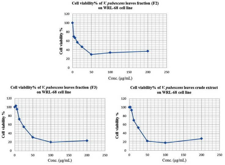
In our study, the effects of plant with fractions (F1 to F7) showed no cytotoxicity and their IC50 were more than 100 μmol/L even at higher concentrations. The moderate cell viability of V. pubescens crude ethanol extracts and V. pubescens fraction F3 exhibited 27%, 23% respectively and the highest viability of V. pubescens fraction F2 was 37% at 200 μg/mL in a dose dependent manner (Fig 1). Thus, fraction F2 of V. pubescens that has a higher viability on WRL 68 was subjected to identify the active compounds using LC-MS.
Fig 1. The effect of different concentrations of V. pubescens fraction (F2) on the viability of WRl-68.
The Data (In triplicate) were expressed as mean ± SEM.
Identification of Active Compounds of the Plant Active Fraction
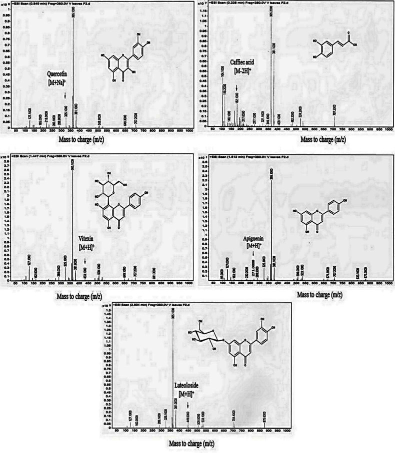
LC-MS was used to identify the active compounds of V. pubescens leaves fraction (F2), and the peaks obtained with their retention time (RT), molecular weight and molecular formula were identified (Table 1 and Fig 2). Our results showed five identified compounds that were investigated based on mass of charge (m/z). They are Caffeic acid 182.100 [M-2H]+, Quercetin 325.100 [M+Na]+, Vitexin 433.100 [M+H]+, Apignenin 271.000 [M+H]+ and luteoloside 449.000 [M+H]+.
Table 1. V. pubescens (F2) compounds identification by LC-MS.
| No | RT (min) | Name of identified compounds | Mass of charge (m/z) | Molecular weight (g/mol) | Molecular formula |
|---|---|---|---|---|---|
| 1 | 0.338 | Caffeic acid | 182.100 [M-2H]+ | 180.16 | C9H8O4 |
| 2 | 0.849 | Quercetin | 325.100 [M+Na]+ | 302.236 | C15H10O7 |
| 3 | 1.447 | Vitexin | 433.100 [M+H]+ | 432.38 | C21H20O10 |
| 4 | 1.612 | Apignenin | 271.000 [M+H]+ | 270.237 | C15H10O5 |
| 5 | 2.894 | luteoloside | 449.000 [M+H]+ | 448.37 | C21H20O11 |
Fig 2. The Mass spectrum (Triple quad QQQ MS ESI+) and identified chemical structure in V. pubescens leaves, F2.
In Vitro Antioxidant Activity of Ethanol Extract of Plants
Ferric reducing antioxidant power (FRAP)
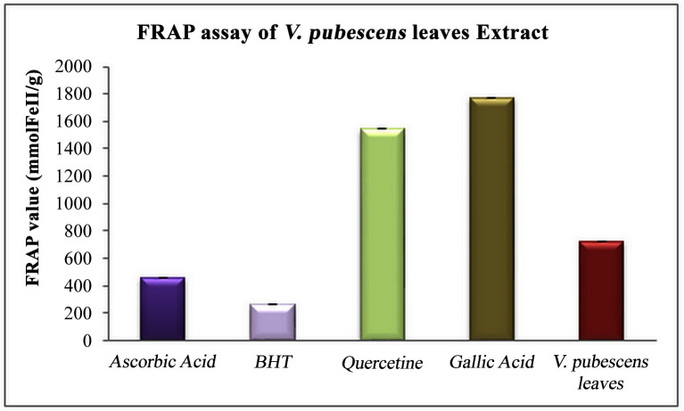
The total antioxidant activity of the ethanol extracts of the plants was calculated by FRAP assay. The reduction of ferric to ferrous ion presented a higher FRAP value for V. pubescens leaf than BHT or ascorbic acid, at 723.0 ± 0.03 μmol Fe (II)/g. The standards applied were BHT, ascorbic acid, quercetin, and gallic acid, whose FRAP values were 261.0 ± 0.009 μmol Fe (II)/g, 457.7 ± 0.005 μmol Fe (II)/g, 1544.3± 0.012 μmol Fe (II)/g, and 1774.3 ± 0.002 μmol Fe (II)/g, respectively (Fig 3).
Fig 3. Total antioxidant activity.
Total antioxidant activity of V. pubescens ethanolic extracts with synthetic reference standards (ascorbic acid, BHT, quercetin, and gallic acid) as determined by FRAP assay. The data are represented as the mean ± SEM.
The free radical scavenging activity (DPPH) assay
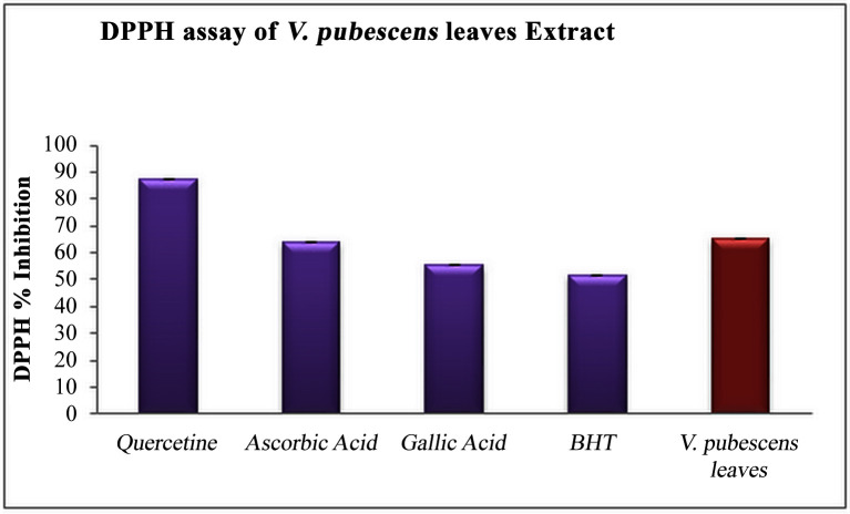
Antioxidant activity and the capability of plant ethanol extracts to scavenge free radicals in vitro were evaluated by the DPPH assay. Fig (4) shows the percentage inhibition of DPPH free-radical scavenging activity of V. pubescens leaves ethanol extract to be 65.32 with an IC50 value of 38.3 ± 0.1 μg/mL. The results were compared to the standards BHT, ascorbic acid, quercetin and gallic acid. The % inhibition levels of the DPPH free-radical scavenging activity of the standards were 51.63, 64.11, 87.52, and 55.47 with IC50 values of 9.1 ± 0.15 μg/mL, 4.9 ± 0.11 μg/mL, 1.8 ± 0.04 μg/mL, and 1.4 ± 0.13 μg/mL, respectively.
Fig 4. DPPH free radical scavenging activity.
DPPH free radical scavenging activity (Inhibition%) of V. pubescens ethanolic extract compared to synthetic reference standards (ascorbic acid, BHT, quercetin, and gallic acid). The data are represented as the mean ± SEM.
In vivo acute toxicity of V. pubescens leaf extract
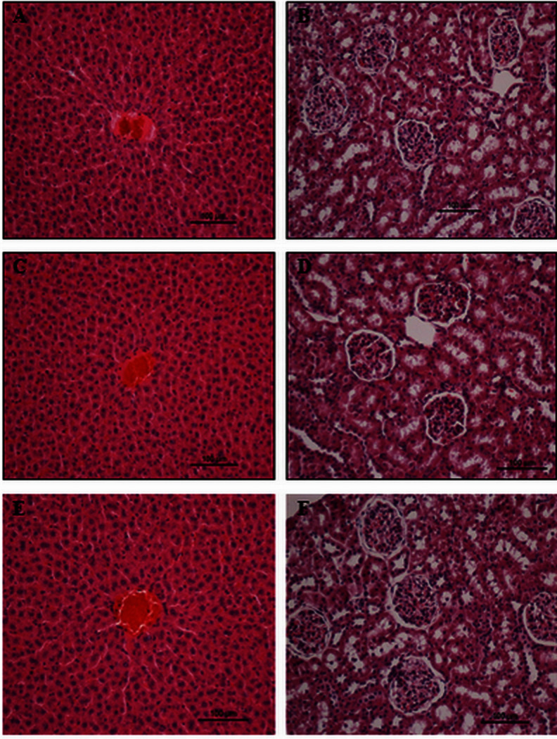
The rats treated with the plant extract demonstrated no mortality or toxic symptoms of this experimentation. There were no body (liver and kidney) weight variations, abnormal physiological changes or behavioral changes at 2 g/kg and 5 g/kg doses throughout 14 days, as illustrated in Table 2. The histological markers of the liver and kidney and their weights were normal based on biochemical analysis and comparable to the control groups, as shown in Fig 5 and Tables 3, 4, 5 and 6. Subsequently, male and female rats manifested no notable signs of toxicity at the orally administered dosages.
Table 2. Effects of V. pubescens leaf extract ethanol extract on the body (liver and kidney) weights of male and female rats.
| V. pubescens extract | Liver Weight | Kidney Weight | ||
|---|---|---|---|---|
| Male | Female | Male | Female | |
| Vehicle (10%Tween20) | 7.23 ± 0.08 | 6.36 ± 0.32 | 1.90± 0.07 | 1.54 ± 0.06 |
| V. pubescens (2 g/kg) | 6.85 ± 0.28 | 6.57 ± 0.28 | 1.59 ± 0.03 | 1.74 ± 0.17 |
| V. pubescens (5 g/kg) | 6.57± 0.30 | 5.40 ± 0.13 | 1.80 ± 0.06 | 1.43 ± 0.07 |
Values expressed as mean ± S.E.M. There are no significant changes between groups.
* Significant value at p <0.05
Fig 5. Liver and kidney histology of the acute toxicity assay.
(A and B) Rats fed with 5 mL/kg of vehicle (10% Tween 20). (C and D) Rats fed with 2 g/kg (5 mL/kg) of V. pubescens extract. (E and F) Rats fed with 5 g/kg (5 mL/kg) of V. pubescens extract. There were 6 rats in each group of experiment. No significant changes were observed between the treated and vehicle control groups (Hematoxylin and Eosin stain).
Table 3. Effects of V. pubescens leaf extract on kidney biochemical parameters in female rats.
| Dose female | Vehicle (10%Tween20) | V. pubescens (2 g/kg) | V. pubescens (5 g/kg) |
|---|---|---|---|
| Sodium (mmol/L) | 147.3 ± 0.76 | 147.3 ± 0.42 | 146.3 ± 0.71 |
| Potassium (mmol/L) | 5.1 ± 0.18 | 4.9 ± 0.09 | 5.0 ± 0.22 |
| Chloride (mmol/L) | 107.0 ± 0.58 | 107.3 ± 0.33 | 108.5 ± 0.62 |
| CO2 (mmol/L) | 25.27 ± 0.52 | 25.45 ± 0.38 | 23.72 ± 0.41 |
| Anion Gap (mmol/L) | 21.8± 0.60 | 21.0 ± 0.52 | 21.0 ± 0.52 |
| Urea (mmol/L) | 5.28 ± 0.18 | 5.88± 0.11 | 5.55 ± 0.10 |
| Creatinine (μmol/L) | 38.5 ± 2.73 | 44.7± 3.17 | 37.8 ± 3.28 |
Values expressed as mean ± S.E.M. There are no significant changes between groups.
* Significant value at p <0.05.
Table 4. Effects of V. pubescens leaf extract on liver biochemical parameters in female rats.
| Dose female | Vehicle (10%Tween20) | V. pubescens (2 g/kg) | V. pubescens (5 g/kg) |
|---|---|---|---|
| Total Protein (g/L) | 80.3 ± 2.29 | 81.2 ± 3.45 | 79.8± 1.68 |
| Albumin (g/L) | 14.3 ± 0.67 | 13.2 ± 0.87 | 12.7± 0.71 |
| Globulin (g/L) | 65.3 ± 1.41 | 62.7± 1.48 | 64.5± 1.12 |
| TB (μmol/L) | 2.7 ± 0.33 | 2.2 ± 0.17 | 2.5 ± 0.22 |
| CB (μmol/L) | 1.5 ± 0.2 | 1 ± 0.00 | 1.5 ± 0.2 |
| ALP (IU/L) | 108.83 ± 9.13 | 108.17 ± 4.33 | 105.00 ± 10.33 |
| ALT (IU/L) | 51.0 ± 0.58 | 47.0 ± 1.37 | 51.0 ± 1.06 |
| AST (IU/L) | 216.7 ± 11.72 | 204.2 ± 4.90 | 205.7 ± 10.10 |
| GGT (IU/L) | 5.7 ± 0.84 | 5.0 ± 0.52 | 4.0 ± 0.37 |
Values expressed as mean ± S.E.M. There are no significant changes between groups. CO2: carbon dioxide;TB: total bilirubin; CB: conjugated bilirubin; AP: alkaline phosphatase; ALT: alanine aminotransferase; AST: aspartate aminotransferase;GGT: G-glutamyltransferase.
* Significant value at p <0.05.
Table 5. Effects of V. pubescens leaf extract on kidney biochemical parameters in male SD rats.
| Dose female | Vehicle (10%Tween20) | V. pubescens (2 g/kg) | V. pubescens (5 g/kg) |
|---|---|---|---|
| Total Protein (g/L) | 80.3 ± 2.29 | 81.2 ± 3.45 | 79.8± 1.68 |
| Albumin (g/L) | 14.3 ± 0.67 | 13.2 ± 0.87 | 12.7± 0.71 |
| Globulin (g/L) | 65.3 ± 1.41 | 62.7± 1.48 | 64.5± 1.12 |
| TB (μmol/L) | 2.7 ± 0.33 | 2.2 ± 0.17 | 2.5 ± 0.22 |
| CB (μmol/L) | 1.5 ± 0.2 | 1 ± 0.00 | 1.5 ± 0.2 |
| ALP (IU/L) | 108.83 ± 9.13 | 108.17 ± 4.33 | 105.00 ± 10.33 |
| ALT (IU/L) | 51.0 ± 0.58 | 47.0 ± 1.37 | 51.0 ± 1.06 |
| AST (IU/L) | 216.7 ± 11.72 | 204.2 ± 4.90 | 205.7 ± 10.10 |
| GGT (IU/L) | 5.7 ± 0.84 | 5.0 ± 0.52 | 4.0 ± 0.37 |
Values expressed as mean ± S.E.M. There are no significant changes between groups.
* Significant value at p <0.05
Table 6. Effects of V. pubescens leaf extract on liver biochemical parameters in male SD rats.
| Dose male | Vehicle (10%Tween20) | V. pubescens (2 g/kg) | V. pubescens (5 g/kg) |
|---|---|---|---|
| Total Protein (g/L) | 68.0 ± 1.26 | 69.5 ± 1.65 | 67.7 ± 0.88 |
| Albumin (g/L) | 12.2 ± 0.48 | 11.8 ± 0.60 | 12.0 ± 0.37 |
| Globulin (g/L) | 56.2 ± 0.75 | 58.2± 1.11 | 56.0 ± 0.77 |
| TB (μmol/L) | 2.5 ± 0.43 | 2.3 ± 0.21 | 2.5 ± 0.22 |
| CB (μmol/L) | 1.7 ± 0.21 | 1.00 ± 0.00 | 1.3 ± 0.21 |
| ALP (IU/L) | 196.33 ± 9.75 | 188.33 ± 1.89 | 193.17 ± 14.46 |
| ALT (IU/L) | 58.3 ± 4.25 | 56.8 ± 0.70 | 56.7 ± 0.84 |
| AST (IU/L) | 227.3 ± 12.52 | 222.2 ± 7.64 | 213.3 ± 5.17 |
| GGT (IU/L) | 3.8 ± 0.31 | 3.7 ± 0.42 | 3.3 ± 0.21 |
Values expressed as mean ± S.E.M. There are no significant changes between groups. CO2: carbon dioxide;TB: total bilirubin; CB: conjugated bilirubin; AP: alkaline phosphatase; ALT: alanine aminotransferase; AST: aspartate aminotransferase;GGT: G-glutamyltransferase.
* Significant value at p <0.05.
Antiulcer Study
Gross evaluation
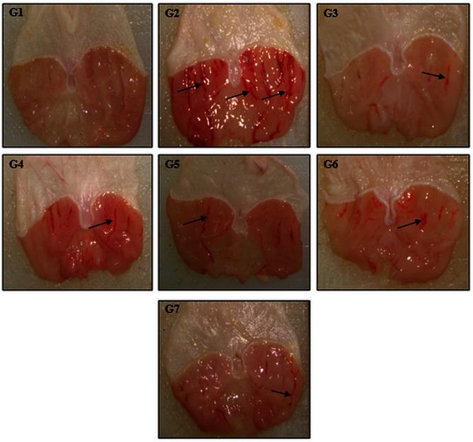
The results established that the pre-treatment of animals with V. pubescens extract and reference drugs significantly reduced the ulcer area of the gastric mucosa compared to the ulcer control group (Table 7 and Fig 6). The ulcer control group demonstrated significantly severe hemorrhagic injury to the gastric mucosa compared to animals pre-treated with the plant extract or omeprazole.
Table 7. Effect of the V. pubescens leaf extracts on the pH of gastric content, mucus weight, ulcer area, and % inhibition of ulcer area in stomach.
| Animal Groups | Pre-treatment 5ml/kg | PH | Mucus weight | Ulcer area | Inhibition % |
|---|---|---|---|---|---|
| Normal control (G1) | 10% Tween20 | 7.17 ± 0.38* | 2.29 ± 0.15* | - | - |
| Ulcer control (G2) | 10% Tween20 | 2.77 ± 0.28 | 0.76 ± 0.15 | 801.60 ± 35.65 | - |
| Omeprazole (G3) | 20mg/kg | 5.72 ± 0.49* | 1.93 ± 0.06* | 96.00 ± 20.42* | 88 |
| V. pubescens leaf extract (G4) | (62.5mg/kg) | 4.99 ± 0.53 | 0.79 ± 0.05 | 336.10 ± 23.22* | 58 |
| V. pubescens leaf extract (G5) | (125mg/kg) | 4.73 ± 0.70 | 0.83 ± 0.05 | 319.57 ± 37.02* | 60 |
| V. pubescens leaf extract (G6) | (250mg/kg) | 4.94 ± 0.49 | 1.47 ± 0.20* | 180.00 ± 20.87* | 77 |
| V. pubescens leaf extract (G7) | (500mg/kg) | 4.75 ± 0.65 | 1.80 ± 0.19* | 166.80 ± 35.05* | 79 |
The results are expressed as mean ± S.EM.
* Indicates significance at p< 0.05 compared to ulcer group.
Fig 6. Macroscopic appearance evaluation of gastric mucosa.
(G1) (Normal control group); (G2) (Ulcer control group); (G3) (Omeprazole); (G4) (62.5 mg/kg), (G5) (125 mg/kg), (G6) (250 mg/kg) and (G7) (500 mg/kg) V. pubescens extract. Black arrow indicates hemorrhagic injury. There were 6 rats in each group of experiment.
Gastric mucus content and acidity
As shown by the results in Table 7, ulcerated animals in G2 (the ulcer control group) yielded the lowest mucus content of the gastric mucosa, while animal groups pre-treated with 500 mg/kg and 250 mg/kg of V. pubescens leaf extract or omeprazole showed significantly increased mucus content compared to the ulcer control group.
Rats pre-treated with V. pubescens leaf extract or omeprazole demonstrated a significant upsurge in the pH of the gastric contents compared to rats pre-treated with vehicle (ulcer control group) (Table 7).
Measurement of gastric antioxidant enzymes and membrane lipid peroxidation (MDA)
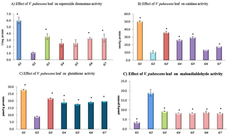
The ulcer control group showed a significant reduction in antioxidant (SOD, CAT and GSH) activities compared with omeprazole or the experimental animal groups (Fig 7).
Fig 7.
Effect of V. pubescens leaf extract on antioxidant activities of (A) superoxide dismutase, (B) catalase, (C) glutathione, (D) malondialdehyde. (G1) (Normal control group); (G) (Ulcer control group); (G3) (Omeprazole); (G4) (62.5 mg/kg), (G5) (125 mg/kg), (G6) (250 mg/kg) and (G7) (500 mg/kg) of V. pubescens extract. All values are expressed as the mean ± SEM. *mean significant at (p <0.05) in rats pre-treated with omeprazole or V. pubescens extract compared to ulcer control group. There were 6 rats in each group of experiment.
The MDA level was markedly greater in the ulcer control group compared with the reference drug (omeprazole) or experimental animals pre-treated with V. pubescens leaf extract (Fig 7).
Histological evaluation of gastric lesions
Hematoxylin and eosin staining and PAS staining
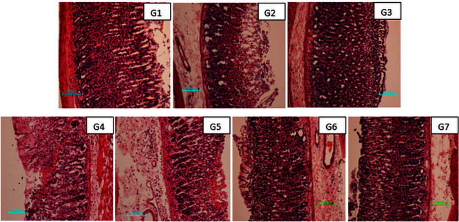
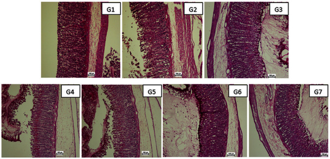
Histological observation of the ulcer control group showed a remarkable disruption of gastric mucosa that penetrated extensively and deeply into the gastric mucosa and also revealed extensive edema and leucocyte infiltration of the submucosal layer compared to the reference group or the experimental animals. Rats pre-treated with omeprazole or plant extracts demonstrated markedly improved protection of the gastric mucosa in a dose-dependent manner and an absence or reduction of edema and leucocyte infiltration of the submucosal layer (Fig 8). The gastric mucosa in the rats pre-treated with omeprazole or the experimental groups demonstrated marked increases in Periodic acid Schiff (PAS) staining intensity compared to the ulcer control group (Fig 9), indicating a higher glycoprotein content of the gastric mucosa compared with the ulcer group.
Fig 8. Effects of V. pubescens on the histology of the stomach wall in ethanol-induced gastric mucosal damage in rats.
(G1) (Normal control group); (G2) (Ulcer control group); (G3) (Omeprazole); (G4) (62.5 mg/kg), (G5) (125 mg/kg), (G6) (250 mg/kg) and (G7) (500 mg/kg) of V. pubescens extract. There were 6 rats in each group of experiment. The rats in the experimental groups (groups 4–7) exhibited markedly better protection of the gastric mucosa, as shown by the reduced ulcer area (white arrow), submucosal edema and leucocyte infiltration (blue arrow) of the submucosal layer (H&E staining magnification 10×).
Fig 9. Effects of V. pubescens on gastric tissue glycoprotein-PAS staining in ethanol-induced gastric injury rats.
(G1) (Normal control group); (G) (Ulcer control group); (G3) (Omeprazole); (G4) (62.5 mg/kg), (G5) (125 mg/kg), (G6) (250 mg/kg) and (G7) (500 mg/kg) of V. pubescens extract. Animals pre-treated with omeprazole or V. pubescens extract exhibited intense glycoprotein staining (white arrow) (PAS stain magnification 20×). There were 6 rats in each group of experiment.
Immunohistochemistry staining
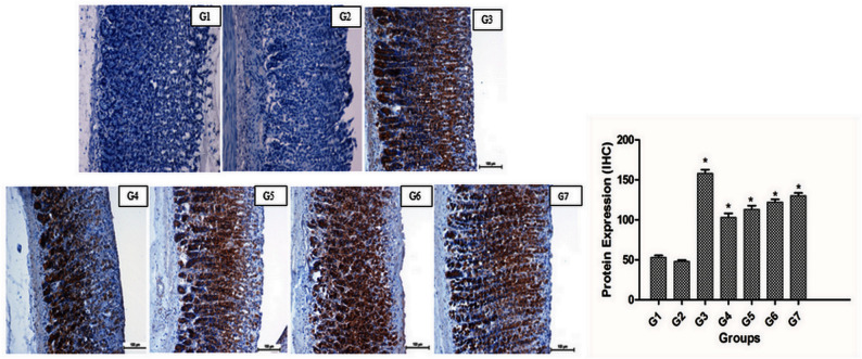
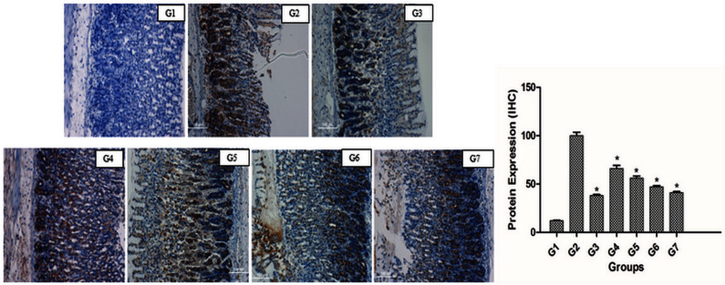
The expression level of HSP70 protein in the gastric mucosa indicated reduced expression in the ulcer control group; however, over-expression of HSP70 protein appeared in rats pre-treated with omeprazole or V. pubescens leaf extract (Fig 10). However, the immunostaining of BAX protein in the gastric wall mucosa showed over-expression in the ulcer control group and reduced expression in rats pre-treated with omeprazole or plant extracts (Fig 11).
Fig 10. Effects of V. pubescens on immunohistochemical staining (HSP70 staining) of stomach wall in ethanol-induced gastric mucosal injury in rats.
(G1) (Normal control group); (G2) (Ulcer control group); (G3) (Omeprazole); (G4) (62.5 mg/kg), (G5) (125 mg/kg), (G6) (250 mg/kg) and (G7) (500 mg/kg) of V. pubescens extract. HSP70 protein was over-expressed in rats pre-treated with omeprazole or V. pubescens extract (brown color shows over-expression of HSP70 protein) (magnification 20×). There were 6 rats in each group of experiment. The Image J program was used to evaluate protein expression. All values are expressed as the means ± the standard error of mean. The mean difference was significant at the p < 0.05 level compared to the cancer control group. Fig 10G1 is excluded from this article's CC BY license. See the accompanying retraction notice for more information.
Fig 11. Effects of V. pubescens on immunohistochemical staining (Bax staining) of stomach wall in ethanol-induced gastric mucosal injury in rats.
(G1) (Normal control group); (G2) (Ulcer control group); (G3) (Omeprazole); (G4) (62.5 mg/kg), (G5) (125 mg/kg), (G6) (250 mg/kg) and (G7) (500 mg/kg) of V. pubescens extract. Bax protein was over-expressed in ulcer control animals (brown) (magnification 20×). There were 6 rats in each group of experiment. The Image J program was used to evaluate protein expression. All values are expressed as the means ± the standard error of mean. The mean difference was significant at the p < 0.05 level compared to the cancer control group.
Discussion
The outcomes of this study revealed that the administration of V. pubescens leaf extract showed no toxicity and no mortality in vivo and no cytotoxicity toward WRL-69 cells in vitro. This result is consistent with other reports [8, 18, 36]. The mitochondrial reduction MTT assay is one of the most frequently used to determine cytotoxicity and cell proliferation. When V. pubescens leaves crude ethanol extract and its fractions with varying concentrations assessed for 48 hrs treatments on hepatic human cell line WRL-68 cells, the cell viabilities were measured using the MTT assay. Amongst all the fractions, number 2 revealed the highest viability used for further identification procedure which the resulted which are similar to identification of Vitex negundo [37].Our investigation demonstrated that V. pubescens possesses good free-radical scavenging and antioxidant activities in vitro. Similarly, the consumption of medicinal plants containing natural antioxidants leads to decreased free radicals and neutralization of their effects, protecting biological molecules from oxidative damage [7, 12, 20, 21]. Several mechanisms are associated with the production of gastric mucosal ulcers. Ethanol directly induces injury to the mucosa of gastric, declining the bicarbonates secretion and the generation of mucus. Ethanol induced injury to the gastrointestinal mucosa begins with the distraction of the vascular endothelium, consequently increasing the vascular permeability and leading to edema and leucocyte infiltration of the submucosal layer [9, 16, 38, 39].
Our findings showed protection of the stomach wall mucosa and reduction of the ulcer area in animals pre-treated with V. pubescens leaf extract. Consistently, numerous authors have reported reduction in the ulcer area of the gastric mucosa, increasing the protection of gastric from ulcers in rats [10, 11, 12, 20]. Ethanol might severely injure the stomach wall mucosa, resulting in elevated neutrophil infiltration into the ulcerated mucosa. Oxygen free radicals originating from penetrated neutrophils in the injured stomach wall impair the outcome of gastric ulcers prevention in rats [11, 40]. Neutrophils are a highly important resource of inflammatory mediators and can release powerful reactive oxygen species that are extremely cytotoxic and encourage tissue injury [41]. Additionally, neutrophil accumulation in the stomach mucosa has been shown to provoke microcirculatory abnormalities [14, 18]. The inhibition of neutrophil permeation throughout inflammation was established to improve gastric ulcer prevention [16].
In this investigation, flattening of the gastric mucosal folds occurred, suggesting that the anti-ulcer outcome of V. pubescens leaf extract strength is associated with a decline in gastric motility. It has been mentioned in literatures that alteration to gastric motility is involved in the avoidance of tentative gastric injury [9, 11, 15, 19]. The outcome of our study exhibited intense staining of the glycoprotein secretions of the gastric wall mucosa glands in rats pre-treated with omeprazole or V. pubescens leaf extract. Mucus secretion is among the important mechanisms of gastric mucosal defense against necrotizing agents [15, 30, 42, 43]. Mucus and bicarbonate secretion might play a significant role in the ulcer-inhibiting process because the mucus/bicarbonate layer protects newly formed cells from acid and peptic injury [17, 37].
Oxidative stress may play a major role in the induction and pathogenesis of stomach ulcers, and antioxidant enzymes have been mentioned to play a main defensive role of protection of the stomach wall mucosa against a variety of necrotic agents [7, 19, 30]. Antioxidant enzymes could inhibit ethanol-induced gastric damage in rat [8], and V. pubescens leaf extract has been demonstrated to contain antioxidants [21]; thus, it is possible that the gastroprotective properties of V. pubescens could be due to its antioxidant properties. Antioxidants are responsible for protecting the gastric mucosa from ulceration [43], as they possess the capability to protect tissue against damages via a radical scavenging mechanism [44]. An earlier study provided evidence that ethanol can cause gastric tissue injury through increasing reactive oxygen species (ROS) development [45]. Consequently, ROS accumulation reduced the GSH level and increased lipid peroxidation [46]. GSH can reduce oxidative stress [47] and perform a significant defensive function against ethanol-induced gastric cell damage [48]. Thus, the detrimental effect of ethanol on the gastric mucosa is clearly linked with decreased GSH levels [49]. Moreover, ethanol affects the properties of the gastric tissue by elevating lipid peroxidation [50], that MDA is the major creation of lipid peroxidation. Thus, MDA acts as a marker of ROS-mediated gastric injuries [51]. This study indicates that the stomach is protected through pre-treatment with V. pubescens by increasing the level of GHS and decrease of MDA level in comparison with ulcer control group.
Our project outcomes indicate that the gastric tissue MDA level was considerably augmented in the ulcer control group, with a significant decrease in the antioxidant enzyme activities of SOD and CAT and of the GSH level in the gastric homogenate. Pre-treatment with V. pubescens significantly reduced the malondialdehyde (MDA) concentration level, an indicator of lipid peroxidation, and also significantly increased the reduced antioxidant enzyme activities in the stomach homogenates, most likely by inhibiting the production of lipid peroxides from fatty acids in the stomach. The reduction in MDA enzyme by the V. pubescens extract in response to the oxidative stress in animals might be because of the antioxidant effect of the extract in the stomach homogenate, where the severe damage to the mucous membranes caused by ethanol is prevented. The significantly decreased levels of MDA in animals fed with the plant extract might be due to the decreased oxidative gastric damage [50]. The findings are consistent with data published elsewhere [9, 17, 31]. Amongst the several pathological trials produced by an inequity among oxidative injury and antioxidant protection systems, lipid peroxidation is a form of oxidative harm that disrupts cell membranes. Similar results have been reported by several researchers [13, 16, 18, 19, 33].
The administration of absolute ethanol cause injuries to the epithelial cells, causing a decrease in protein concentrations. SOD and CAT are the main scavenging enzymes that eliminate radicals in vivo [18]. A decline in the activity of these antioxidant enzymes lead the additional accessibility of superoxide radicals, such as superoxide anions and hydrogen peroxide.
Ethanol-generated ROS by unfolding and aggregation of proteins cause damage of proteins. HSP70 is an important endogenous cytoprotective factor. The cells are protected from oxidative stress by HSP70 proteins and allowed refold of the partially denatured proteins. In this study, upsurge of HSP70 could suggest that V. pubescens protected the stomach through the increase of HSP70 by increasing mucosal blood flow under stress conditions. The induction of HSP70 seems to affect the mucosal protection. In agreement with the results of the current investigation, several studies have reported the upsurge of HSP70 protein to protect the stomach from necrotizing agents [9, 13, 16, 30]. Under stressful and thermal conditions, the HSP70 protein is induced and performs its cytoprotective repair role through its molecular chaperone activity [52, 53]. Ethanol damages the gastric mucosa and produces lesions, and based on our experiments, V. pubescens treatment in rats exerts its protective role through significant HSP70 induction to reduce lesion development [9, 14, 18].
Following mitochondrial injury and apoptosis activation, Bax, a key pro-apoptotic protein, is translocated to the mitochondria from the cytoplasm [54].
Immunohistochemical analysis showed that V. pubescens extract significantly inhibited increase Bax protein expression. Therefore, these results demonstrate that V. pubescens extract exhibits s significant protective efficiency against injury in rat’s stomach, which is related to decrease of Bax protein. Numerous studies on this effect have been reported by many investigators [9, 13, 16, 19, 30].
Conclusion
Based on the results of this study, V. pubescens leaf extract exhibited significant and dose-dependent anti-ulcer protection against ethanol-induced gastric lesions in the rat model. The gastroprotective effect of V. pubescens was associated with the effective direct radical scavenging activity, increased SOD, catalase and GSH levels, depression of lipid peroxidation, HSP70 protein up-regulation and decreased Bax protein.
Data Availability
All relevant data are within the paper and its Supporting Information files.
Funding Statement
The authors have no support or funding to report.
References
- 1.Diniz LR, Vieira CF, Santos EC, Lima GC, Aragão KK, Vasconcelos RP, et al. Gastroprotective effects of the essential oil of Hyptis crenata Pohl ex Benth. on gastric ulcer models. J Ethnopharmacol. 2013; 51:179–187. doi: 10.1016/j.jep.2013.07.026 [DOI] [PubMed] [Google Scholar]
- 2.Shaker E, Mahmoud H, and Mona S. Anti-inflammatory and anti-ulcer activity of the extract from Alhagi maurorum (camelthorn). Food Chem Toxicol. 2010; 48:2785–2790. doi: 10.1016/j.fct.2010.07.007 [DOI] [PubMed] [Google Scholar]
- 3.Ji C, Fan D, Li W, Guo L, Liang Z, Xu R, Zhang J. Evaluation of the anti-ulcerogenic activity of the antidepressants duloxetine, amitriptyline, fluoxetine and mirtazapine in different models of experimental gastric ulcer in rats. Eur. J. Pharmacol. 2012; 691:46–51. doi: 10.1016/j.ejphar.2012.06.041 [DOI] [PubMed] [Google Scholar]
- 4.Tuluce Y, Ozkol H, Koyuncu I, Ine H. Gastroprotective effect of small centaury (Centaurium erythraea L) on aspirin-induced gastric damage in rats. Toxicol. Ind. Health. 2011; 27:760–768. doi: 10.1177/0748233710397421 [DOI] [PubMed] [Google Scholar]
- 5.Da Silva L., Allemand A, Mendes D, dos Santos A, André E, de Souza L, et al. Ethanolic extract of roots from Arctium lappa L. accelerates the healing of acetic acid-induced gastric ulcer n rats: Involvement of the antioxidant system. Food Chem Toxicol. 2013; 51, 179–187. doi: 10.1016/j.fct.2012.09.026 [DOI] [PubMed] [Google Scholar]
- 6.Ham M, Kaunitz J. Gastroduodenal mucosal defense. Curr Opin Gastroenterol. 2008; 24(6):665–73. doi: 10.1097/MOG.0b013e328311cd93 [DOI] [PubMed] [Google Scholar]
- 7.Abdelwahab S, Taha M, Abdulla M, Nordin N, Hadi H, Mohan S, et al. Gastroprotective mechanism of Bauhinia thonningii Schum. J Ethnopharmacol. 2013; 148(1): 277–286. doi: 10.1016/j.jep.2013.04.027 [DOI] [PubMed] [Google Scholar]
- 8.Moghadamtousi S, Rouhollahi E, Karimian H, Fadaeinasab M, Abdulla M. The gastroprotective activity of Annona muricata leaves against ethanol-induced gastric injury in rats via hsp70/Bax involvement. Drug Des Dev Ther. 2014; 8: 2099–2111. doi: 10.214/DDDT.S0096 [DOI] [PMC free article] [PubMed] [Google Scholar]
- 9.Hajrezaie M, Salehen N, Karimian H, Zahedifard M, Shams k, Al Batran R, et al. Biochanin a gastroprotective effects in ethanol-induced gastric mucosal ulceration in rats. PLoS One. 2015; 10(3): e0121529. doi: 10.1371/journal.pone.0121529 [DOI] [PMC free article] [PubMed] [Google Scholar] [Retracted]
- 10.Indran M, Abdulla M, Kuppusamy U. Protective effect of Carica papaya L leaf extract against alcohol induced acute gastric damage and blood oxidative stress in rats. West Indian Med J. 2008; (4): 323–326. [PubMed] [Google Scholar]
- 11.Abdulla M.A, Ali H M Noor S M, Ismail S. Evaluation of the anti-ulcer activities of Morus alba extracts in experimentally-induced gastric ulcer in rats. Biomed Res-India. 2009; 20(1). 35–39. [Google Scholar]
- 12.Wasman S, Mahmood A, Chua L, Alshawsh M A, & Hamdan S. Antioxidant and gastroprotective activities of Andrographis paniculata (Hempedu Bumi) in Sprague Dawley rats. Indian J Exp Biol. 2011; 49 (10): 767–772. [PubMed] [Google Scholar]
- 13.AlRashdi AS, Salama SM, Alkiyumi SS, Abdulla MA, Hadi AH, Abdelwahab SI, et al. Mechanisms of gastroprotective effects of ethanolic leaf extract of Jasminum sambac against HCl/Ethanol-induced gastric mucosal injury in rats.Evid Based Compl Alternat Med. 2012. 10.1155/2012/786426. doi: 10.1155/2012/786426 [DOI] [PMC free article] [PubMed] [Google Scholar]
- 14.Ismail I, Golbabapour S, Hassandarvish P, Hajrezaie M, Abdul Majid N, Kadir FA, et al. Gastroprotective activity of Polygonum chinense aqueous leaf extract on ethanol-induced hemorrhagic mucosal lesions in rats. Evid Based Compl Alternat Med. 2012. doi: 10.1155/2012/404012 [DOI] [PMC free article] [PubMed] [Google Scholar]
- 15.Taha MM, Salga MS, Ali H M, Abdulla M A, Abdelwahab S I, Hadi A H A. Gastroprotective activities of Tunera diffusa wild. ex Schult. Reviited: Role of arbutin. J. Ethnopharmacol. 2012; 141(1): 273–281. doi: 10.1016/j.jep.2012.02.030 [DOI] [PubMed] [Google Scholar]
- 16.Al Batran R, Al-Bayaty F, Al-Obaidi MM, Abdualkader AM, Hadi HA, Ali H M, et al. In vivo antioxidant and antiulcer activity of Parkia speciosa ethanolic leaf extract against ethanol-induced gastric ulcer in rats. PloS one. 2013; 8:e64751. doi: 10.1371/journal.pone.0064751 [DOI] [PMC free article] [PubMed] [Google Scholar] [Retracted]
- 17.Golbabapour S, Hajrezaie M, Hassandarvish P, Abdul Majid N, Hadi A, Nordin N, et al. acute toxicity and gastroprotective role of M. pruriens in ethanol-induced gastric mucosal injuries in rats. Int J of Biomed Res. 2013. doi: 10.1155/2013/974185 [DOI] [PMC free article] [PubMed] [Google Scholar]
- 18.Nordin N, Salama S, Golbabapour S, Hajrezaie M, Hassandarvish P, Kamalidehghan B et al. Anti-ulcerogenic effect of methanolic extracts from Enicosanthellum pulchrum (King) Heusden against ethanol-induced acute gastric lesion in animal models. PloS one. 2014; 9(11): e111925. doi: 10.1371/journal.pone.0111925 [DOI] [PMC free article] [PubMed] [Google Scholar] [Retracted]
- 19.Rouhollahi E, Moghadamtousi S, Hamdi O, Fadaeinasab M, Hajirezaie M, Awang K, et al. Evaluation of acute toxicity and gtroprotective activity of Curcuma purpuasrascens Bl, rhizome against ethanol-induced gastric mucosal injury in rats, BMC Compl Altern Med. 2014; 14: 378–390. doi: 10.1186/1472-6882-14-378 [DOI] [PMC free article] [PubMed] [Google Scholar]
- 20.Sidahmed HMA, Azizan AHS, Mohan S, Abdulla MA, Abdelwahab SI, Taha MME, et al. Gastroprotective effect of desmosdumotin C isolated from Mitrella kentii against ethanol-induced gastric mucosal hemorrhage in rats: possible involvement of glutathione, heat-shock protein-70, sulfhydryl compounds, nitric oxide, and anti-Helicobacter pylori activity. BMC Compl Altern Med. 2013; 13:183–200. doi: 10.1186/1472-6882-13-183 [DOI] [PMC free article] [PubMed] [Google Scholar] [Retracted]
- 21.Meena A, Niranjan U, Rao M, Padhi M, Babu R. A review of the important chemical constituents andmedicinal uses of Vitex genus. Asian J Tradit Med. 2011; 6 (2):54–60. [Google Scholar]
- 22.Ong H, Nordiana M. Malay ethno-medico botany in Machang, Kelantan, Malaysia. Fitoterapia. 1999; 70:502–513. doi: 10.1016/s0367-326x(99)00077-5 [DOI] [Google Scholar]
- 23.Batubara I, Mitsunaga T, and Ohashi H. Screening antiacne potency of Indonesian medicinal plants: antibacterial, lipase inhibition, and antioxidant activities. Wood Sci. 2009; 55:230–235. doi: 10.1007/s10086-008-1021-1 [DOI] [Google Scholar]
- 24.Oramahi H, and Yoshimura T. Antifungal and antitermitic activities of wood vinegar from Vitex pubescens Vahl. Wood Sci. 2013; 59:344–350. doi: 10.1007/s10086-013-1340-8 [DOI] [Google Scholar]
- 25.Hor SY, Ahmad M, Farsi E, Lim CP, Asmawi MZ, Yam M F. Acute and subchronic oral toxicity of Coriolus versicolor standardized water extract in Sprague-Dawley rats.J Ethnopharmacol. 2011; 137:1067–1076. doi: 10.1016/j.jep.2011.07.007 [DOI] [PubMed] [Google Scholar]
- 26.Ng W, Yazan L, Ismail M. Thymoquinone from Nigella sativa was more potent than cisplatin in eliminating of SiHa cells via apoptosis with down-regulation of Bcl-2 protein. Toxicol In Vitro. 2011; 25:1392–1398. doi: 10.1016/j.tiv.2011.04.030 [DOI] [PubMed] [Google Scholar]
- 27.Benzie I, Strain J. The ferric reducing ability of plasma (FRAP) as a measure of “antioxidant power”: the FRAP assay. Anal Biochem. 1996; 239:70–76. doi: 10.1006/abio.1996.0292 [DOI] [PubMed] [Google Scholar]
- 28.Gorinstein S, Martin-Belloso O, Katrich E, Lojek A, Číž M, Gligelmo-Miguel N, et al. Comparison of the contents of the main biochemical compounds and the antioxidant activity of some Spanish olive oils as determined by four different radical scavenging tests. J Nutr Biochem. 2003; 14:154–159. doi: 10.1016/s0955-2863(02)00278-4 [DOI] [PubMed] [Google Scholar]
- 29.OECD, 2005. OECD Guideline for testing of chemicals. Enviro Med J. 2005; 5. [Google Scholar]
- 30.Hajrezaie M., Golbabapour S, Hassandarvish P, Nura Suleiman Gwaram NS, Hadi AH, Ali HM, et al. Acute toxicity and gastroprotection studies of a new Schiff base derived copper (II) complex against ethanol-induced acute gastric lesions in rats. PloS one. 2012; 7(12): e51537. doi: 10.1371/journal.pone.0051537 [DOI] [PMC free article] [PubMed] [Google Scholar] [Retracted]
- 31.Ibrahim M, Ali H, Abdullah M, Hassandarvish P. Acute Toxicity and gastroprotective effect of the Schiff base ligand 1H-Indole-3-ethylene-5-nitrosalicylaldimine and its Nickel (II) complex on ethanol induced gastric lesions in rats. Molecules. 2012; 17:12449–12459. doi: 10.3390/molecules171012449 [DOI] [PMC free article] [PubMed] [Google Scholar]
- 32.Gwaram N, Musalam L, Ali H, Abdulla M. Synthesis of 2’-(5-Chloro-2-Hydroxybenzylidene) benzenesulfanohydrazide Schiff base and its anti-ulcer activity in ethanol-induced gastric mucosal lesions in rats. Trop J Pharm Res. 2012; 11:251–257. doi: 10.4314/tjpr.v11i2.11 [DOI] [Google Scholar]
- 33.Sidahmed H, Hashim N, Abdulla M, Ali H, Mohan S, Abdelwahab SI, et al. Antisecretory, Gastroprotective, Antioxidant and Anti-Helicobcter Pylori Activity of Zerumbone from Zingiber Zerumbet (L.) Smith. PloS One. 2015; 10(3): e0121060. doi: 10.1371/journal.pone.0121060 [DOI] [PMC free article] [PubMed] [Google Scholar] [Retracted]
- 34.Insights AR. Quantitative analysis of histological staining and fluorescence using imagej. Anat Rec. 2013; 296:378–381. doi: 10.1002/ar.22641 [DOI] [PubMed] [Google Scholar]
- 35.Gornall A, Bardawill C, David M. Determination of serum proteins by means of the biuret reaction. J Biol Chem. 1949; 177: 751–766. [PubMed] [Google Scholar]
- 36.Chung YC, Chou ST, Jhan JK, Liao JW, Chen SJ. In vitro and in vivo safety of aqueous extracts of Graptopetalum paraguayense E. Walther.J Ethnopharmacol. 2012; 140:91–97. doi: 10.1016/j.jep.2011.12.033 [DOI] [PubMed] [Google Scholar]
- 37.Huang S, Chang S, Mu S, Jiang H, Wang T, Kao J, et al. Imiquimod activates p53-dependent apoptosis in a human basal cell carcinoma cell line. J Dermato Sci. 2016; 81, 182–91. doi: 10.1016/j.jdermsci.2015.12.011 [DOI] [PubMed] [Google Scholar]
- 38.Halabi M, Shakir R, Bardi D, AlWajeeh N, Ablat A, Hassandarvish P, et al. Gastroprotective activity of ethyl-4-[(3,5-di-tert-butyl-2hydroxybenzylidene) amino]benzoate against ethanol induced gastric mucosal ulcer in rats. PloS one. 2014; 9(5): e95908. doi: 10.1371/journal.pone.0095908 [DOI] [PMC free article] [PubMed] [Google Scholar] [Retracted]
- 39.Ketuly K, Hadi A, Golbabapour S, Hajrezaie M, Hassandarvish P, Ali H. M, et al. Acute toxicity and gastroprotection studies with a newly synthesized steroid. PloS one. 2013; 8(3): e59296. doi: 10.1371/journal.pone.0059296 [DOI] [PMC free article] [PubMed] [Google Scholar] [Retracted]
- 40.Kobayashi T, Ohta Y, Yoshino J, Nakazawa S. Teprenone promotes the healing of acetic acid-induced chronic gastric ulcers in rats by inhibiting neutrophil infiltration and lipid peroxidation in ulcerated gastric tissues. Pharmacol Res. 2001; 43: 23–30. doi: 10.1006/phrs.2000.0748 [DOI] [PubMed] [Google Scholar]
- 41.Cheng C, Koo M. Effect of Centella asiatica on ethanol induced gastric mucosal lesions in rats. Life Sci. 2000; 67, 2647–2653. doi: 10.1016/s0024-3205(00)00848-1 [DOI] [PubMed] [Google Scholar]
- 42.Salga M, Ali H, Abdulla M, Abdelwahab S. Gastroprotective activity and mechanism of novel dichloride-zinc (II)-4-2(2-(5-methoxybenzylidenamino)ethyl) piperazin-1-iumphenolate complex on ethanol-induced gastric ulceration. Chem Biol Interact. 2012; 195: 144–153. doi: 10.1016/j.cbi.2011.11.008 [DOI] [PubMed] [Google Scholar]
- 43.Tandon R, Khanna R, Dorababu M, Goel R. Oxidative stress and antioxidants status in peptic ulcer and gastric carcinoma. Indian J Physiol Pharmacol. 2004; 48(1):115–118. [PubMed] [Google Scholar]
- 44.Tachakittirungrod S, Okonogi S, Chowwanapoonpohn S. Study on antioxidant activity of certain plants in Thailand: mechanism of antioxidant action of guava leaf extract. Food Chem. 2007; 103(2):381–388. doi: 10.1016/j.foodchem.2006.07.034 [DOI] [Google Scholar]
- 45.Chen SH, Liang YC, Chao JC, Tsai LH, Chang CC, Wang CC, et al. Protective effects of Ginkgo biloba extract on the ethanol-induced gastric ulcer in rats. World J Gastroenterol. 2005; 11(24):3746–3750. doi: 10.3748/wjg.v11.i24.3746 [DOI] [PMC free article] [PubMed] [Google Scholar]
- 46.Kurose I, Higuchi H, Miura S, Saito H, Watanabe N, Hokari R,et al. Oxidative stress‐mediated apoptosis of hepatocytes exposed to acute ethanol intoxication. Hepatology. 1997; 25(2):368–378. doi: 10.1002/hep.510250219 [DOI] [PubMed] [Google Scholar]
- 47.Yoshikawa T, Naito Y, Kishi A, Tomii T, Kaneko T, Iinuma S, et al. Role of active oxygen, lipid peroxidation, and antioxidants in the pathogenesis of gastric mucosal injury induced by indomethacin in rats. Gut. 1993; 34(6):732–737 doi: 10.1136/gut.34.6.732 [DOI] [PMC free article] [PubMed] [Google Scholar]
- 48.Sugimoto T. Protective role of intracellular glutathione against ethanol-induced damage in cultured rat gastric mucosal cells. Gastroenterology. 1990; 98, 1452–1459. doi: 10.1016/0016-5085(90)91075-h [DOI] [PubMed] [Google Scholar]
- 49.Victor BE, Schmidt KL, Smith GS, Miller TA. Protection against ethanol injury in the canine stomach: role of mucosal glutathione. Am J Physiol Gastrointest Liver Physiol. 1991; 261(6):G966–G973. [DOI] [PubMed] [Google Scholar]
- 50.Rozza A, Moraes T, Kushima H, Tanimoto A, Marques M, Bauab TM et al. Gastroprotective mechanisms of Citrus lemon (Rutaceae) essential oil and its majority compounds limonene and β-pinene: involvement of heat-shock protein-70, vasoactive intestinal peptide, glutathione, sulfhydryl compounds, nitric oxide and prostaglandin E2. Chem Biol Int. 2011; 189(1):82–89. doi: 10.1016/j.cbi.2010.09.031 [DOI] [PubMed] [Google Scholar]
- 51.Kwiecien S, Brzozowski T, Konturek SJ. Effects of reactive oxygen species action on gastric mucosa in various models of mucosal injury. J Physiol Pharmacol. 2002; 53:39–50. doi: 10.1016/j.cbi.2010.09.031 [DOI] [PubMed] [Google Scholar]
- 52.Riabowol K, Mizzen L, Welch W. Heat shock is lethal to fibroblasts microinjected with antibodies against HSP 70. Science. 1998; 242: 433–436. doi: 10.1126/science.3175665 [DOI] [PubMed] [Google Scholar]
- 53.Beckmann R, Mizzen L, Welch W. Interaction of Hsp 70 with newly synthesized proteins: implications for protein folding and assembly. 1990; 248(4957):850–854. doi: 10.1126/science.2188360 [DOI] [PubMed] [Google Scholar]
- 54.Chao D, Korsmeyer S. BCL-2 family: regulators of cell death. Annu Rev Immunol. 1998; 16, 385–419. doi: 10.1146/annurev.immunol.16.1.395 [DOI] [PubMed] [Google Scholar]
Associated Data
This section collects any data citations, data availability statements, or supplementary materials included in this article.
Data Availability Statement
All relevant data are within the paper and its Supporting Information files.


