Abstract
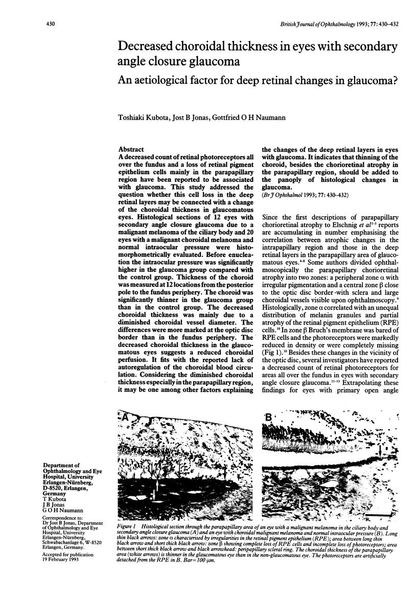
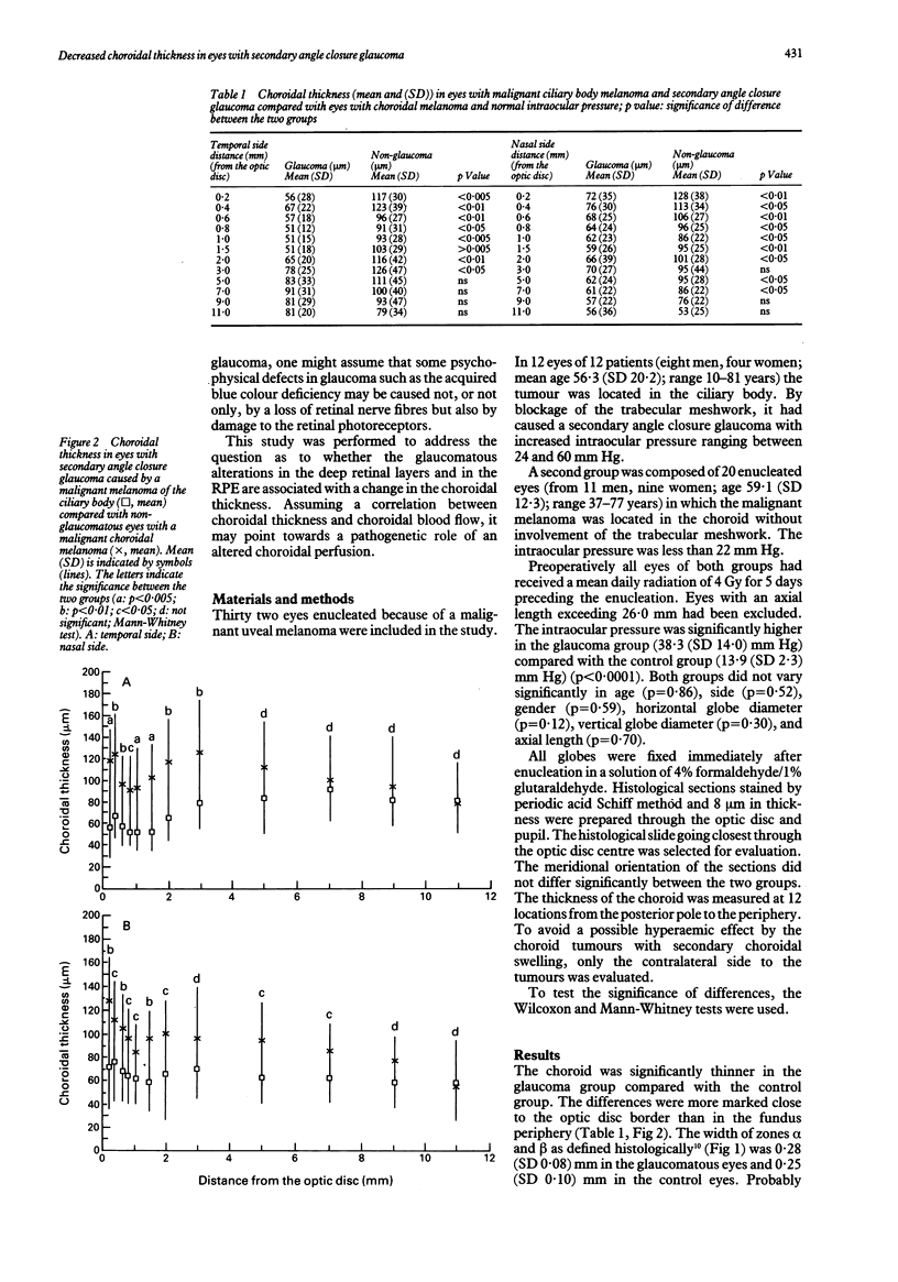
A decreased count of retinal photoreceptors all over the fundus and a loss of retinal pigment epithelium cells mainly in the parapapillary region have been reported to be associated with glaucoma. This study addressed the question whether this cell loss in the deep retinal layers may be connected with a change of the choroidal thickness in glaucomatous eyes. Histological sections of 12 eyes with secondary angle closure glaucoma due to a malignant melanoma of the ciliary body and 20 eyes with a malignant choroidal melanoma and normal intraocular pressure were histomorphometrically evaluated. Before enucleation the intraocular pressure was significantly higher in the glaucoma group compared with the control group. Thickness of the choroid was measured at 12 locations from the posterior pole to the fundus periphery. The choroid was significantly thinner in the glaucoma group than in the control group. The decreased choroidal thickness was mainly due to a diminished choroidal vessel diameter. The differences were more marked at the optic disc border than in the fundus periphery. The decreased choroidal thickness in the glaucomatous eyes suggests a reduced choroidal perfusion. It fits with the reported lack of autoregulation of the choroidal blood circulation. Considering the diminished choroidal thickness especially in the parapapillary region, it may be one among other factors explaining the changes of the deep retinal layers in eyes with glaucoma. It indicates that thinning of the choroid, besides the chorioretinal atrophy in the parapapillary region, should be added to the panoply of histological changes in glaucoma.
Full text
PDF


Images in this article
Selected References
These references are in PubMed. This may not be the complete list of references from this article.
- Alm A., Bill A. Blood flow and oxygen extraction in the cat uvea at normal and high intraocular pressures. Acta Physiol Scand. 1970 Sep;80(1):19–28. doi: 10.1111/j.1748-1716.1970.tb04765.x. [DOI] [PubMed] [Google Scholar]
- Alm A., Bill A. Ocular and optic nerve blood flow at normal and increased intraocular pressures in monkeys (Macaca irus): a study with radioactively labelled microspheres including flow determinations in brain and some other tissues. Exp Eye Res. 1973 Jan 1;15(1):15–29. doi: 10.1016/0014-4835(73)90185-1. [DOI] [PubMed] [Google Scholar]
- Cristini G., Cennamo G., Daponte P. Choroidal thickness in primary glaucoma. Ophthalmologica. 1991;202(2):81–85. doi: 10.1159/000310179. [DOI] [PubMed] [Google Scholar]
- Dollery C. T., Henkind P., Kohner E. M., Paterson J. W. Effect of raised intraocular pressure on the retinal and choroidal circulation. Invest Ophthalmol. 1968 Apr;7(2):191–198. [PubMed] [Google Scholar]
- FRANCOIS J., NEETENS A. VASCULARITY OF THE EYE AND THE OPTIC NERVE IN GLAUCOMA. Arch Ophthalmol. 1964 Feb;71:219–225. doi: 10.1001/archopht.1964.00970010235017. [DOI] [PubMed] [Google Scholar]
- Hayreh S. S. Blood supply of the optic nerve head and its role in optic atrophy, glaucoma, and oedema of the optic disc. Br J Ophthalmol. 1969 Nov;53(11):721–748. doi: 10.1136/bjo.53.11.721. [DOI] [PMC free article] [PubMed] [Google Scholar]
- Hayreh S. S. Optic disc changes in glaucoma. Br J Ophthalmol. 1972 Mar;56(3):175–185. doi: 10.1136/bjo.56.3.175. [DOI] [PMC free article] [PubMed] [Google Scholar]
- Jonas J. B., Königsreuther K. A., Naumann G. O. Optic disc histomorphometry in normal eyes and eyes with secondary angle-closure glaucoma. II. Parapapillary region. Graefes Arch Clin Exp Ophthalmol. 1992;230(2):134–139. doi: 10.1007/BF00164651. [DOI] [PubMed] [Google Scholar]
- Jonas J. B., Nguyen X. N., Gusek G. C., Naumann G. O. Parapapillary chorioretinal atrophy in normal and glaucoma eyes. I. Morphometric data. Invest Ophthalmol Vis Sci. 1989 May;30(5):908–918. [PubMed] [Google Scholar]
- Panda S., Jonas J. B. Decreased photoreceptor count in human eyes with secondary angle-closure glaucoma. Invest Ophthalmol Vis Sci. 1992 Jul;33(8):2532–2536. [PubMed] [Google Scholar]
- Primrose J. Early signs of the glaucomatous disc. Br J Ophthalmol. 1971 Dec;55(12):820–825. doi: 10.1136/bjo.55.12.820. [DOI] [PMC free article] [PubMed] [Google Scholar]
- Pum D., Ahnelt P. K., Grasl M. Iso-orientation areas in the foveal cone mosaic. Vis Neurosci. 1990 Dec;5(6):511–523. doi: 10.1017/s0952523800000687. [DOI] [PubMed] [Google Scholar]
- Ueno M., Naumann G. O. Uveal damage in secondary glaucoma. A morphometric study. Graefes Arch Clin Exp Ophthalmol. 1989;227(4):380–383. doi: 10.1007/BF02169417. [DOI] [PubMed] [Google Scholar]
- Wilensky J. T., Kolker A. E. Peripapillary changes in glaucoma. Am J Ophthalmol. 1976 Mar;81(3):341–345. doi: 10.1016/0002-9394(76)90251-8. [DOI] [PubMed] [Google Scholar]


