

Abstract
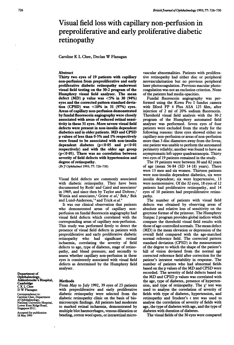
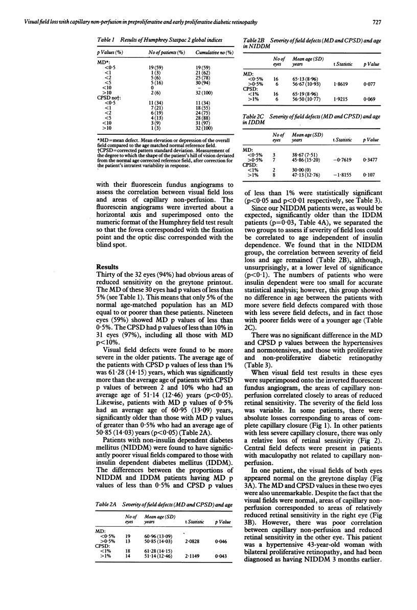
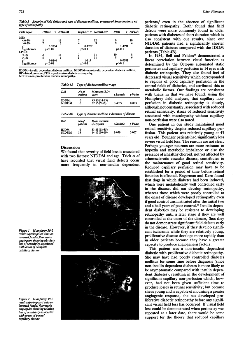
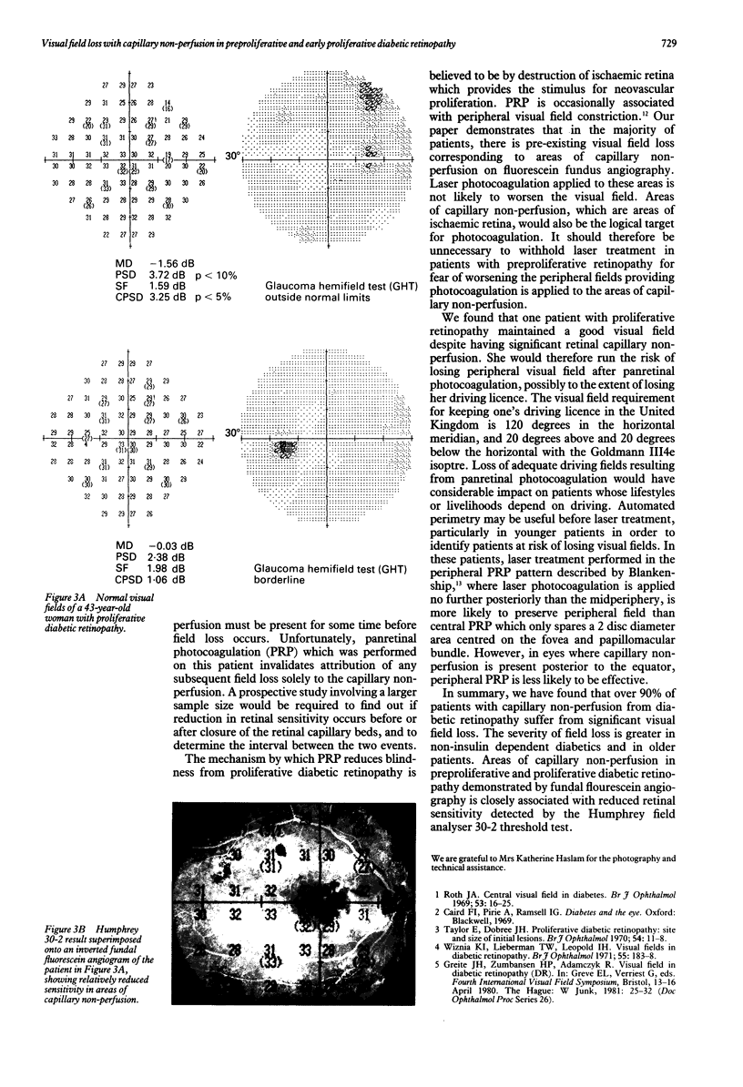
Thirty two eyes of 19 patients with capillary non-perfusion from preproliferative and early proliferative diabetic retinopathy underwent visual field testing on the 30-2 program of the Humphrey visual field analyser. The mean defect (MD) p value was < 5% in 30 (94%) eyes and the corrected pattern standard deviation (CPSD) was < 10% in 31 (97%) eyes. Areas of capillary non-perfusion demonstrated by fundal fluorescein angiography were closely associated with areas of reduced retinal sensitivity in these 31 eyes. More severe visual field defects were present in non-insulin dependent diabetics and in older patients. MD and CPSD p values of less than 0.5% and 1% respectively were found to be associated with non-insulin dependent diabetes (p < 0.05 and p < 0.01 respectively) and with the older age group (p < 0.05). There was no correlation between severity of field defects with hypertension and degree of retinopathy.
Full text
PDF
Images in this article
Selected References
These references are in PubMed. This may not be the complete list of references from this article.
- Bek T. Coexistence of localized scotomata and neovascularizations in proliferative diabetic retinopathy. Acta Ophthalmol (Copenh) 1990 Aug;68(4):421–427. doi: 10.1111/j.1755-3768.1990.tb01670.x. [DOI] [PubMed] [Google Scholar]
- Bek T., Lund-Andersen H. Localised blood-retinal barrier leakage and retinal light sensitivity in diabetic retinopathy. Br J Ophthalmol. 1990 Jul;74(7):388–392. doi: 10.1136/bjo.74.7.388. [DOI] [PMC free article] [PubMed] [Google Scholar]
- Bell J. A., Feldon S. E. Retinal microangiopathy. Correlation of OCTOPUS perimetry with fluorescein angiography. Arch Ophthalmol. 1984 Sep;102(9):1294–1298. doi: 10.1001/archopht.1984.01040031044020. [DOI] [PubMed] [Google Scholar]
- Blankenship G. W. A clinical comparison of central and peripheral argon laser panretinal photocoagulation for proliferative diabetic retinopathy. Ophthalmology. 1988 Feb;95(2):170–177. doi: 10.1016/s0161-6420(88)33212-4. [DOI] [PubMed] [Google Scholar]
- Engerman R. L., Kern T. S. Progression of incipient diabetic retinopathy during good glycemic control. Diabetes. 1987 Jul;36(7):808–812. doi: 10.2337/diab.36.7.808. [DOI] [PubMed] [Google Scholar]
- Hamilton A. M., Townsend C., Khoury D., Gould E., Blach R. K. Xenon arc and argon laser photocoagulation in the treatment of diabetic disc neovascularization. Part 1. Effect on disc vessels, visual fields, and visual acuity. Trans Ophthalmol Soc U K. 1981;101(1):87–92. [PubMed] [Google Scholar]
- Roth J. A. Central visual field in diabetes. Br J Ophthalmol. 1969 Jan;53(1):16–25. doi: 10.1136/bjo.53.1.16. [DOI] [PMC free article] [PubMed] [Google Scholar]
- Trick G. L., Trick L. R., Kilo C. Visual field defects in patients with insulin-dependent and noninsulin-dependent diabetes. Ophthalmology. 1990 Apr;97(4):475–482. doi: 10.1016/s0161-6420(90)32557-5. [DOI] [PubMed] [Google Scholar]
- Wisznia K. I., Lieberman T. W., Leopold I. H. Visual fields in diabetic retinopathy. Br J Ophthalmol. 1971 Mar;55(3):183–188. doi: 10.1136/bjo.55.3.183. [DOI] [PMC free article] [PubMed] [Google Scholar]