

Abstract
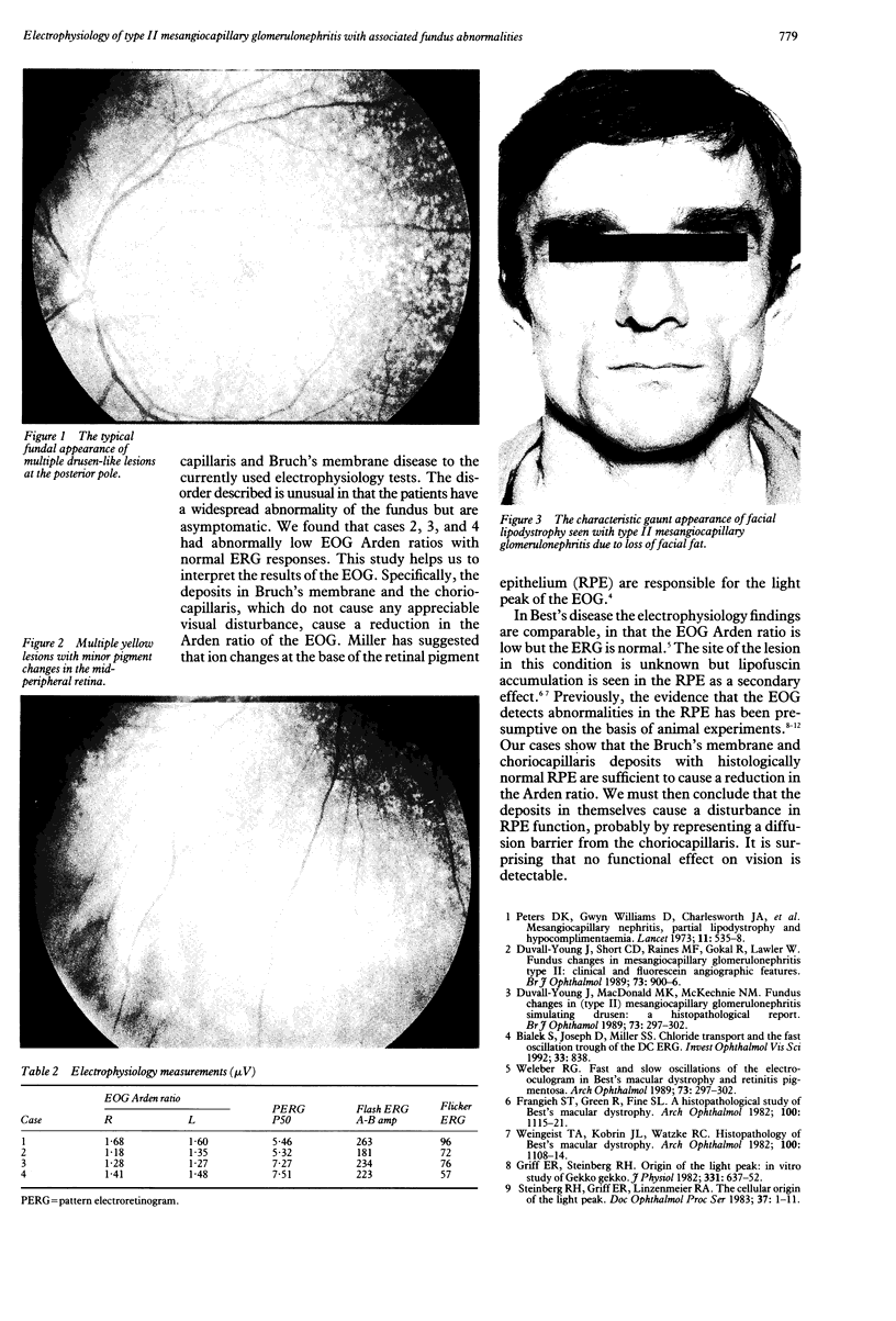
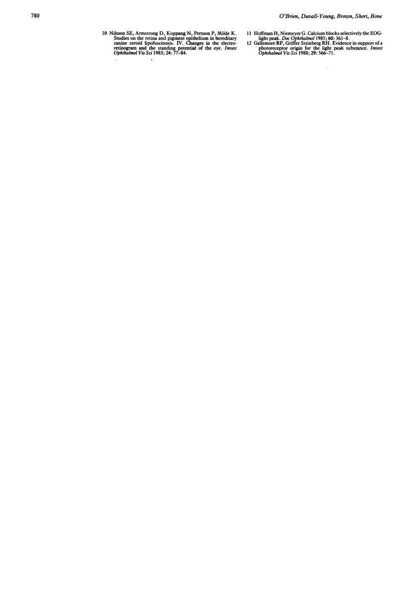
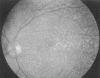
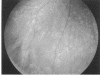
The retinal electrophysiology is reported in four patients with type II mesangiocapillary glomerulonephritis and partial lipodystrophy with associated fundus abnormalities and no visual symptoms. The histological hallmark of the condition is that of widespread electron dense deposits in the renal glomerulus and in the choriocapillaris and Bruch's membrane of the eye. Three of the four patients had the typical fundal appearance of multiple, yellow, drusen-like lesions at the posterior pole of the eye with normal visual acuity. These three patients had abnormally low Arden ratios on electro-oculography with normal electroretinography responses. This is the first clinical model of disease known to be isolated to the choriocapillaris and Bruch's membrane causing an electro-oculographic abnormality without any clinically detectable deficit in visual function.
Full text
PDF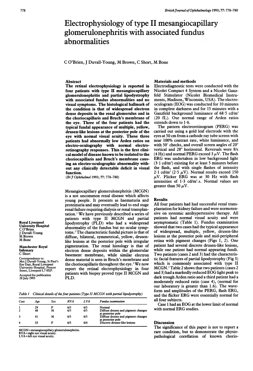
Images in this article
Selected References
These references are in PubMed. This may not be the complete list of references from this article.
- Duvall-Young J., MacDonald M. K., McKechnie N. M. Fundus changes in (type II) mesangiocapillary glomerulonephritis simulating drusen: a histopathological report. Br J Ophthalmol. 1989 Apr;73(4):297–302. doi: 10.1136/bjo.73.4.297. [DOI] [PMC free article] [PubMed] [Google Scholar]
- Duvall-Young J., Short C. D., Raines M. F., Gokal R., Lawler W. Fundus changes in mesangiocapillary glomerulonephritis type II: clinical and fluorescein angiographic findings. Br J Ophthalmol. 1989 Nov;73(11):900–906. doi: 10.1136/bjo.73.11.900. [DOI] [PMC free article] [PubMed] [Google Scholar]
- Frangieh G. T., Green W. R., Fine S. L. A histopathologic study of Best's macular dystrophy. Arch Ophthalmol. 1982 Jul;100(7):1115–1121. doi: 10.1001/archopht.1982.01030040093017. [DOI] [PubMed] [Google Scholar]
- Gallemore R. P., Griff E. R., Steinberg R. H. Evidence in support of a photoreceptoral origin for the "light-peak substance". Invest Ophthalmol Vis Sci. 1988 Apr;29(4):566–571. [PubMed] [Google Scholar]
- Griff E. R., Steinberg R. H. Origin of the light peak: in vitro study of Gekko gekko. J Physiol. 1982 Oct;331:637–652. doi: 10.1113/jphysiol.1982.sp014395. [DOI] [PMC free article] [PubMed] [Google Scholar]
- Hofmann H., Niemeyer G. Calcium blocks selectively the EOG-light peak. Doc Ophthalmol. 1985 Oct 15;60(4):361–368. doi: 10.1007/BF00158925. [DOI] [PubMed] [Google Scholar]
- Nilsson S. E., Armstrong D., Koppang N., Persson P., Milde K. Studies on the retina and the pigment epithelium in hereditary canine ceroid lipofuscinosis. IV. Changes in the electroretinogram and the standing potential of the eye. Invest Ophthalmol Vis Sci. 1983 Jan;24(1):77–84. [PubMed] [Google Scholar]
- Peters D. K., Charlesworth J. A., Sissons J. G., Williams D. G., Boulton-Jones J. M., Evans D. J., Kourilsky O., Morel-Maroger L. Mesangiocapillary nephritis, partial lipodystrophy, and hypocomplementaemia. Lancet. 1973 Sep 8;2(7828):535–538. doi: 10.1016/s0140-6736(73)92351-9. [DOI] [PubMed] [Google Scholar]
- Weingeist T. A., Kobrin J. L., Watzke R. C. Histopathology of Best's macular dystrophy. Arch Ophthalmol. 1982 Jul;100(7):1108–1114. doi: 10.1001/archopht.1982.01030040086016. [DOI] [PubMed] [Google Scholar]