

Abstract
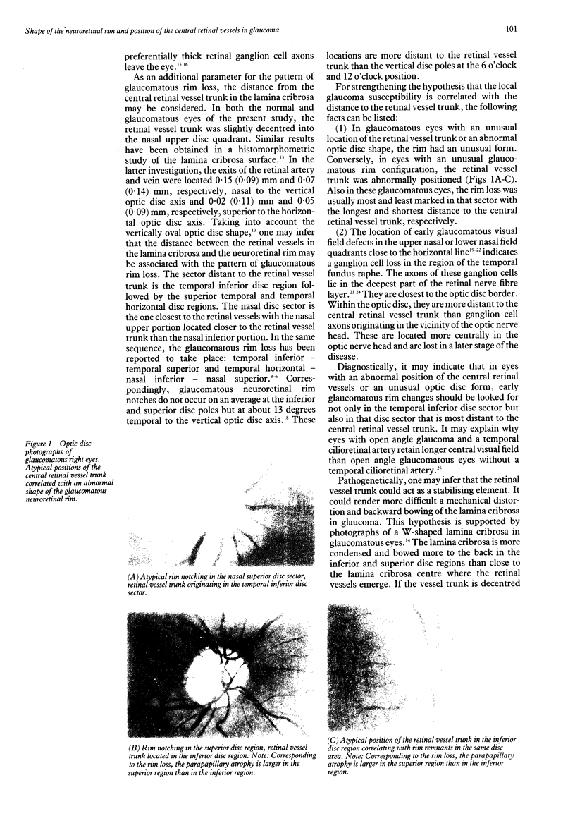
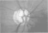
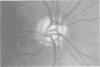
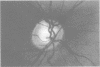
Glaucomatous neuroretinal rim loss can occur in a sequence of sectors with the temporal inferior disc sector as the first and the nasal superior disc sector as the last to be affected. This study evaluated whether the position of the central retinal vessel trunk is correlated with this pattern of glaucomatous rim loss. Morphometrically stereo colour optic disc photographs of 157 glaucomatous eyes and 67 normal eyes were checked. In the normal and glaucomatous eyes, the central retinal vessel trunk was located eccentrically in the upper nasal quadrant of the optic disc. Taking into account the vertically oval disc shape, the distance to the central vessel trunk was largest for the temporal inferior disc region and shortest for the nasal superior disc area. An abnormal form of the glaucomatous neuroretinal rim was found in eyes with an atypical location of the retinal vessel trunk. Also in these glaucomatous eyes, the rim loss was usually most and least marked in that sector with the longest and shortest distance, respectively, to the central retinal vessel trunk. One could infer that the sequence of rim loss in glaucoma is dependent upon the distance of the region to the central retinal vessel trunk; the further away the region from the retinal vessel trunk, the more likely it is to be affected by rim loss. This suggest that the distance from the central retinal vessels is one factor among others that is correlated with the regional vulnerability of the neuroretinal rim to the glaucomatous process.
Full text
PDF
Images in this article
Selected References
These references are in PubMed. This may not be the complete list of references from this article.
- Bengtsson B. The variation and covariation of cup and disc diameters. Acta Ophthalmol (Copenh) 1976 Dec;54(6):804–818. doi: 10.1111/j.1755-3768.1976.tb01801.x. [DOI] [PubMed] [Google Scholar]
- Britton R. J., Drance S. M., Schulzer M., Douglas G. R., Mawson D. K. The area of the neuroretinal rim of the optic nerve in normal eyes. Am J Ophthalmol. 1987 Apr 15;103(4):497–504. doi: 10.1016/s0002-9394(14)74271-0. [DOI] [PubMed] [Google Scholar]
- Caprioli J., Miller J. M. Optic disc rim area is related to disc size in normal subjects. Arch Ophthalmol. 1987 Dec;105(12):1683–1685. doi: 10.1001/archopht.1987.01060120081030. [DOI] [PubMed] [Google Scholar]
- Caprioli J., Sears M., Miller J. M. Patterns of early visual field loss in open-angle glaucoma. Am J Ophthalmol. 1987 Apr 15;103(4):512–517. doi: 10.1016/s0002-9394(14)74273-4. [DOI] [PubMed] [Google Scholar]
- Drance S. M. The early field defects in glaucoma. Invest Ophthalmol. 1969 Feb;8(1):84–91. [PubMed] [Google Scholar]
- Gramer E., Gerlach R., Krieglstein G. K., Leydhecker W. Zur Topographie früher glaukomatöser Gesichtsfeldausfälle bei der Computerperimetrie. Klin Monbl Augenheilkd. 1982 Jun;180(6):515–523. doi: 10.1055/s-2008-1055138. [DOI] [PubMed] [Google Scholar]
- Hart W. M., Jr, Becker B. The onset and evolution of glaucomatous visual field defects. Ophthalmology. 1982 Mar;89(3):268–279. doi: 10.1016/s0161-6420(82)34798-3. [DOI] [PubMed] [Google Scholar]
- Jonas J. B., Fernández M. C., Stürmer J. Pattern of glaucomatous neuroretinal rim loss. Ophthalmology. 1993 Jan;100(1):63–68. doi: 10.1016/s0161-6420(13)31694-7. [DOI] [PubMed] [Google Scholar]
- Jonas J. B., Gusek G. C., Naumann G. O. Optic disc morphometry in chronic primary open-angle glaucoma. I. Morphometric intrapapillary characteristics. Graefes Arch Clin Exp Ophthalmol. 1988;226(6):522–530. doi: 10.1007/BF02169199. [DOI] [PubMed] [Google Scholar]
- Jonas J. B., Gusek G. C., Naumann G. O. Optic disc, cup and neuroretinal rim size, configuration and correlations in normal eyes. Invest Ophthalmol Vis Sci. 1988 Jul;29(7):1151–1158. [PubMed] [Google Scholar]
- Jonas J. B., Gusek G. C., Naumann G. O. Optic disk morphometry in high myopia. Graefes Arch Clin Exp Ophthalmol. 1988;226(6):587–590. doi: 10.1007/BF02169209. [DOI] [PubMed] [Google Scholar]
- Jonas J. B., Mardin C. Y., Schlötzer-Schrehardt U., Naumann G. O. Morphometry of the human lamina cribrosa surface. Invest Ophthalmol Vis Sci. 1991 Feb;32(2):401–405. [PubMed] [Google Scholar]
- Jonas J. B., Müller-Bergh J. A., Schlötzer-Schrehardt U. M., Naumann G. O. Histomorphometry of the human optic nerve. Invest Ophthalmol Vis Sci. 1990 Apr;31(4):736–744. [PubMed] [Google Scholar]
- Kirsch R. E., Anderson D. R. Clinical recognition of glaucomatous cupping. Am J Ophthalmol. 1973 Mar;75(3):442–454. doi: 10.1016/0002-9394(73)91153-7. [DOI] [PubMed] [Google Scholar]
- Lee S. S., Schwartz B. Role of the temporal cilioretinal artery in retaining central visual field in open-angle glaucoma. Ophthalmology. 1992 May;99(5):696–699. doi: 10.1016/s0161-6420(92)31908-6. [DOI] [PubMed] [Google Scholar]
- Lieberman M. F., Maumenee A. E., Green W. R. Histologic studies of the vasculature of the anterior optic nerve. Am J Ophthalmol. 1976 Sep;82(3):405–423. doi: 10.1016/0002-9394(76)90489-x. [DOI] [PubMed] [Google Scholar]
- Minckler D. S. The organization of nerve fiber bundles in the primate optic nerve head. Arch Ophthalmol. 1980 Sep;98(9):1630–1636. doi: 10.1001/archopht.1980.01020040482019. [DOI] [PubMed] [Google Scholar]
- Pederson J. E., Anderson D. R. The mode of progressive disc cupping in ocular hypertension and glaucoma. Arch Ophthalmol. 1980 Mar;98(3):490–495. doi: 10.1001/archopht.1980.01020030486010. [DOI] [PubMed] [Google Scholar]
- Quigley H. A., Addicks E. M., Green W. R., Maumenee A. E. Optic nerve damage in human glaucoma. II. The site of injury and susceptibility to damage. Arch Ophthalmol. 1981 Apr;99(4):635–649. doi: 10.1001/archopht.1981.03930010635009. [DOI] [PubMed] [Google Scholar]
- Quigley H. A., Addicks E. M. Regional differences in the structure of the lamina cribrosa and their relation to glaucomatous optic nerve damage. Arch Ophthalmol. 1981 Jan;99(1):137–143. doi: 10.1001/archopht.1981.03930010139020. [DOI] [PubMed] [Google Scholar]
- Quigley H. A., Sanchez R. M., Dunkelberger G. R., L'Hernault N. L., Baginski T. A. Chronic glaucoma selectively damages large optic nerve fibers. Invest Ophthalmol Vis Sci. 1987 Jun;28(6):913–920. [PubMed] [Google Scholar]
- Radius R. L., Anderson D. R. The course of axons through the retina and optic nerve head. Arch Ophthalmol. 1979 Jun;97(6):1154–1158. doi: 10.1001/archopht.1979.01020010608021. [DOI] [PubMed] [Google Scholar]
- Radius R. L. Regional specificity in anatomy at the lamina cribrosa. Arch Ophthalmol. 1981 Mar;99(3):478–480. doi: 10.1001/archopht.1981.03930010480020. [DOI] [PubMed] [Google Scholar]
- Sanchez R. M., Dunkelberger G. R., Quigley H. A. The number and diameter distribution of axons in the monkey optic nerve. Invest Ophthalmol Vis Sci. 1986 Sep;27(9):1342–1350. [PubMed] [Google Scholar]
- Tuulonen A., Airaksinen P. J. Initial glaucomatous optic disk and retinal nerve fiber layer abnormalities and their progression. Am J Ophthalmol. 1991 Apr 15;111(4):485–490. doi: 10.1016/s0002-9394(14)72385-2. [DOI] [PubMed] [Google Scholar]