Abstract
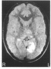
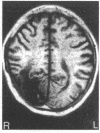
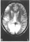
The representation of the field of vision in the human striate cortex is based on the Holmes map in which about 25% of the surface area of the striate cortex is allocated to the central 15 degrees of vision. Following the introduction of computed tomography of the brain, the accuracy of the Holmes map was apparently confirmed by clinical/radiological correlation, but a revision has been proposed by Horton and Hoyt based on a magnetic resonance imaging study of three patients with visual field defects due to striate lesions. They propose that the central cortical representation of vision occupies a much larger area. This study reviews the perimetric and imaging findings in a larger series of patients with striate cortical disease and provides support for the revised representation. The clinical phenomenon of macular sparing and its relation to representation of the macula at the occipital pole is also discussed.
Full text
PDF
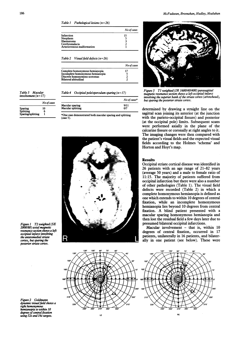
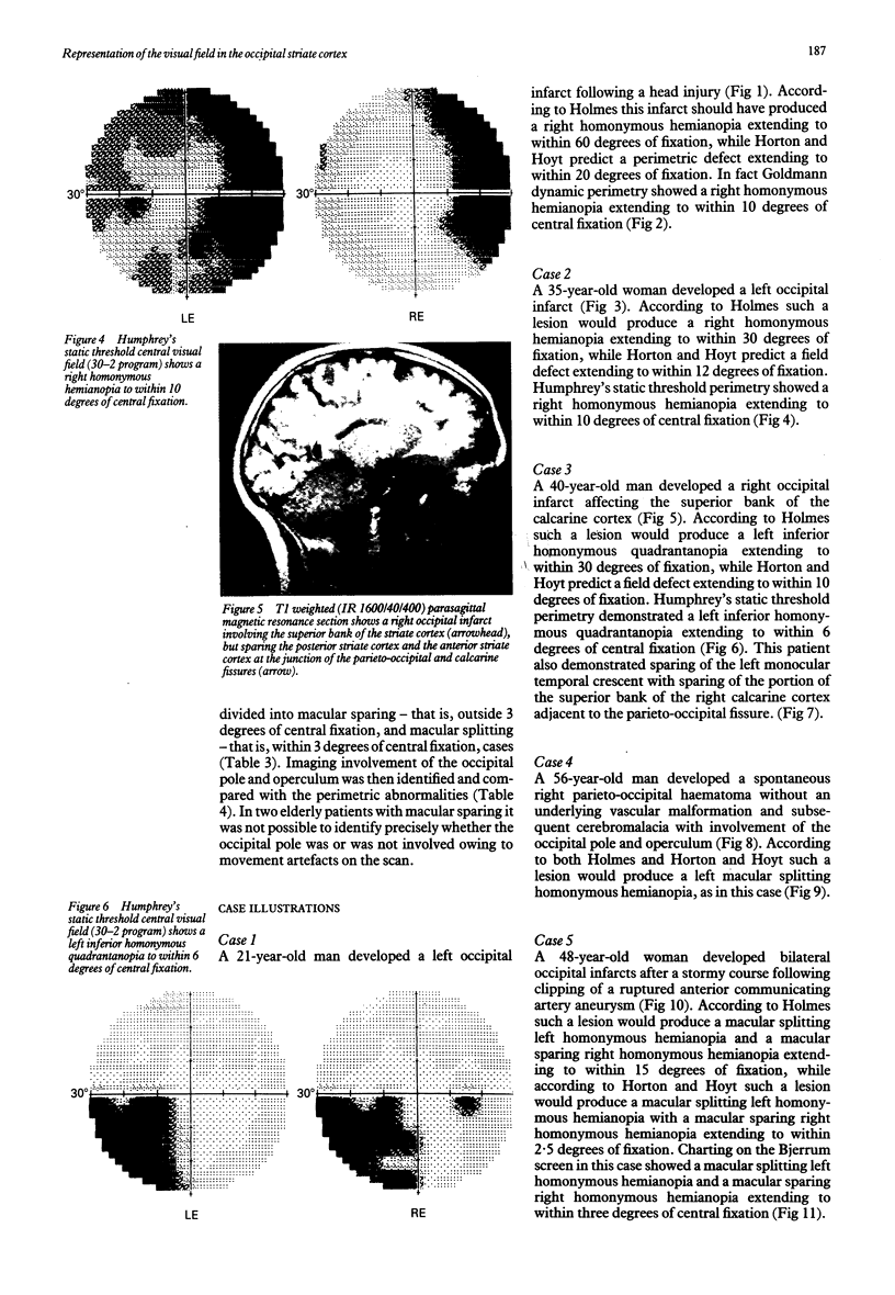
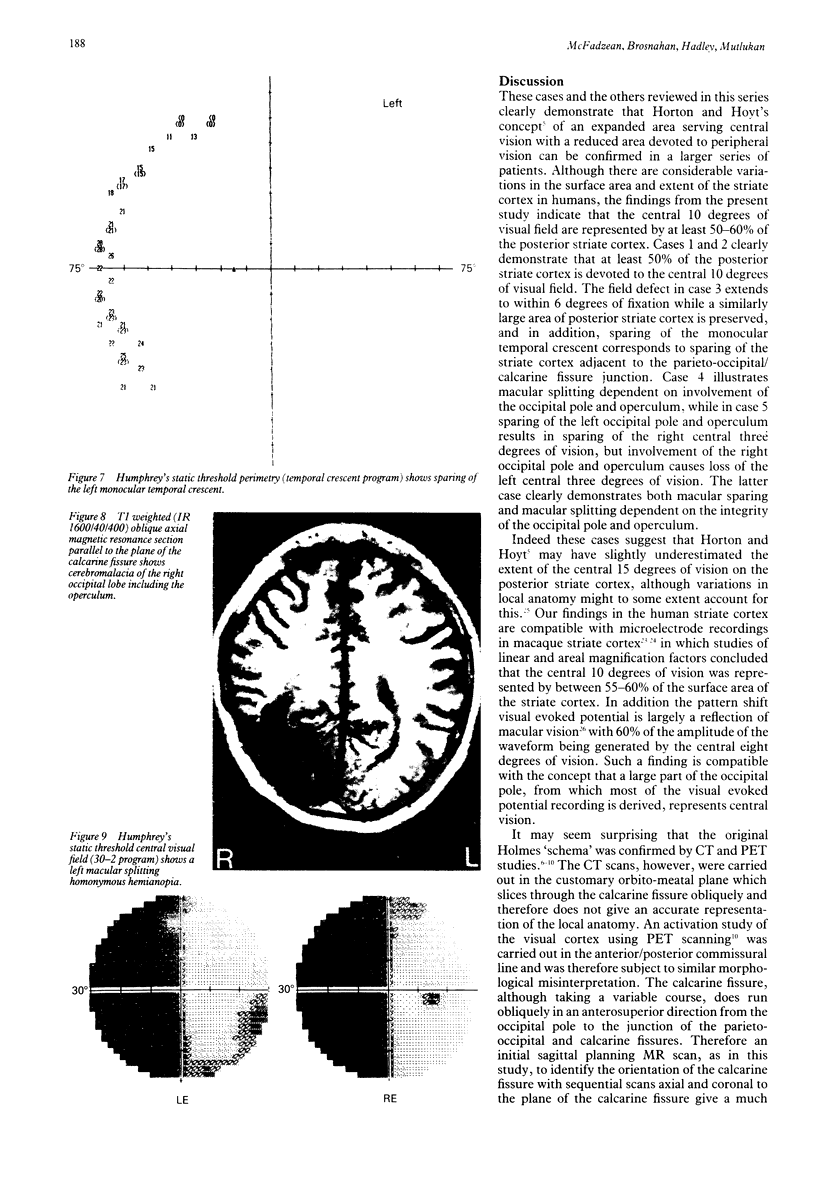
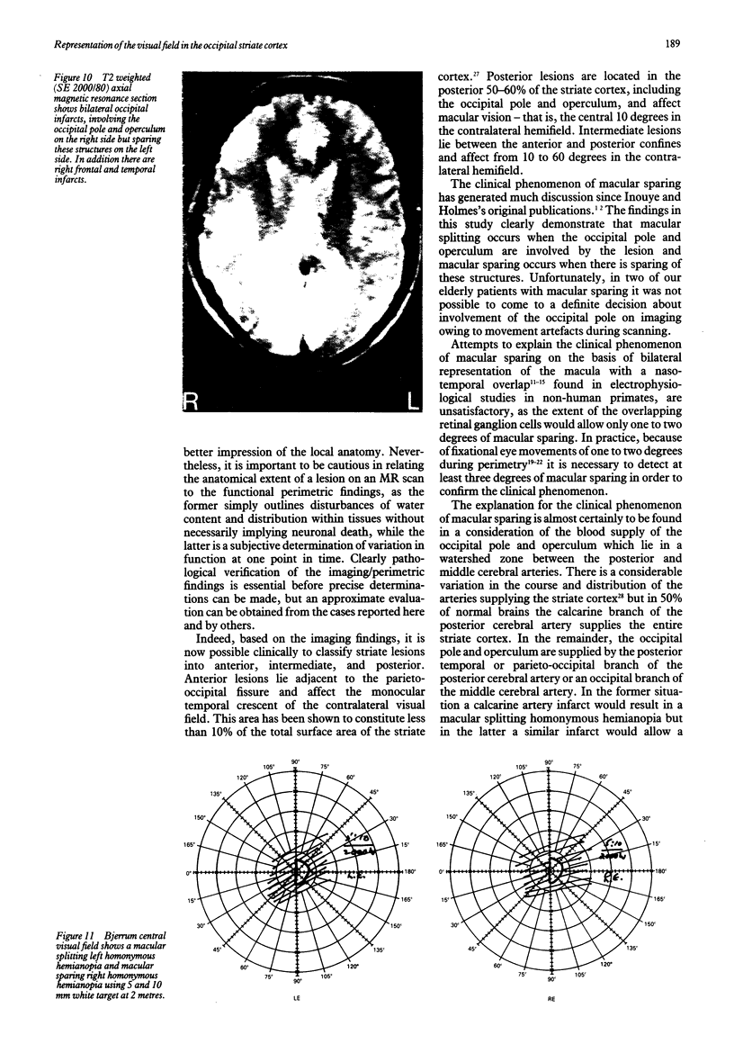
Images in this article
Selected References
These references are in PubMed. This may not be the complete list of references from this article.
- Belliveau J. W., Kennedy D. N., Jr, McKinstry R. C., Buchbinder B. R., Weisskoff R. M., Cohen M. S., Vevea J. M., Brady T. J., Rosen B. R. Functional mapping of the human visual cortex by magnetic resonance imaging. Science. 1991 Nov 1;254(5032):716–719. doi: 10.1126/science.1948051. [DOI] [PubMed] [Google Scholar]
- Bunt A. H., Minckler D. S. Foveal sparing. New anatomical evidence for bilateral representation of the central retina. Arch Ophthalmol. 1977 Aug;95(8):1445–1447. doi: 10.1001/archopht.1977.04450080155021. [DOI] [PubMed] [Google Scholar]
- Bunt A. H., Minckler D. S., Johanson G. W. Demonstration of bilateral projection of the central retina of the monkey with horseradish peroxidase neuronography. J Comp Neurol. 1977 Feb 15;171(4):619–630. doi: 10.1002/cne.901710412. [DOI] [PubMed] [Google Scholar]
- DANIEL P. M., WHITTERIDGE D. The representation of the visual field on the cerebral cortex in monkeys. J Physiol. 1961 Dec;159:203–221. doi: 10.1113/jphysiol.1961.sp006803. [DOI] [PMC free article] [PubMed] [Google Scholar]
- Dell'Osso L. F., Abel L. A., Daroff R. B. "Inverse latent" macro square-wave jerks and macro saccadic oscillations. Ann Neurol. 1977 Jul;2(1):57–60. doi: 10.1002/ana.410020109. [DOI] [PubMed] [Google Scholar]
- Dow B. M., Vautin R. G., Bauer R. The mapping of visual space onto foveal striate cortex in the macaque monkey. J Neurosci. 1985 Apr;5(4):890–902. doi: 10.1523/JNEUROSCI.05-04-00890.1985. [DOI] [PMC free article] [PubMed] [Google Scholar]
- Fox P. T., Miezin F. M., Allman J. M., Van Essen D. C., Raichle M. E. Retinotopic organization of human visual cortex mapped with positron-emission tomography. J Neurosci. 1987 Mar;7(3):913–922. doi: 10.1523/JNEUROSCI.07-03-00913.1987. [DOI] [PMC free article] [PubMed] [Google Scholar]
- Fukuda Y., Sawai H., Watanabe M., Wakakuwa K., Morigiwa K. Nasotemporal overlap of crossed and uncrossed retinal ganglion cell projections in the Japanese monkey (Macaca fuscata). J Neurosci. 1989 Jul;9(7):2353–2373. doi: 10.1523/JNEUROSCI.09-07-02353.1989. [DOI] [PMC free article] [PubMed] [Google Scholar]
- Herishanu Y. O., Sharpe J. A. Normal square wave jerks. Invest Ophthalmol Vis Sci. 1981 Feb;20(2):268–272. [PubMed] [Google Scholar]
- Holmes G. DISTURBANCES OF VISION BY CEREBRAL LESIONS. Br J Ophthalmol. 1918 Jul;2(7):353–384. doi: 10.1136/bjo.2.7.353. [DOI] [PMC free article] [PubMed] [Google Scholar]
- Horton J. C., Dagi L. R., McCrane E. P., de Monasterio F. M. Arrangement of ocular dominance columns in human visual cortex. Arch Ophthalmol. 1990 Jul;108(7):1025–1031. doi: 10.1001/archopht.1990.01070090127054. [DOI] [PubMed] [Google Scholar]
- Horton J. C., Hoyt W. F. The representation of the visual field in human striate cortex. A revision of the classic Holmes map. Arch Ophthalmol. 1991 Jun;109(6):816–824. doi: 10.1001/archopht.1991.01080060080030. [DOI] [PubMed] [Google Scholar]
- Kattah J. C., Dennis P., Kolsky M. P., Schellinger D., Cohan S. L. Computed tomography in patients with homonymous visual field defects--A. Clinico-radiologic correlation. Comput Tomogr. 1981 Oct-Dec;5(4):301–312. doi: 10.1016/0363-8235(81)90037-5. [DOI] [PubMed] [Google Scholar]
- Kwong K. K., Belliveau J. W., Chesler D. A., Goldberg I. E., Weisskoff R. M., Poncelet B. P., Kennedy D. N., Hoppel B. E., Cohen M. S., Turner R. Dynamic magnetic resonance imaging of human brain activity during primary sensory stimulation. Proc Natl Acad Sci U S A. 1992 Jun 15;89(12):5675–5679. doi: 10.1073/pnas.89.12.5675. [DOI] [PMC free article] [PubMed] [Google Scholar]
- Leventhal A. G., Ault S. J., Vitek D. J. The nasotemporal division in primate retina: the neural bases of macular sparing and splitting. Science. 1988 Apr 1;240(4848):66–67. doi: 10.1126/science.3353708. [DOI] [PubMed] [Google Scholar]
- McAuley D. L., Russell R. W. Correlation of CAT scan and visual field defects in vascular lesions of the posterior visual pathways. J Neurol Neurosurg Psychiatry. 1979 Apr;42(4):298–311. doi: 10.1136/jnnp.42.4.298. [DOI] [PMC free article] [PubMed] [Google Scholar]
- Smith C. G., Richardson W. F. The course and distribution of the arteries supplying the visual (striate) cortex. Am J Ophthalmol. 1966 Jun;61(6):1391–1396. doi: 10.1016/0002-9394(66)90475-2. [DOI] [PubMed] [Google Scholar]
- Spector R. H., Glaser J. S., David N. J., Vining D. Q. Occipital lobe infarctions: Perimetry and computed tomography. Neurology. 1981 Sep;31(9):1098–1106. doi: 10.1212/wnl.31.9.1098. [DOI] [PubMed] [Google Scholar]
- Stensaas S. S., Eddington D. K., Dobelle W. H. The topography and variability of the primary visual cortex in man. J Neurosurg. 1974 Jun;40(6):747–755. doi: 10.3171/jns.1974.40.6.0747. [DOI] [PubMed] [Google Scholar]
- Stone J., Leicester J., Sherman S. M. The naso-temporal division of the monkey's retina. J Comp Neurol. 1973 Aug;150(3):333–348. doi: 10.1002/cne.901500306. [DOI] [PubMed] [Google Scholar]
- Tootell R. B., Switkes E., Silverman M. S., Hamilton S. L. Functional anatomy of macaque striate cortex. II. Retinotopic organization. J Neurosci. 1988 May;8(5):1531–1568. doi: 10.1523/JNEUROSCI.08-05-01531.1988. [DOI] [PMC free article] [PubMed] [Google Scholar]
- Van Essen D. C., Newsome W. T., Maunsell J. H. The visual field representation in striate cortex of the macaque monkey: asymmetries, anisotropies, and individual variability. Vision Res. 1984;24(5):429–448. doi: 10.1016/0042-6989(84)90041-5. [DOI] [PubMed] [Google Scholar]
- Winterson B. J., Collewijn H. Microsaccades during finely guided visuomotor tasks. Vision Res. 1976;16(12):1387–1390. doi: 10.1016/0042-6989(76)90156-5. [DOI] [PubMed] [Google Scholar]
- Yiannikas C., Walsh J. C. The variation of the pattern shift visual evoked response with the size of the stimulus field. Electroencephalogr Clin Neurophysiol. 1983 Apr;55(4):427–435. doi: 10.1016/0013-4694(83)90131-1. [DOI] [PubMed] [Google Scholar]


