Abstract
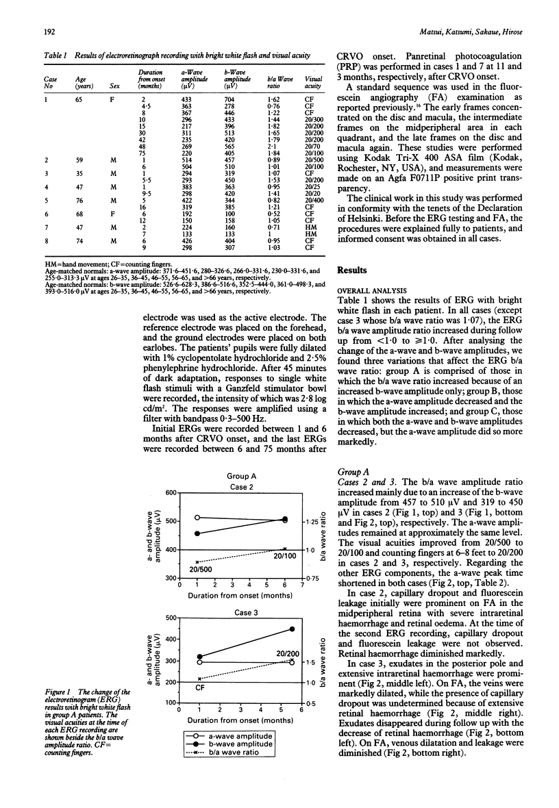
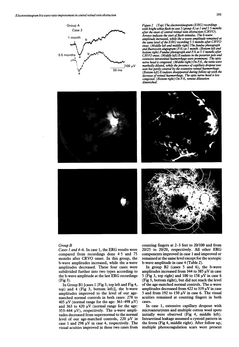
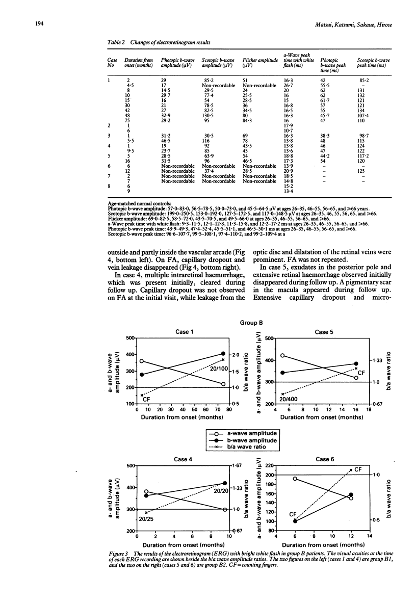
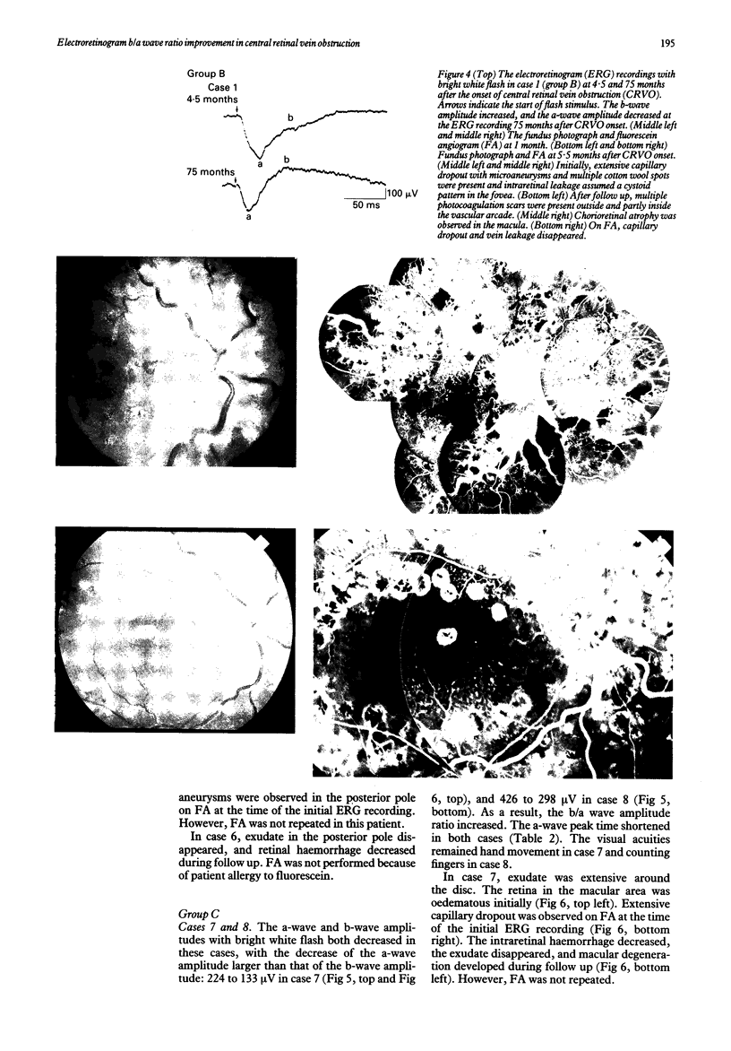
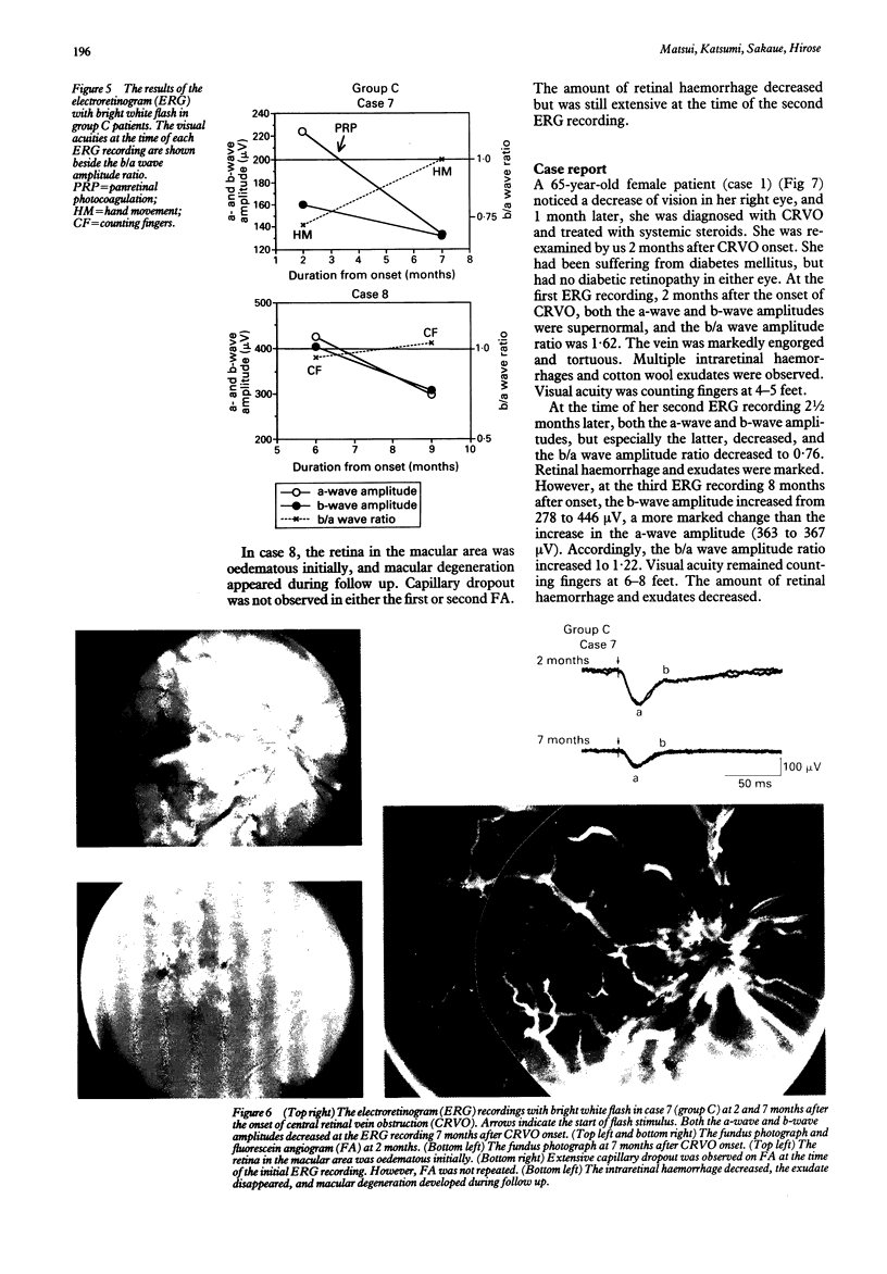
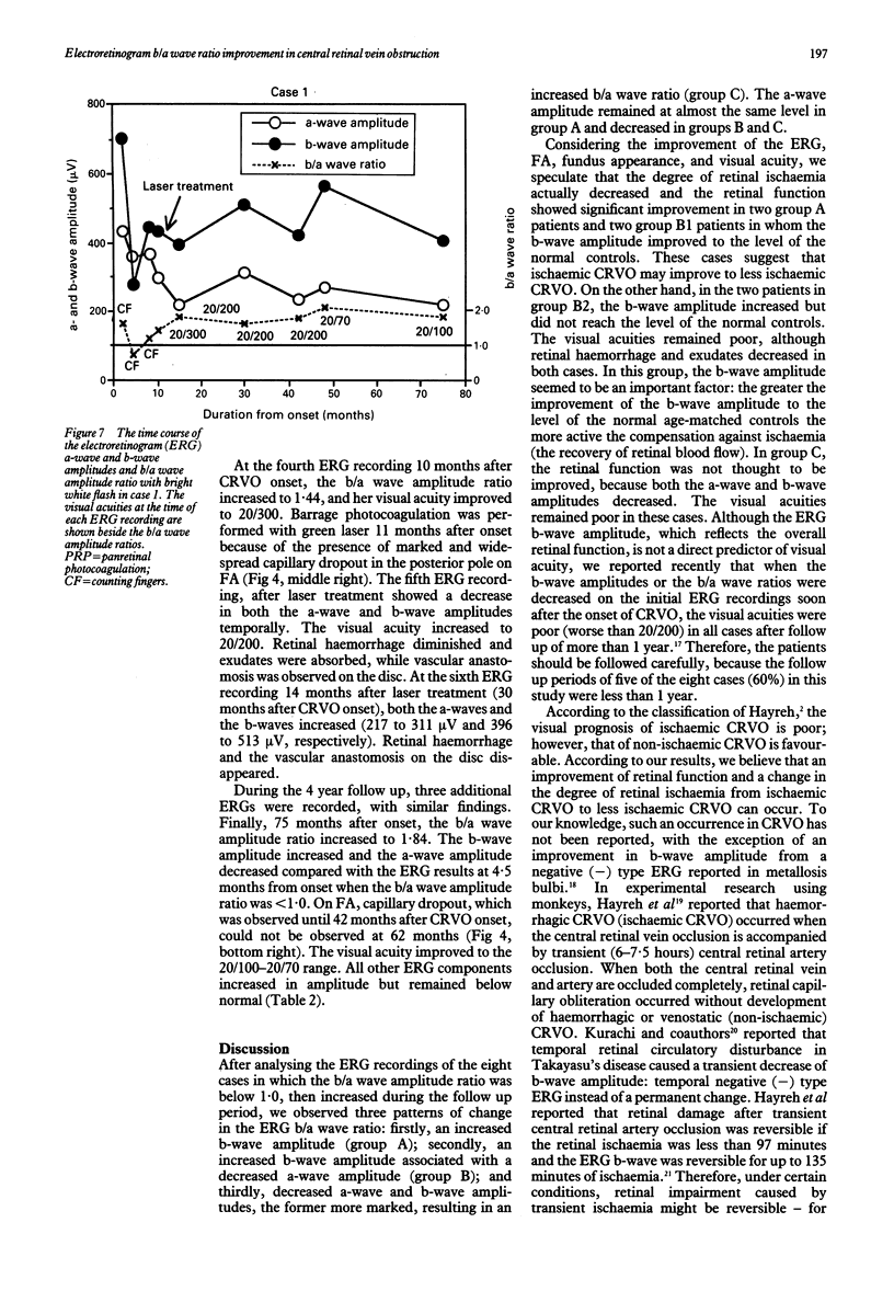
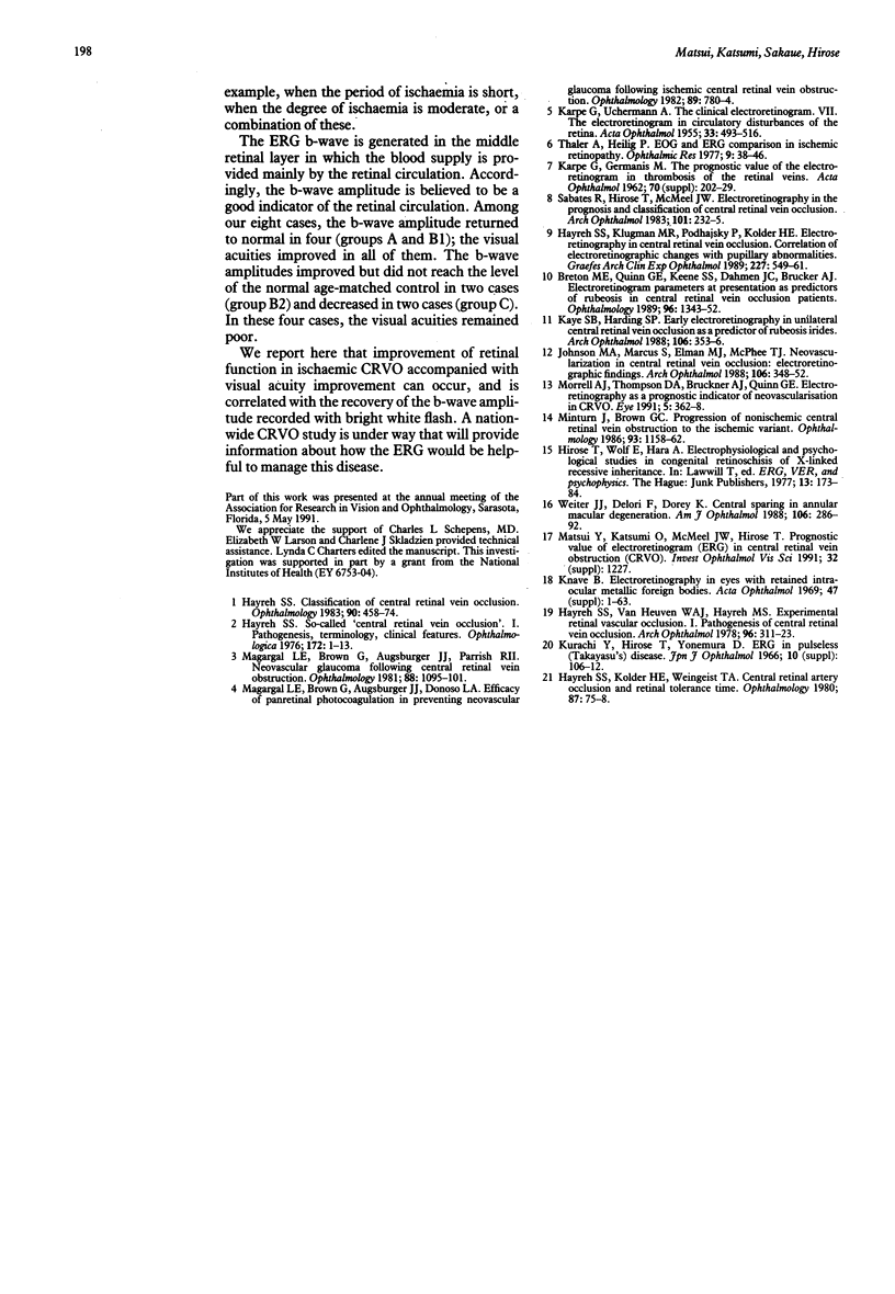
The electroretinogram (ERG), especially the b/a wave ratio, is considered a good indicator of retinal ischaemia in central retinal vein obstruction (CRVO). Seven CRVO patients who showed b/a wave ratio improvement from < 1.0 [negative type (-) ERG] to > or = 1.0 and one from 1.07 to 1.53 were studied. Three mechanisms of change were observed: firstly, the b-wave amplitude increased without an a-wave amplitude decrease (group A, n = 2); secondly, the b-wave amplitude increased with an a-wave amplitude decrease (group B, n = 4); and, thirdly, both decreased, but the a-wave amplitude decreased more markedly (group C, n = 2). In group A, the visual acuities improved markedly. In group B, the visual acuities improved in two cases in which the b-wave amplitude reached the normal range; the visual acuities did not improve in two cases in which the b-wave amplitude did not reach the normal range. In group C, the visual acuities remained poor. The negative (-) ERG or significantly reduced b/a wave ratio is associated with ischaemic CRVO and did not occur because of the filtering effect of the haemorrhage, which may reduce the stimulus light for the ERG. Improvement of the reduced b/a wave ratio with an increased b-wave amplitude was accompanied by improvements in fundus appearance and visual acuity in CRVO. The results suggest that the retinal ischaemia in CRVO, as revealed by the ERG and fluorescein angiogram, may be reversible in some cases.
Full text
PDF
Images in this article
Selected References
These references are in PubMed. This may not be the complete list of references from this article.
- Breton M. E., Quinn G. E., Keene S. S., Dahmen J. C., Brucker A. J. Electroretinogram parameters at presentation as predictors of rubeosis in central retinal vein occlusion patients. Ophthalmology. 1989 Sep;96(9):1343–1352. doi: 10.1016/s0161-6420(89)32742-4. [DOI] [PubMed] [Google Scholar]
- Hayreh S. S. Classification of central retinal vein occlusion. Ophthalmology. 1983 May;90(5):458–474. doi: 10.1016/s0161-6420(83)34530-9. [DOI] [PubMed] [Google Scholar]
- Hayreh S. S., Klugman M. R., Podhajsky P., Kolder H. E. Electroretinography in central retinal vein occlusion. Correlation of electroretinographic changes with pupillary abnormalities. Graefes Arch Clin Exp Ophthalmol. 1989;227(6):549–561. doi: 10.1007/BF02169451. [DOI] [PubMed] [Google Scholar]
- Hayreh S. S., Kolder H. E., Weingeist T. A. Central retinal artery occlusion and retinal tolerance time. Ophthalmology. 1980 Jan;87(1):75–78. doi: 10.1016/s0161-6420(80)35283-4. [DOI] [PubMed] [Google Scholar]
- Hayreh S. S. So-called "central retinal vein occlusion". I. Pathogenesis, terminology, clinical features. Ophthalmologica. 1976;172(1):1–13. doi: 10.1159/000307579. [DOI] [PubMed] [Google Scholar]
- Hayreh S. S., van Heuven W. A., Hayreh M. S. Experimental retinal vascular occlusion. I. Pathogenesis of central retinal vein occlusion. Arch Ophthalmol. 1978 Feb;96(2):311–323. doi: 10.1001/archopht.1978.03910050179015. [DOI] [PubMed] [Google Scholar]
- Johnson M. A., Marcus S., Elman M. J., McPhee T. J. Neovascularization in central retinal vein occlusion: electroretinographic findings. Arch Ophthalmol. 1988 Mar;106(3):348–352. doi: 10.1001/archopht.1988.01060130374025. [DOI] [PubMed] [Google Scholar]
- KARPE G., GERMANIS M. The prognostic value of the electroretinogram in thrombosis of the retinal veins. Acta Ophthalmol Suppl. 1962;Suppl 70:202–229. doi: 10.1111/j.1755-3768.1962.tb00323.x. [DOI] [PubMed] [Google Scholar]
- KARPE G., UCHERMANN A. The clinical electroretinogram. VII. The electroretinogram in circulatory disturbances of the retina. Acta Ophthalmol (Copenh) 1955;33(5):493–516. doi: 10.1111/j.1755-3768.1955.tb03325.x. [DOI] [PubMed] [Google Scholar]
- Kaye S. B., Harding S. P. Early electroretinography in unilateral central retinal vein occlusion as a predictor of rubeosis iridis. Arch Ophthalmol. 1988 Mar;106(3):353–356. doi: 10.1001/archopht.1988.01060130379026. [DOI] [PubMed] [Google Scholar]
- Magargal L. E., Brown G. C., Augsburger J. J., Donoso L. A. Efficacy of panretinal photocoagulation in preventing neovascular glaucoma following ischemic central retinal vein obstruction. Ophthalmology. 1982 Jul;89(7):780–784. doi: 10.1016/s0161-6420(82)34724-7. [DOI] [PubMed] [Google Scholar]
- Magargal L. E., Brown G. C., Augsburger J. J., Parrish R. K., 2nd Neovascular glaucoma following central retinal vein obstruction. Ophthalmology. 1981 Nov;88(11):1095–1101. doi: 10.1016/s0161-6420(81)34901-x. [DOI] [PubMed] [Google Scholar]
- Minturn J., Brown G. C. Progression of nonischemic central retinal vein obstruction to the ischemic variant. Ophthalmology. 1986 Sep;93(9):1158–1162. doi: 10.1016/s0161-6420(86)33599-1. [DOI] [PubMed] [Google Scholar]
- Morrell A. J., Thompson D. A., Gibson J. M., Kritzinger E. E., Drasdo N. Electroretinography as a prognostic indicator or neovascularisation in CRVO. Eye (Lond) 1991;5(Pt 3):362–368. doi: 10.1038/eye.1991.58. [DOI] [PubMed] [Google Scholar]
- Sabates R., Hirose T., McMeel J. W. Electroretinography in the prognosis and classification of central retinal vein occlusion. Arch Ophthalmol. 1983 Feb;101(2):232–235. doi: 10.1001/archopht.1983.01040010234010. [DOI] [PubMed] [Google Scholar]
- Weiter J. J., Delori F., Dorey C. K. Central sparing in annular macular degeneration. Am J Ophthalmol. 1988 Sep 15;106(3):286–292. doi: 10.1016/0002-9394(88)90363-7. [DOI] [PubMed] [Google Scholar]


