

Abstract
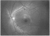
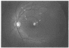
Glaucoma can be associated with a diffuse or localised loss of the retinal nerve fibre layer (RNFL). This study evaluated the wedge shaped localised RNFL defects. Red free wide angle RNFL photographs of 421 patients with glaucoma and 193 normal subjects were examined. Localised RNFL defects were described for one eye of the normal group and for 20% of the patients with glaucoma. They were usually located in the inferior temporal and superior temporal fundus regions. Within the glaucoma group, localised RNFL defects occurred most often (p < 0.05) in normal pressure glaucoma, followed by primary open angle glaucoma, and finally secondary open angle glaucoma. They were positively associated with disc haemorrhages. The localised RNFL defects had a high specificity to indicate optic nerve damage. The nerve fibre layer defects occurring more likely in mild rather than advanced glaucoma, they were helpful in the diagnosis of early glaucoma. The association between localised RNFL defects and disc haemorrhages and the varying frequency of localised RNFL defects in different types of glaucoma may be important diagnostically and pathogenetically.
Full text
PDF


Images in this article

Selected References
These references are in PubMed. This may not be the complete list of references from this article.
- Airaksinen P. J., Drance S. M., Douglas G. R., Mawson D. K., Nieminen H. Diffuse and localized nerve fiber loss in glaucoma. Am J Ophthalmol. 1984 Nov;98(5):566–571. doi: 10.1016/0002-9394(84)90242-3. [DOI] [PubMed] [Google Scholar]
- Airaksinen P. J., Drance S. M. Neuroretinal rim area and retinal nerve fiber layer in glaucoma. Arch Ophthalmol. 1985 Feb;103(2):203–204. doi: 10.1001/archopht.1985.01050020055018. [DOI] [PubMed] [Google Scholar]
- Airaksinen P. J., Heijl A. Visual field and retinal nerve fibre layer in early glaucoma after optic disc haemorrhage. Acta Ophthalmol (Copenh) 1983 Apr;61(2):186–194. doi: 10.1111/j.1755-3768.1983.tb01412.x. [DOI] [PubMed] [Google Scholar]
- Airaksinen P. J., Mustonen E., Alanko H. I. Optic disc haemorrhages precede retinal nerve fibre layer defects in ocular hypertension. Acta Ophthalmol (Copenh) 1981 Oct;59(5):627–641. doi: 10.1111/j.1755-3768.1981.tb08728.x. [DOI] [PubMed] [Google Scholar]
- Airaksinen P. J., Nieminen H. Retinal nerve fiber layer photography in glaucoma. Ophthalmology. 1985 Jul;92(7):877–879. doi: 10.1016/s0161-6420(85)33941-6. [DOI] [PubMed] [Google Scholar]
- Burk R. O., Rohrschneider K., Noack H., Völcker H. E. Are large optic nerve heads susceptible to glaucomatous damage at normal intraocular pressure? A three-dimensional study by laser scanning tomography. Graefes Arch Clin Exp Ophthalmol. 1992;230(6):552–560. doi: 10.1007/BF00181778. [DOI] [PubMed] [Google Scholar]
- Drance S. M., Airaksinen P. J., Price M., Schulzer M., Douglas G. R., Tansley B. W. The correlation of functional and structural measurements in glaucoma patients and normal subjects. Am J Ophthalmol. 1986 Nov 15;102(5):612–616. doi: 10.1016/0002-9394(86)90533-7. [DOI] [PubMed] [Google Scholar]
- Drance S. M. Mechanisms of optic nerve damage in glaucoma. Fortschr Ophthalmol. 1988;85(6):611–613. [PubMed] [Google Scholar]
- Hoyt W. F., Frisén L., Newman N. M. Fundoscopy of nerve fiber layer defects in glaucoma. Invest Ophthalmol. 1973 Nov;12(11):814–829. [PubMed] [Google Scholar]
- Hoyt W. F., Schlicke B., Eckelhoff R. J. Fundoscopic appearance of a nerve-fibre-bundle defect. Br J Ophthalmol. 1972 Aug;56(8):577–583. doi: 10.1136/bjo.56.8.577. [DOI] [PMC free article] [PubMed] [Google Scholar]
- Iwata K., Nanba K., Abe H. [Typical slit-like retinal nerve fiber layer defect and corresponding scotoma (author's transl)]. Nippon Ganka Gakkai Zasshi. 1981 Oct 10;85(10):1791–1803. [PubMed] [Google Scholar]
- Jonas J. B., Fernández M. C., Naumann G. O. Correlation of the optic disc size to glaucoma susceptibility. Ophthalmology. 1991 May;98(5):675–680. doi: 10.1016/s0161-6420(91)32234-6. [DOI] [PubMed] [Google Scholar]
- Jonas J. B., Fernández M. C., Naumann G. O. Glaucomatous parapapillary atrophy. Occurrence and correlations. Arch Ophthalmol. 1992 Feb;110(2):214–222. doi: 10.1001/archopht.1992.01080140070030. [DOI] [PubMed] [Google Scholar]
- Jonas J. B., Gusek G. C., Guggenmoos-Holzmann I., Naumann G. O. Size of the optic nerve scleral canal and comparison with intravital determination of optic disc dimensions. Graefes Arch Clin Exp Ophthalmol. 1988;226(3):213–215. doi: 10.1007/BF02181183. [DOI] [PubMed] [Google Scholar]
- Jonas J. B., Gusek G. C., Naumann G. O. Optic disc morphometry in chronic primary open-angle glaucoma. I. Morphometric intrapapillary characteristics. Graefes Arch Clin Exp Ophthalmol. 1988;226(6):522–530. doi: 10.1007/BF02169199. [DOI] [PubMed] [Google Scholar]
- Jonas J. B., Gusek G. C., Naumann G. O. Optic disc, cup and neuroretinal rim size, configuration and correlations in normal eyes. Invest Ophthalmol Vis Sci. 1988 Jul;29(7):1151–1158. [PubMed] [Google Scholar]
- Jonas J. B., Gusek G. C., Naumann G. O. Optic disk morphometry in high myopia. Graefes Arch Clin Exp Ophthalmol. 1988;226(6):587–590. doi: 10.1007/BF02169209. [DOI] [PubMed] [Google Scholar]
- Jonas J. B., Mardin C. Y., Schlötzer-Schrehardt U., Naumann G. O. Morphometry of the human lamina cribrosa surface. Invest Ophthalmol Vis Sci. 1991 Feb;32(2):401–405. [PubMed] [Google Scholar]
- Jonas J. B., Nguyen N. X., Naumann G. O. The retinal nerve fiber layer in normal eyes. Ophthalmology. 1989 May;96(5):627–632. doi: 10.1016/s0161-6420(89)32838-7. [DOI] [PubMed] [Google Scholar]
- Jonas J. B., Nguyen N. X., Strahwald H., Naumann G. O. Die retinale Nervenfaserschicht in Normal- und Glaukomaugen. I. Semiquantitative Daten von 398 Glaukomaugen. Klin Monbl Augenheilkd. 1989 Jun;194(6):437–446. doi: 10.1055/s-2008-1046398. [DOI] [PubMed] [Google Scholar]
- Jonas J. B., Schiro D. Visibility of the normal retinal nerve fiber layer correlated with rim width and vessel caliber. Graefes Arch Clin Exp Ophthalmol. 1993 Apr;231(4):207–211. doi: 10.1007/BF00918842. [DOI] [PubMed] [Google Scholar]
- Jonas J. B., Xu L. Parapapillary chorioretinal atrophy in normal-pressure glaucoma. Am J Ophthalmol. 1993 Apr 15;115(4):501–505. doi: 10.1016/s0002-9394(14)74453-8. [DOI] [PubMed] [Google Scholar]
- Kitazawa Y., Shirato S., Yamamoto T. Optic disc hemorrhage in low-tension glaucoma. Ophthalmology. 1986 Jun;93(6):853–857. doi: 10.1016/s0161-6420(86)33658-3. [DOI] [PubMed] [Google Scholar]
- Littmann H. Zur Bestimmung der wahren Grösse eines Objektes auf dem Hintergrund des lebenden Auges. Klin Monbl Augenheilkd. 1982 Apr;180(4):286–289. doi: 10.1055/s-2008-1055068. [DOI] [PubMed] [Google Scholar]
- Nanba K., Schwartz B. Nerve fiber layer and optic disc fluorescein defects in glaucoma and ocular hypertension. Ophthalmology. 1988 Sep;95(9):1227–1233. doi: 10.1016/s0161-6420(88)33024-1. [DOI] [PubMed] [Google Scholar]
- Quigley H. A., Addicks E. M. Quantitative studies of retinal nerve fiber layer defects. Arch Ophthalmol. 1982 May;100(5):807–814. doi: 10.1001/archopht.1982.01030030811018. [DOI] [PubMed] [Google Scholar]
- Quigley H. A., Addicks E. M. Regional differences in the structure of the lamina cribrosa and their relation to glaucomatous optic nerve damage. Arch Ophthalmol. 1981 Jan;99(1):137–143. doi: 10.1001/archopht.1981.03930010139020. [DOI] [PubMed] [Google Scholar]
- Quigley H. A., Katz J., Derick R. J., Gilbert D., Sommer A. An evaluation of optic disc and nerve fiber layer examinations in monitoring progression of early glaucoma damage. Ophthalmology. 1992 Jan;99(1):19–28. doi: 10.1016/s0161-6420(92)32018-4. [DOI] [PubMed] [Google Scholar]
- Quigley H. A., Miller N. R., George T. Clinical evaluation of nerve fiber layer atrophy as an indicator of glaucomatous optic nerve damage. Arch Ophthalmol. 1980 Sep;98(9):1564–1571. doi: 10.1001/archopht.1980.01020040416003. [DOI] [PubMed] [Google Scholar]
- Radius R. L. Thickness of the retinal nerve fiber layer in primate eyes. Arch Ophthalmol. 1980 Sep;98(9):1625–1629. doi: 10.1001/archopht.1980.01020040477018. [DOI] [PubMed] [Google Scholar]
- Sommer A., Katz J., Quigley H. A., Miller N. R., Robin A. L., Richter R. C., Witt K. A. Clinically detectable nerve fiber atrophy precedes the onset of glaucomatous field loss. Arch Ophthalmol. 1991 Jan;109(1):77–83. doi: 10.1001/archopht.1991.01080010079037. [DOI] [PubMed] [Google Scholar]
- Sommer A., Miller N. R., Pollack I., Maumenee A. E., George T. The nerve fiber layer in the diagnosis of glaucoma. Arch Ophthalmol. 1977 Dec;95(12):2149–2156. doi: 10.1001/archopht.1977.04450120055003. [DOI] [PubMed] [Google Scholar]
- Sommer A., Quigley H. A., Robin A. L., Miller N. R., Katz J., Arkell S. Evaluation of nerve fiber layer assessment. Arch Ophthalmol. 1984 Dec;102(12):1766–1771. doi: 10.1001/archopht.1984.01040031430017. [DOI] [PubMed] [Google Scholar]
- Tuulonen A., Airaksinen P. J. Initial glaucomatous optic disk and retinal nerve fiber layer abnormalities and their progression. Am J Ophthalmol. 1991 Apr 15;111(4):485–490. doi: 10.1016/s0002-9394(14)72385-2. [DOI] [PubMed] [Google Scholar]
- Tuulonen A., Airaksinen P. J. Optic disc size in exfoliative, primary open angle, and low-tension glaucoma. Arch Ophthalmol. 1992 Feb;110(2):211–213. doi: 10.1001/archopht.1992.01080140067029. [DOI] [PubMed] [Google Scholar]