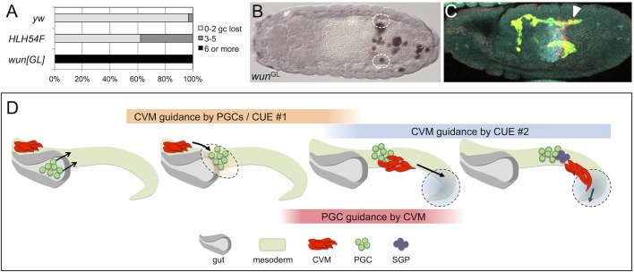
Fig. 8.
CVM cell and PGC migrations rely on multiple cues, including guidance from each other. (A) Quantification of the penetrance and severity of germ cell (gc) loss in wild-type embryos (yw, n=65), zygotic wunGL mutants (i.e. zygotic wun wun2, n=30) and HLH54F mutants (n=72). (B) Anti-Vasa staining of a wunGL homozygous embryo scored in A, showing many lost germ cells outside of the presumptive gonads (dashed ovals). (C) A M+Z− Tre1 mutant embryo showing CVM cell migration (arrowhead) relative to germ cells. (D) Model depicting how the migration of CVM cells could be guided by multiple cues. The overall successful migration of CVM cells in the absence of germ cells suggests that, besides the PGCs (Cue#1), another cue (Cue#2) is present in the vicinity of the pmg that acts in concert with the PGCs to initiate movement of the CVM cells towards the anterior and onto the TVM.

