

Abstract
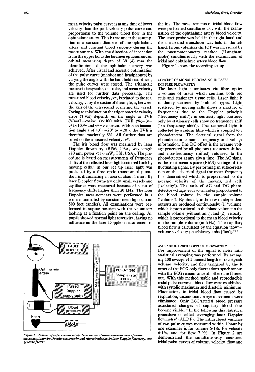
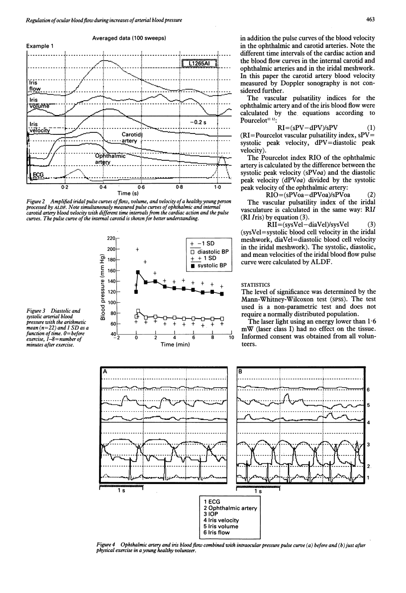
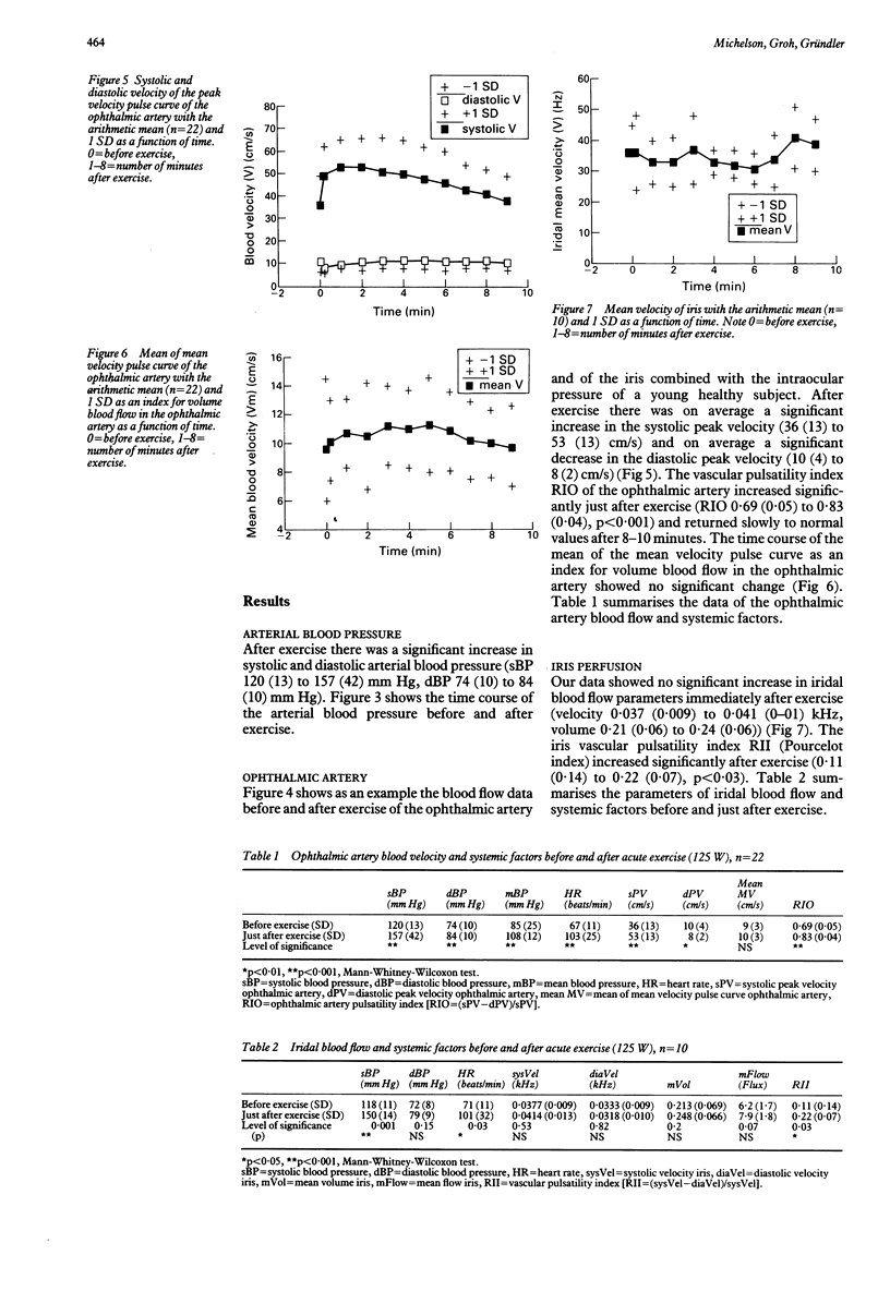
The blood flow in the uvea in cats and monkeys during acute increases of arterial blood pressure is well controlled by a sympathetic mechanism protecting the eye from overperfusion. Ocular macrocirculation (ophthalmic artery) and ocular microcirculation (iris) were examined in 22 healthy subjects during acute increases of arterial blood pressure induced by physical exercise (125 W). With a data acquisition and storage software in real time mode several parameters of ocular perfusion and systemic functions were measured simultaneously. Blood flow parameters were measured in the ophthalmic artery by pulsed Doppler sonography and in the iris by laser Doppler flowmetry. Systolic, diastolic, and mean velocities of the ophthalmic artery peak velocity pulse curve, the ophthalmic artery mean velocity pulse curve, and the iris velocity pulse curve were estimated off line. The ophthalmic artery mean velocity pulse curve resembles the integrated velocity of all erythrocytes in the vessel including the slowly running cells near the vessel wall. The iris velocity pulse curve was calculated by a special statistic procedure (ALDF). After exercise there was a significant increase in systolic and diastolic blood pressure and heart rate. The pulse curve of the ophthalmic artery showed significantly increased systolic and decreased diastolic velocities. The vascular resistance of the branches of the ophthalmic artery increased significantly. The iridal vasculature showed no significant change in blood cell velocity but an increased vascular resistance. It was observed that the elevated perfusion pressure was associated with an increased vascular resistance and a constant mean blood velocity in the ophthalmic artery and iridal vessels. The parallel elevation of vascular resistance and blood pressure during exercise may be the reason for a constant blood flow in the ophthalmic artery and the iris. This may be accounted for by a sympathetic mechanism for protecting the eye from overperfusion.
Full text
PDF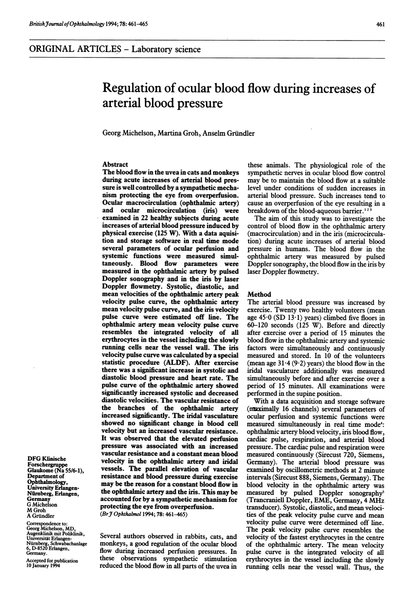
Selected References
These references are in PubMed. This may not be the complete list of references from this article.
- Alm A., Bill A. Ocular and optic nerve blood flow at normal and increased intraocular pressures in monkeys (Macaca irus): a study with radioactively labelled microspheres including flow determinations in brain and some other tissues. Exp Eye Res. 1973 Jan 1;15(1):15–29. doi: 10.1016/0014-4835(73)90185-1. [DOI] [PubMed] [Google Scholar]
- Alm A., Bill A. The effect of stimulation of the cervical sympathetic chain on retinal oxygen tension and on uveal, retinal and cerebral blood flow in cats. Acta Physiol Scand. 1973 May;88(1):84–94. doi: 10.1111/j.1748-1716.1973.tb05436.x. [DOI] [PubMed] [Google Scholar]
- Alm A. The effect of sympathetic stimulation on blood flow through t,e uvea, retina and optic nerve in monkeys (Macacca irus). Exp Eye Res. 1977 Jul;25(1):19–24. doi: 10.1016/0014-4835(77)90241-x. [DOI] [PubMed] [Google Scholar]
- Bill A., Linder M., Linder J. The protective role of ocular sympathetic vasomotor nerves in acute arterial hypertension. Bibl Anat. 1977;(16 Pt 2):30–35. [PubMed] [Google Scholar]
- Ernest J. T. The effect of systolic hypertension on rhesus monkey eyes after ocular sympathectomy. Am J Ophthalmol. 1977 Sep;84(3):341–344. doi: 10.1016/0002-9394(77)90676-6. [DOI] [PubMed] [Google Scholar]
- Harris A., Malinovsky V. E., Cantor L. B., Henderson P. A., Martin B. J. Isocapnia blocks exercise-induced reductions in ocular tension. Invest Ophthalmol Vis Sci. 1992 Jun;33(7):2229–2232. [PubMed] [Google Scholar]
- Legarth J., Nolsoe C. Doppler blood velocity waveforms and the relation to peripheral resistance in the brachial artery. J Ultrasound Med. 1990 Aug;9(8):449–453. doi: 10.7863/jum.1990.9.8.449. [DOI] [PubMed] [Google Scholar]
- Michelson G., Gierth K., Priem R., Laumer R. Blood velocity in the ophthalmic artery in normal subjects and patients with endophthalmitis. Invest Ophthalmol Vis Sci. 1990 Sep;31(9):1919–1923. [PubMed] [Google Scholar]
- Steinmeier R., Fahlbusch R., Powers A. D., Dötterl A., Buchfelder M. Pituitary microcirculation: physiological aspects and clinical implications. A laser-Doppler flow study during transsphenoidal adenomectomy. Neurosurgery. 1991 Jul;29(1):47–54. [PubMed] [Google Scholar]