Abstract
Purpose
The aim of the study is to compare subciliary incision and ‘sutureless’ transconjunctival incision in the treatment of infraorbital rim fractures.
Materials and method
In this prospective study, 40 patients with fractures of the infraorbital rim were selected and divided into 2 groups using random sampling technique. Group A patients were treated using ‘sutureless’ transconjunctival technique and group B patients were treated using subciliary approach. The following parameters were compared a) time taken, intraoperative ease of access, exposure achieved; b) clinical outcome and postoperative complications; c) Aesthetic outcome at intervals of 15 days, 1 month and 3 months.
Results
Total time taken for completion of surgery was lesser in group A patients. The presence of subconjunctival ecchymosis (at 1 month interval) and neurological deficit was found to be statistically significant (P<0.05) in the ‘subciliary’ group of patients. The transconjunctival approach showed better esthetic results and fewer post-operative complications.
Conclusion
The subciliary approach gives good exposure of the infra-orbital rim and is better suited to reduce extensively displaced fractures of the infra-orbital rim. The transconjunctival approach is comparatively faster, gives better esthetic results and fewer post-operative complications but is technique sensitive and requires an additional lateral canthotomy in cases where more exposure is needed.
Keywords: Subciliary incision, Transconjunctival incision, Infraorbital rim fractures
Introduction
Orbit is particularly susceptible to fractures because of its prominence in the facial skeleton [1]. It encloses the ocular globe and periorbital tissues, due to which injuries in this region have profound functional as well as aesthetic implications. The choice of approach is guided by the following goals: good intra-operative visibility, minimal post-operative scar formation and good esthetic results [2].
Varieties of surgical approaches to the infraorbital rim exist and can be conveniently categorized as transcutaneous or transconjunctival. Although it may seem that the difference lies only at the level of the incision from the ciliary margin, the anatomy of the region and the plane of dissection also influence the final aesthetic result [3].
The subciliary incision was popularized by Converse in 1944 and is perhaps the most commonly used ‘transcutaneous’ approach [3]. The transconjunctival incision initially developed by Bourguet in 1928 for cosmetic procedures, was utilized by Converse and Tessier for the treatment of fractures and congenital malformations [4]. Transconjunctival incisons can be pre-septal or post-septal with the former more commonly used for treatment of fractures [3, 5].
Both the approaches are used extensively and have their own set of advantages and disadvantages and complications [6–10]. We through this study attempt to establish clear superiority of one approach over the other, if any.
The aim of this study is to comprehensively evaluate, analyse and compare the following:
Intra-operative parameters (ease of access and exposure achieved),
Postoperative complications
The aesthetic outcome
Though a few studies have tried to compare the two approaches, none of the studies to our knowledge have tried to compare the subciliary approach with the ‘sutureless’ transconjunctival technique and on the basis of so many parameters with a follow-up of 3 months.
Materials and Methods
The study group Consisted of 40 patients (20 to 60 years age groups) with fracture of the infra-orbital rim who reported to the department of Oral and Maxillofacial Surgery of our institution.
Patients with comminuted fracture of the infraorbital rim or extensive soft tissue injury in the periorbital region were not considered. The study was granted approval by the institutional review board and the ethical committee. A written informed consent was obtained from all patients. Forty random numbers were generated from a random sampling table and were then alternatively assigned into two groups—group A and group B. The patients were then asked to choose from the 40 random numbers that were generated, depending upon the number they chose; they were divided into the two groups.
Group A- sutureless transconjunctival group
Group B- subciliary group
Initial pre-operative assessment was done for all patients. The surgeries were performed by a single operator.The following parameters were assessed:
Time taken, intraoperative ease of access, exposure achieved
Postoperative complications (chronic lid edema, scleral show, ectropion, corneal or conjunctival damage, entropion, buttonhole laceration of the eyelid, eye function, shape and position of the eyelid and neurological deficit).
Aesthetic outcome at Intervals of 15 days, 30 days and 3 months.
Evaluation of the study parameters were done before the surgical intervention, immediately after the procedure and at Intervals of 24 hours, 48 hours, 15 days, 30 days and 3 months postoperatively. The contra-lateral eye and previous photographs (taken anytime 6 months before trauma) were used to compare and assess pre-operative and post operative parameters. Evaluations were carried out by a single independent investigator in all the cases. The patients were also asked to assess (satisfaction/any gross aesthetic abnormality) their appearance at the one month post-op appointment.
Criteria for Evaluation
Periorbital Tissues
The affected eye was examined for presence of oedema, circumorbital-ecchymosis and subconjuctival haemorrhage.
Eyelids
The eyelids were observed for changes in shape, inclination, ptosis and associated lacerations. Intraoperatively any incidence of laceration of tarsal plate or buttonhole laceration of lower eyelid was recorded.
Eye
The affected eye was examined for diplopia, ocular movements, exopthalmos, enopthalmos, and ophthalmic injury and was compared with the contra-lateral eye. Pupillary reaction to light was assessed. Initial evaluation of the patient was done to compare the size and shape of the pupils.
The visual field in both eyes was appraised by directing the patient to cover one eye with the heel of his/her hand and then gaze on the examiner’s fingers with the other eye. The examiner then directed the patient to count the number of fingers held up in various quadrants. The same procedure was repeated on the contra-lateral eye. When limited extraocular movement was evident, muscle entrapment was differentiated from neurological deficit by performing a forced duction test. The conjunctiva was thoroughly examined for any laceration, ecchymosis.
Ectropion
Ectropion was graded as:
Mild:
slight lifting of the eyelid from the globe; Moderate: slight eversion and shortening of the vertical height of the lid; Severe: combination of severe shortening and true eversion of the affected eyelid.
Entropion
Inversion of the lower eyelid was recorded for its presence or absence.
Neurological Deficits
Presence or absence of paresthesia and functionality of the extraocular muscles were checked. Anaesthesia or paresthesia in the region of distribution of infraorbital nerve was assessed by cotton wool application method. The patients were examined bilaterally with the non traumatized side serving as control. Eyes of the patients were kept closed during the examination.
Statistical Parameters
Null Hypothesis
This is to be considered when there is no significant difference in the proportion of positive results of different parameters in Subciliary and Transconjunctival groups i.e. ρ1 = ρ2
Level of Significance
α = 0.05
Statistical Technique Used
Z-test for proportions
Decision Criterion
The decision criterion is to reject the null hypothesis if the p value is less than 0.05. Otherwise the null hypothesis is accepted.
Surgical Technique
Subciliary Incision
Tarsorraphy was performed in all cases. The first skin crease in the infraorbital region was taken as a landmark. In cases of edema or soft tissue injury, skin creases in the contra-lateral side were evaluated and used as the guideline (Fig. 1).
Fig. 1.
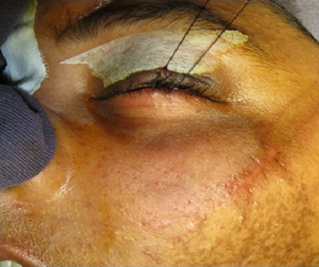
Pre-operative view after tarsorraphy prior to placing a subciliary incison
Subciliary incision was placed approximately 2 mm below the eyelashes and extended laterally as necessary using a no 15 blade (Fig. 2). Initial incision was placed over the skin only. The plane of dissection was superficial to the orbital septum to prevent damage to its integrity and deep to the orbicularis oculi muscle. Once the periosteum was exposed, blunt dissection was continued approximately 5 mm inferiorly before incising. Care was taken to identify the infraorbital nerve and vessels and precautions were taken to avoid traumatizing them (Fig. 3). Fracture was reduced, fixed and stabilized appropriately.
Fig. 2.
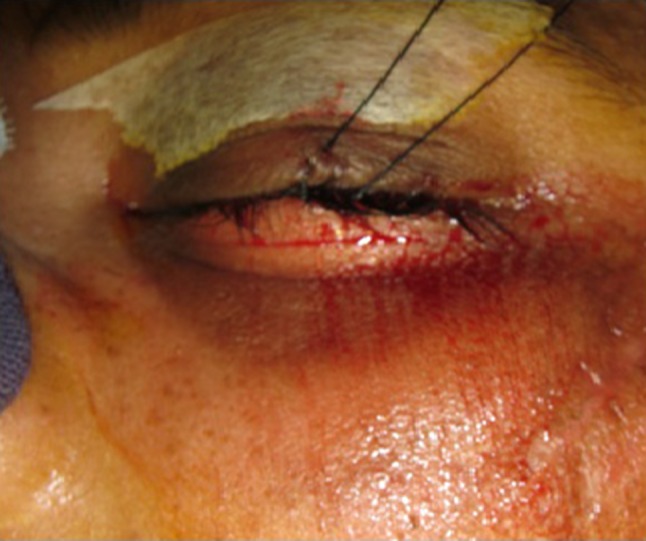
Subciliary incision placed
Fig. 3.
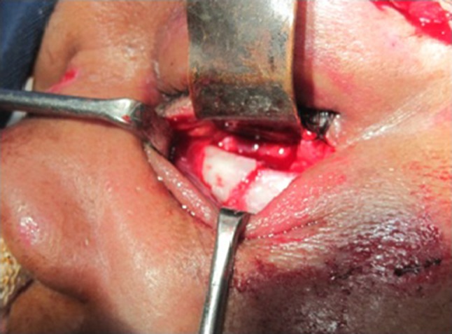
Exposure achieved. Fractured infra-orbital rim exposed
Care was taken to re-approximate the periosteum. Periosteal closure was done using 5-0 Polyglactin sutures (Vicryl®). Closure of the muscle was attempted only when the muscular plane could be correctly identified. Skin closure was done using 4-0 polypropylene (Prolene®) (Fig. 4).
Fig. 4.
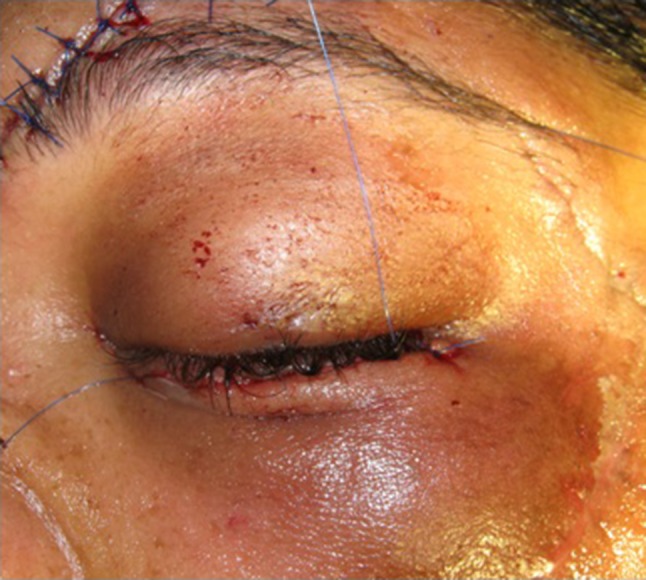
Closure
‘Sutureless’ Pre-septal Transconjunctival Incison
For protection of the globe during the procedure, a corneal shield/lens was placed. Eye was lubricated using viscous carboxy-methyl cellulose eye gel. The lower eyelid was everted and retracted using two traction sutures. A small incision was placed 3 mm below the tarsal plate on the medial aspect in line with the punctum and extended laterally in the same plane as far as the line of the lateral canthus (Fig. 5). The conjunctiva was then divided using fine dissectors. Tissues were separated on a plane superficial to the orbital septum but deep to the orbicularis oculi muscle and a pre-septal plane of dissection was ensured. Care was taken not to distort the periosteum and to clearly define it across the entire width of the inferior orbital rim without damaging the integrity of the septum. A lateral canthotomy was opted for if it was felt that the exposure of the fracture site was inadequate (Fig. 6).
Fig. 5.
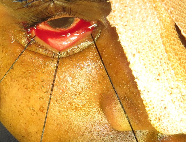
Traction sutures and placement of transconjunctival incision
Fig. 6.

Exposure achieved to access the fractured infra-orbital rim
A periosteal closure was not considered mandatory. However meticulous care was taken to passively reposition the periosteum. Palpebral conjunctival closure was also not considered to be necessary (Fig. 7). The lateral canthotomy incision was closed using 4-0 polypropylene suture.
Fig. 7.
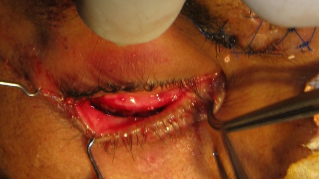
Conjunctival redraping post fixation
Post-operative Care
Patients were put on antibiotic eyedrops (Moxifloxacin 0.5 %, 1–2 drops 4th hourly for 3 days postoperatively) and steroid eyedrops (Betamethasone 0.1 %, 1–2 drops 4th hourly for 2 days post-operatively). Patients were also put on a tapering intravenous dosage regimen of steroid (Dexamethasone/8 mg/IV).
Results
Out of the 40 patients selected for the study, 36 were males and 4 were females and road traffic accidents accounted for the trauma in 36 (90 %) of the total number of patients. The age of the patients’ ranged from 20 to 60 years with the average age being 37 years.
The average time taken from incision to exposure of fracture was 12 minutes for the subciliary approach, 15 minutes for transconjunctival approach and 20 minutes for transconjunctival approach with lateral canthotomy. Closure of subciliary incision post fracture repair took 20 minutes.
Mild post-operative ectropion was noticed in two patients in the subciliary group at 2 weeks interval. 2 out of 20 patients in the transconjunctival group developed entropion. One patient in the subciliary group complained of blurred vision which resolved on its own on the second post-op day. One patient in the transconjunctival group complained of diplopia which persisted for 2 weeks. This was probably due to orbital floor fracture and not because of the incision used. No statistically significant difference was found in the incidence of intraoperative lid lacerations or button hole defects in the two groups. (Tables 1, 2).
Table 1.
Number of patients with positive findings after undergoing fracture reduction using the subciliary approach
| Conditions/complications observed | Time intervals at which patients were reviewed | |||||
|---|---|---|---|---|---|---|
| Pre-op | 24 h | 48 h | 2 weeks | 1 month | 3 months | |
| Subciliary approach (Number of patients out of 20 with positive finding) | ||||||
| Periorbital edema | 20 | 20 | 06 | – | – | – |
| Subconjuctival ecchymosis | 18 | 20 | 20 | 18 | 04 | – |
| Eye function and globe position | 04 | 01 | – | – | – | – |
| Ectropion | – | 02 (Mild) | 02 (Mild) | 02 (Mild) | – | – |
| Entropion | – | – | – | – | – | – |
| Neurological deficit | 20 | 20 | 20 | 12 | 04 | – |
Table 2.
Number of patients with positive findings after undergoing fracture reduction using the transconjunctival approach
| Conditions/complications observed | Time intervals at which patients were reviewed | |||||
|---|---|---|---|---|---|---|
| Pre-op | 24 h | 48 h | 2 weeks | 1 month | 3 months | |
| Transconjunctival approach (Number of patients out of 20 with positive finding) | ||||||
| Periorbital edema | 16 | 18 | 06 | – | – | – |
| Subconjunctival ecchymosis | 20 | 20 | 18 | 10 | – | – |
| Eye function and globe position | 02 | 1 | 1 | 1 | – | – |
| Ectropion | – | – | – | – | – | – |
| Entropion | – | 01 | 01 | 01 | 01 | 01 |
| Neurological deficit | 16 | 16 | 10 | 02 | – | – |
No significant difference was observed between subciliary and transconjunctival groups with respect to the proportion of samples with the positive finding of periorbital odema, eyelid abnormalities, abnormalities in eye function and globe position, presence of ectropion or entropion at any of the time intervals (P > 0.05). The 2 patients with mild ectropion in the subciliary group were taught digital palpebral massaging which helped to resolve the complication. No entropion was observed in the subciliary group of patients. One patient in the transconjunctival group who required an additional lateral canthotomy developed trichiasis and had to undergo epilation as a corrective measure.
The difference in the proportion of samples with presence of subconjuctival ecchymosis at 1 month between Subciliary and Transconjunctival groups was found to be statistically significant (P < 0.01) with four patients in the subciliary group showing persistent ecchymosis at that interval. No significant difference was observed at other time intervals (P > 0.05).
All patients in the subciliary group complained of some degree of paresthesia the second post-operative day. Four patients in the subciliary group had persistent neurological deficit which lasted for a month. Only ten patients in the transconjunctival group complained of paresthesia on the second post-op day and none of them had any persistent neurological deficit which persisted beyond 2 weeks. These resulted in statistically significant difference in patients of the two groups with neurological deficit at the 48 h interval (P < 0.05) and at 2 weeks (P < 0.05).
The patients were asked to rate the appearance of the surgical site/scar at pre-specified intervals as satisfactory/unsatisfactory. Sixteen out of 20 patients treated with the subcilary approach were satisfied with the esthetic outcome, a month after surgery where as all patients treated with the transconjunctival approach were satisfied with the esthetic outcome at the same interval (Figs. 8, 9), (Table 3).
Fig. 8.
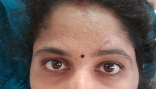
One month post-operative view
Fig. 9.
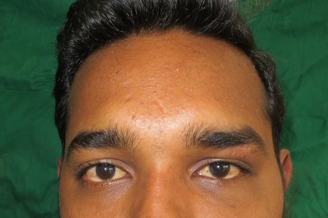
One month post operative classic view
Table 3.
Aesthetic outcome of the incision site
| Intervals | Satisfactory | Unsatisfactory | Percentage |
|---|---|---|---|
| Subciliary approach | |||
| 15 days | 14 | 06 | 70 |
| 1 month | 16 | 04 | 80 |
| 3 months | 18 | 02 | 90 |
| Transconjunctival approach | |||
| 15 days | 16 | 04 | 80 |
| 1 month | 20 | 00 | 100 |
| 3 months | 20 | 00 | 100 |
Discussion
Transcutaneous approaches or transconjunctival incisions offer good access to the operative field but differ in terms of the simplicity of the procedure, time needed to gain access and aesthetic results. Subciliary as well as transconjunctival approaches have their own advantages and disadvantages with each approach having proponents who vouch for them [11–15].
The causes of lower eyelid retraction after orbital floor surgery are multifactorial. Factors predisposing to eyelid retraction and ectropion following orbital fracture repair include inadequate skin and tissue management, anterior lamellar insufficiency, hematoma, eyelid edema, adhesions of the orbital septum, scar contracture, horizontal laxity of the eyelid margin, weakening of the pretarsal muscle, and wide dissection of the anterior periosteum. Any event, whether surgically iatrogenic or traumatic, that contributes to contracture of the orbital septum will cause the drape-like orbital septum to contract and, thereby, to pull the lower eyelid down from its normal position. The risk of developing postoperative eyelid retraction also varies with the type of approach used to expose the inferior orbital rim and floor. [10, 15]
Few authors [2, 3, 10, 15] have compared subciliary approach with transconjunctival approach but no study has tried the comparison with ‘sutureless’ transconjunctival technique on the basis of so many pre-defined parameters with a follow-up of 3 months. We observed that main drawbacks of the subciliary approach include the post-operative scarring and the risk for potential complications. The main disadvantages of the transconjunctival approach are its technique sensitivity, a relatively higher percentage of lower eyelid malpositioning when combined with a lateral canthotomy and relatively limited exposure when used alone.
We noted that although the exposure of the fractured bone took longer while using the transconjunctival approach, the total time taken was more with the subciliary approach because of the meticulous closure needed for the latter. The ‘sutureless’ transconjunctival technique offers a simpler, effective and faster alternative method with excellent post-operative healing of the conjunctiva, no shortening of the fornix and fewer complications like infection, lid malposition (Fig. 10) [16]. The incidence of post-operative complications was lower in the transconjunctival group. Number of cases with persisting neurologic deficit and subconjunctival ecchymosis post surgery in the subcilary group was statistically significant. Persistent paresthesia can probably be attributed to comparatively increased manipulation of tissues adjacent to the infraorbital foramen while using the subciliary technique. Aesthetic satisfaction was better in transconjunctival approach with patients reporting better acceptance of post op appearance.
Fig. 10.
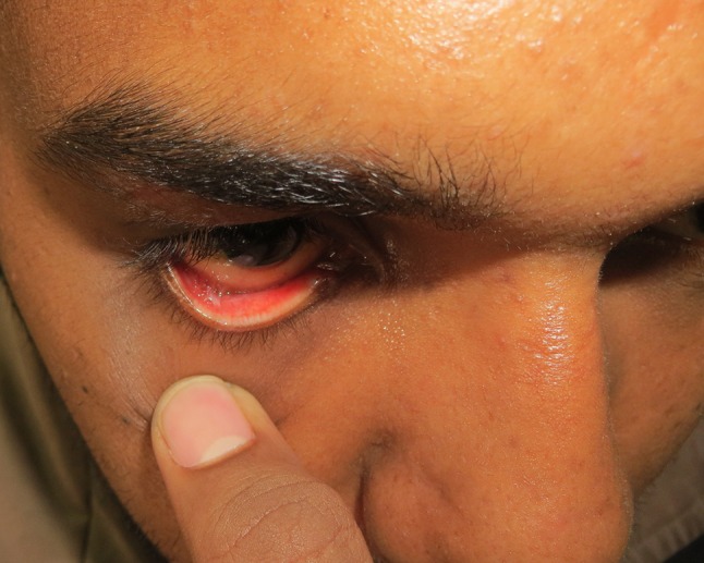
One month postoperative view showing conjunctival healing and sulcular depth
Conclusion
Both the approaches have potential for sequelae and complications. The approach must be based, in part, on the surgeon’s particular abilities in terms of preferred incision and also on the potential complications. To summarize, the subciliary approach gives good exposure of the infra-orbital rim and is better suited to reduce extensively displaced fractures of the infra-orbital rim. The transconjunctival approach is comparatively faster, gives better esthetic results and fewer post-operative complications but is technique sensitive and requires an additional lateral canthotomy in cases where more exposure is needed.
Acknowledgments
The authors have received no grant for the completion of the study and declare that they have no conflict of interest to declare. Approval to conduct the study was obtained by the ethical committee of the institutional review board. In addition, a written consent was obtained from every patient who was included in the study. Appropriate permission was also obtained to use clinical photographs for the purpose of further academic research and publication in academic journals.
Funding
No funds were sought or received for the conduction and completion of this study
Compliance with Ethical Standards
Conflict of interest
All the seven authors have no conflicts of interests to declare.
Human and Animal Rights
No animals were used in this study. All procedures performed in studies involving human participants were in accordance with the ethical standards of the institutional and/or national research committee and with the 1964 Helsinki declaration and its later amendments or comparable ethical standards. Approval to conduct the study was obtained by the ethical committee of the institutional review board.
Informed Consent
Informed consent was obtained from all individual participants included in the study. Additional informed consent was obtained from all individual participants for whom identifying information is included in this article. Appropriate permission was also obtained to use clinical photographs for the purpose of further academic research and publication in academic journals.
References
- 1.Wang S, Xiao J, Liu L, Lin Y, et al. Orbital floor reconstruction: a retrospective study of 21 cases. Surg Oral Med Oral Pathol Oral Radiol Endod. 2008;106:324–330. doi: 10.1016/j.tripleo.2007.12.022. [DOI] [PubMed] [Google Scholar]
- 2.De Riu G, Meloni SM, Gobbi R, Soma D, Baj A, Tullio A. Subcilliary versus swinging eyelid approach to the orbital floor. J Cran-Maxillary Surg. 2008;36:439–442. doi: 10.1016/j.jcms.2008.07.005. [DOI] [PubMed] [Google Scholar]
- 3.Subramanian B, Krishnamurthy S, Kumar S, Saravanan B, Padmanabhan M. Comparison of various approaches for exposure of infraorbital rim fractures of zygoma. J Maxillofac Oral Surg. 2009;8(2):99–102. doi: 10.1007/s12663-009-0026-7. [DOI] [PMC free article] [PubMed] [Google Scholar]
- 4.Palmer FR, Rice DH, Churukian MM (1993) Transconjunctival blepharoplasty—complications and their avoidance: a retrospective analysis and review of the literature. Arch Otolaryngol Head and Neck Surg 119:993–999 [DOI] [PubMed]
- 5.Lee PK, Lee JH, Choi YS, Oh DY, et al. Single transconjunctival incision and two point fixation for the treatment of noncomminuted zygomatic complex fracture. J Korean Med Sci. 2006;21:1080–1085. doi: 10.3346/jkms.2006.21.6.1080. [DOI] [PMC free article] [PubMed] [Google Scholar]
- 6.Salgarelli AC, Consolo U. The Orbicularis Oculi Muscle flap in subciliary access for management of orbital trauma: technical note. J Oral Maxillofac Surg. 2002;60:470–472. doi: 10.1053/joms.2002.31243. [DOI] [PubMed] [Google Scholar]
- 7.Werther JR. Cutaneous approaches to the lower lid and orbit. J Oral Maxillofac Surg. 1998;56:60–65. doi: 10.1016/S0278-2391(98)90917-X. [DOI] [PubMed] [Google Scholar]
- 8.Langsdon PR, Rohman GT, Hixon R, Stumpe M, Metzinger S. Upper lid transconjunctival versus transutaneous approach for fracture repair of the lateral orbital rim. Ann Plast Surg. 2010;65(1):52–55. doi: 10.1097/SAP.0b013e3181c1fe14. [DOI] [PubMed] [Google Scholar]
- 9.Patel PC, Sobota BT, Yates JA, Millman B (1996) Comparison of transconjunctival versus subciliary incisions for orbital fractures. Otolaryngol–Head Neck Surg 115:P161
- 10.Appling WD, Patrinely JR, Salzer TA (1993) Transconjunctival approach versus subcillary skin muscle flap approach for orbital fracture repair. Arch Otolaryngol Head and Neck Surg 119:1000–1007 [DOI] [PubMed]
- 11.de Melo Crosara J, da Rosa ELS, Alves e Silva MRM (2009) Comparision of cutaneous incision to approach infraorbital rim and orbital floor. Braz J Oral Sci 8:88–91
- 12.Manganello Souza LC, de Freitas RR. Transconjunctival approach to zygomatic and orbital floor fractures. Int J Oral Maxillofac Surg. 1997;26:31–34. doi: 10.1016/S0901-5027(97)80843-0. [DOI] [PubMed] [Google Scholar]
- 13.Goyal A, Tyagi I, Jain S, Syal R, Singh AP, Kapila R (2010) Transconjunctival incision for total maxillectomy—an alternative for subcilliary incision. Br J Oral Maxillofac Surg 49:442–446 [DOI] [PubMed]
- 14.Wesley R. Transconjunctival approaches to lower lid and orbit. J Oral Maxillofac Surg. 1998;56:66–69. doi: 10.1016/S0278-2391(98)90918-1. [DOI] [PubMed] [Google Scholar]
- 15.Salgarelli AC, Bellini P, Landini B, Multinu A, Consolo U. A comparative study of different approaches in the treatment of orbital trauma: an experience based on 274 cases. Oral Maxillofac Surg. 2010;14:23–27. doi: 10.1007/s10006-009-0176-2. [DOI] [PubMed] [Google Scholar]
- 16.Lane KA, Bilyk J, Taub D, Pribitkin Edmund. “Sutureless” repair of orbital floor and rim fractures. Ophthalmology. 2009;116:135–138. doi: 10.1016/j.ophtha.2008.08.042. [DOI] [PubMed] [Google Scholar]


