Abstract
Introduction
The term oro-antral fistula is understood to mean of fistular canal covered with epithelia which may or may not be filled with granulation tissue or polyposis of the sinus mucous membrane. With the presence of a fistula the sinus is permanently open, which enables the passage of microbial flora of the oral cavity into the maxillary sinus and the occurrence of inflammation with all possible consequences. Every now and then various researchers have proposed innumerable techniques to treat this defect. Starting from simple tissue flaps to autogenous grafts to alloplastic materials, an array of procedures have been evaluated in literature but the most promising technique still needs to be evaluated. Consequently, after reviewing an array of such procedures, our present study focussed on a new technique for the closer of oro-antral fistulas using autogenous auricular cartilage graft supported by buccal advancement flap.
Material and method
A total of 20 patients of oro-antral fistula were included in the study and after excising the fistular tract a double layer closure was done by placing auricular cartilage over the defect followed by buccal mucoperiosteal flap. The graft was harvested using posterior auricular approach. Assessment of patients was done at the end of 1 week, 3 weeks, 6 weeks, and 3 months.
Conclusion
We found that the autogenous auricular cartilage graft is an effective sealing material in oro-antral fistula closure. We recommend this technique for the defect size ≤10 mm2 in which future dental implant placement is sought as it allows easy sinus lifting procedure.
Keywords: Oroantral fistula, Auricular cartilage graft, Buccal advancement flap
Introduction
Oro-antral communication (OAC) and oro antral fistula (OAF) are two closely related terms used to describe the pathological communication between the oral cavity and the maxillary sinus. Only notable difference between these two entities is epithelization of communicating tract. The initial unnatural non epithelized continuation between antrum and oral cavity is referred as OAC while later when the same tract gets epithelized it is regarded as OAF that may be filled by granulation tissue or by polyposis of the sinus membrane. The air current which passes from the sinus through the alveoli into the oral cavity during expiration also facilitates the formation of a fistular canal, which connects the sinus with the oral cavity [1]. Depending on the location it can be classified as alveolo-sinusal, palatal-sinusal and vestibulo-sinusal [2].
At birth maxillary sinus is present as a small cavity; its growth begins in the 3rd month of foetal life, and ends between the 18th and 20th years of life. It increases at the same rate as the growth of jaws and permanent teeth. Because of the small volume of the sinus the risk of the occurence of oro-antral communication in children and adolescents is less. In adults the volume of the sinus amounts to 20–25 ml. Although beneficial, functionally and anatomically, a major shortcoming encountered during the course of pneumatisation is the incorporation of roots of various maxillary posterior teeth within the pneumatic space of maxilla, i.e. the maxillary sinus. This anatomical relationship serves obvious disadvantages.
Most common cause of oro-antral communications is the extraction of maxillary posterior teeth. That might attain closure on its own or may get complicated further to get epithelized and result in a fistulous tract. Other causes that may cause OAF are following facial trauma, tumours, cysts, minor surgery and following pathologies in the maxilla, dehiscence of floor of the sinus secondary to periapical lesions, forcing a tooth/tooth root into the sinus cavity during attempted removal, chronic osteomyelitis, gumma, infected maxillary implant dentures, malignant granuloma or may even be iatrogenic in nature.
Incidence of OAF has been reported to commonly occur following extractions of maxillary first molar, followed by the second molar, third molar, and bicuspid because of anatomic proximity of root apices of these teeth and maxillary antrum [3, 4]; the incidence ranges from 0.31 to 4.7 % [4]. Since the largest part of the upper jaw is taken up by the maxillary sinus by 20th year of life therefore incidence of OAF has been reported to occur most frequently after third decade of life [4–6].
It has been described in literature that most OAC with defect size <2 mm in diameter may close spontaneously in absence of infection but in more than 3–4 mm defect opening persists and requires closure [7], which has also been seen in our study. To prevent chronic sinusitis and the development of fistulas, it is generally accepted that all of these defects should be closed within 24–48 h [8] otherwise reluctantly there is a risk that an epithelialized oroantral fistula with resultant maxillary sinusitis may develop [9].
Various surgical modalities are available for closure of OAF like soft tissue flaps including buccal flap, rotational pedicled palatal flap, buccal pad fat flap, palatinal island flaps, vestibular flaps; autogenous bone grafts; third molar transplantation by Yoshimasa et al. [10] and bone substitute technique by Ogunsalu [11] have been described in literature. Although any technique can be used for closure of OAF and good results can be obtained but every technique has some shortcomings and pitfalls. So the choice of surgical technique used for closure of OAF varies under different clinical conditions and also depends on the outcome desired like choice for bone or bone substitute grafting technique if dental implant has to be placed in near future.
Since the use of nasal septal cartilage [12] for closure of OAF has also been described, here in this article we present our experience of closure of OAF using autogenous auricular cartilage graft supported by buccal advancement flap as an alternate technique.
Materials and Methods
A total of 20 patients of oro-antral fistula attending the Department of Oral and Maxillofacial Surgery, Faculty of Dental Sciences, King George’s Medical University, Lucknow were included in the present study. Patients suffering from any renal or hepatic disease, blood dyscrasia, any known hypersensitivities, allergies or idiosyncratic reactions to any medications were excluded from the study. The diagnosis of OAF was established by the nose-blowing test and introduction of a silver probe into the antrum through the fistula (Figs. 1, 2). In all patients, antibiotics, decongestant nasal drops, steam inhalation were given for 7 days preoperatively and irrigation of sinus was done with normal saline and no radiographic evidence of maxillary sinusitis was ensured before surgery.
Fig. 1.
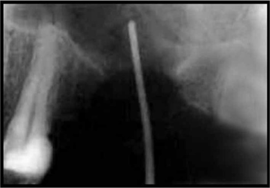
Intra-oral periapical view X ray showing oroantral fistula in relation with 26 region
Fig. 2.
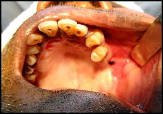
Intra-oral photograph showing oroantral fistula in relation with 26 region
Under local anesthesia initially recipient site was prepared. A circular incision with a 2 mm margin was made around the oro-antral fistula, and the epithelial tract and any inflammatory tissue within the opening were completely excised (Fig. 3) and trapezoidal shape buccal sliding flap as described by Berger [13] was raised. After recipient site preparation, local anesthesia was infiltrated in post-auricular sulcus and conchal fossa for harvesting auricular cartilage graft (Fig. 4). A semicircular incision was made posteriorly over the conchal cartilage, but not through perichondrium. A blunt dissection was used to expose the conchal cartilage with overlying perichondrium intact and attached to the graft. The index finger was placed in the conchal fossa laterally, guiding the knife medially to cut the conchal cartilage while leaving an adequate rim along the fossa. Desired amount of cartilage depending upon the size of the defect was removed with circular incision (Figs. 5, 6). Hemostasis was achieved and graft site was closed with 6–0 nylon by vertical mattress suture (Fig. 7). Harvested auricular graft was than adapted on the perforation site and sutured with bone/surrounding tissue with 3/0 polyglactin for stabilization (Fig. 8). The buccal mucoperiosteal flap was then advanced and sutured with palatal mucosa with 3/0 silk (Fig. 9).
Fig. 3.
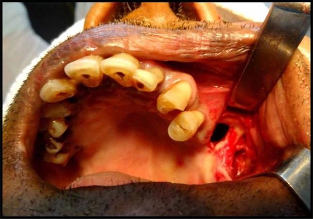
Oroantral fistula site after removal of fistulous tract
Fig. 4.
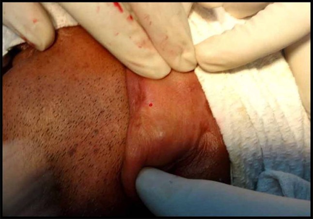
Donor site—ear
Fig. 5.
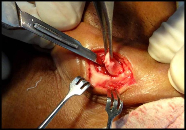
Harvesting of auricular cartilage
Fig. 6.
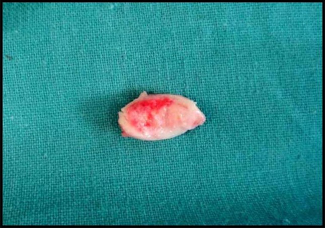
Harvested autogenous auricular cartilage graft
Fig. 7.
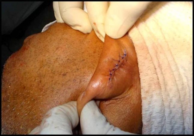
Photograph showing closure at graft site
Fig. 8.
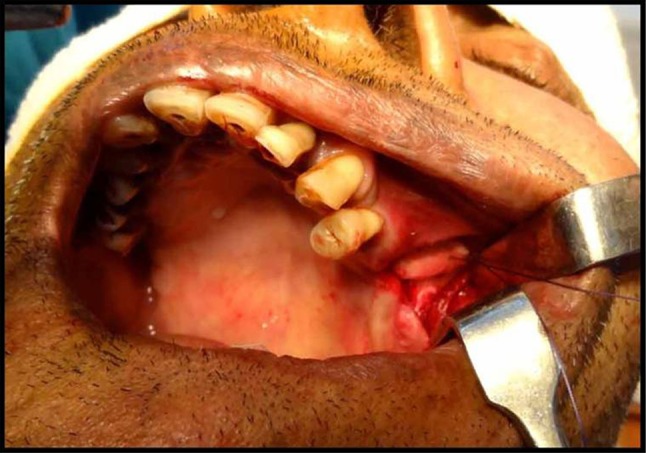
Auricular cartilage at the defect site
Fig. 9.
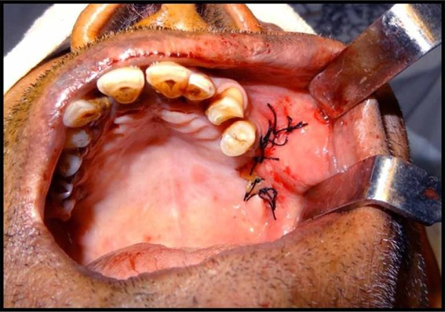
Closure of oro antral fistula
After the surgery, the patients were instructed to avoid activities that may produce pressure changes between the nasal passages and oral cavity for at least 2 weeks, such as sucking on a straw, blowing the nose, and sneezing with a closed mouth and were advised to avoid smoking. Antibiotics and nasal decongestants were given for 1 week postoperatively. Sutures were removed 10 days after the surgery. All the patients were assessed at the end of 1 week, 3 weeks, 6 weeks, and 3 months under following parameters: pain, infection, swelling, healing, graft acceptance or rejection (Figs. 10, 11).
Fig. 10.
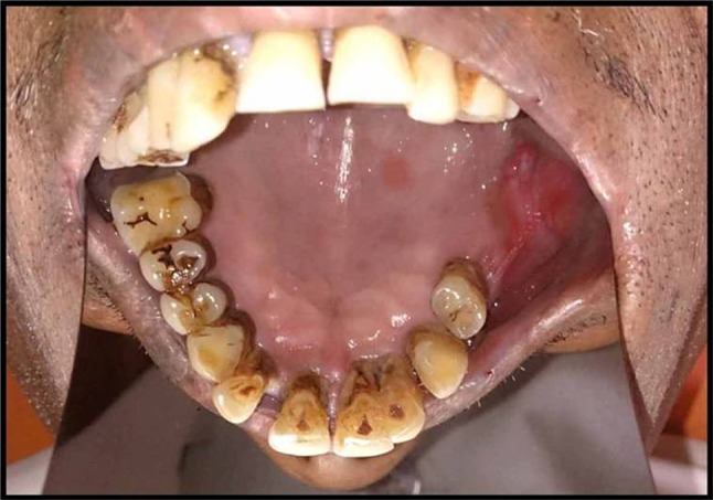
Follow up after 15 days
Fig. 11.
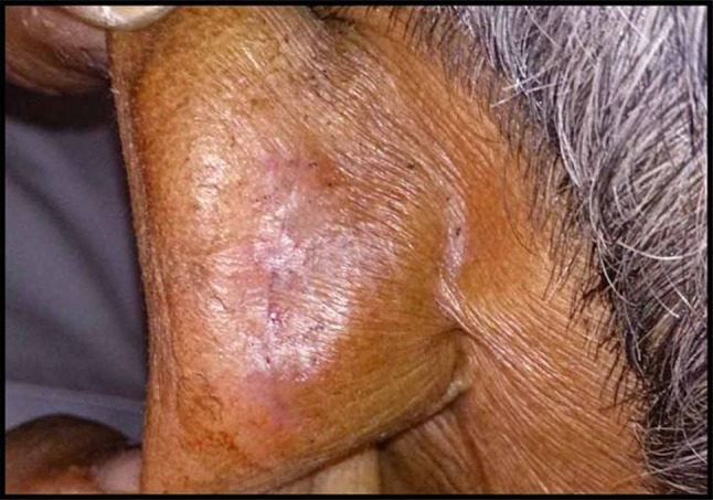
Follow up graft site after 15 days
Results
A total of 20 patients of age ranging from 18 to 65 years undergoing surgery for closure of oro-antral fistula irrespective of sex, caste and creed were included in the present study.
Among the 20 patients included in the study 3 (15 %) were 18–35 years of age, 9 (45 %) were 36–50 years old and remaining 8 (40 %) were 51–65 years of age. In 80 % of cases in our study, OAF developed after tooth extraction followed by cyst in 15 % of cases.
Most common site of oro-antral fistula in our study was maxillary 1st molars in (n = 9) 45 % followed by maxillary 2nd molar (n = 6) 30 %, maxillary 2nd premolar (n = 3) 15 % and maxillary 1st premolar (n = 2)10 % cases (Table 1).
Table 1.
Distribution of patients
| Age group (in years) | Sex | Site of AOF | Size of defect | |||||||||
|---|---|---|---|---|---|---|---|---|---|---|---|---|
| 18–35 | 36–50 | 51–65 | Male | Female | 1st PM | 2nd PM | 1st molar | 2nd molar | <5 mm | 5–10 mm | >10 mm | |
| No. of Patients | 3 | 9 | 8 | 11 | 9 | 2 | 3 | 9 | 6 | 5 | 13 | 2 |
| Total | 20 | 20 | 20 | 20 | ||||||||
In the study group size of defect was between 5 and 10 mm2 in (n = 13) 65 % subjects and size of defect was <5 mm in (n = 5) 25 % subjects, while only (n = 2)10 % subjects were having size >10 mm2. Post-operative healing at graft site was satisfactory in all the patients at interval of 1 and 3 months follow up, but at the surgical site, out of the 20 patients 18 healed uneventfully while in 2 patients graft was extruded and required removal. Although post operative infection was observed in 3 patients (15 %) at the surgical site but under the antibiotic coverage and postoperative care 2 patients showed uneventful healing at 3 months follow up and only 1 patient (5 %) was among those two patients who needed graft removal (Table 2).
Table 2.
Incidence of post-operative infection, pain and healing (n = no of patients)
| Time interval | Satisfactory objective healing | Postoperative infection | Postoperative pain (Mean ± SD on VAS scale) | |||
|---|---|---|---|---|---|---|
| Graft | Recipient | Graft site | recipient site | Graft site | Recipient site | |
| % | % | % | % | |||
| 1 week | 90 (n = 18) | 85 (n = 17) | 5 (n = 1) | 15 (n = 3) | 3.30 ± 0.66 | 3.55 ± 0.76 |
| 15 days | 95 (n = 19 | 90 (n = 18) | – | – | 2.05 ± 1.00 | 3.00 ± 0.79 |
| 1 month | 100 (n = 20) | 90 (n = 18) | – | – | 0.90 ± 0.72 | 1.20 ± 0.83 |
| 3 months | 100 (n = 20) | 90 (n = 18) | – | – | 0.20 ± 0.62 | 0.10 ± 0.31 |
Discussion
Maxillary sinus, also known as Antrum of Highmore after the English anatomist Nathaniel Highmore who first described the sinus in seventeenth century, achieves its maximum size after nearly second decade of life, therefore, the risk of the occurrence of oro-antral communication in children and adolescents is less because of the smaller volume of the sinus. Lin et al. [6] claimed that females exhibit larger sinuses than males and therefore are at greater risk of oro-antral fistula. But in our study frequency of occurrence of OAF was nearly the same in both sexes. Our study comprised of a total of 20 patients in the age group of 18–65 years. This age group was selected keeping in the view the maximum degree of pneumatisation that takes place uptil this age and also happens to be the prevalence age for maximum posterior teeth extraction. The mean age of the subjects included was 46 years with 55 % male patients and 45 % female patients.
Punwutikorn et al. [4] reported that extraction of maxillary posterior teeth was the most common cause of OAF. Our study is also consistent with study of Punwutikorn et al. [4] where removal of the upper molar teeth was the most common anetiological factor for creation of oro-antral fistula. In 80 % cases oro-antral fistula was created after the extraction and 1st molar was the most commonly involved tooth in 45 % cases followed by 2nd molar in 30 % cases.
Controversy prevails relating to spontaneous closure of oro-antral fistula. Some authors, such as Martensson [14] considered that there is less possibility of spontaneous healing when the oroantral fistula has been present for 3 to 4 weeks, or when its diameter is greater than 5 mm. In contrast to this, Hanazawe et al. [7], reported that an oroantral fistula of less than 2 mm diameter has the possibility of spontaneous healing, while in the case of a diameter of more than 3 mm, spontaneous healing is hampered because of the possibility of inflammation of the sinus or periodontal region. At our centre we found that only smaller openings of 1–2 mm diameter may heal spontaneously and that too in absence of sinus infection only. Since a chronic communication between the oral cavity and the maxillary sinus can represent an access route for infective organisms into the sinus, hence it is advisable that any communication between the maxillary sinus and the oral cavity lasting for more than 3 weeks should be surgically closed in order to avoid further sinus problems. Also a high success rate of 95 % after immediate repairs of the acute oroantral defect has been reported in literature [2].
Many techniques have been described for the closure of oroantral fistula, including soft tissue flaps like buccal or palatal alveolar flaps and their modifications: Rehrman buccal advancement flap, Moczair buccal sliding advancement flap, Buccal transpositional flap, Rotational advancement palatal flap, Palatal island flap, Hinged or inversion flap, Straight advancement palatal flap. In addition to the above mentioned techniques autogenous bone grafts, gold foils and recently fascia lata and dura mater have also been used and described in literature. In recent years, the use of a pedicled buccal fat pad in closure of large oro-antral openings [7] has become popular. Distant flaps from the extremities or forehead [15] or tongue flaps [16] have been described earlier.
Soft tissue grafting alone may fail in case of large or chronic fistulas, needing implant rehabilitation or pre-implant surgical procedures, such as sinus lifting. The routine use of soft tissue as the sole method of closure of oro-antral fisula may lead to fusion of the oral mucosa and the schneiderian membrane and making elevation of the sinus membrane without disrupting it impossible. Autogenous auricular cartilage supported by buccal advancement flap not only leads to a proper anatomic closure, but auricular cartilage acts as a separator barrier between the sinus membrane and the oral mucosa, preventing their fusion which allows successful healing and aids in sinus lifting procedures and thus preclude the need for use of any alloplastic barrier membrane. Similar studies were reported by Isler et al. [17]. They used palatinal rotational flap along with auricular cartilage graft for closure of oroantral fistula and observed the ideal healing on 2 months follow up.
Auricular cartilage is biocompatible, resistant to infection, non-resorbable, easily manipulated, structurally sound, non-carcinogenic, easy to obtain and cost-effective. It acts like a separator barrier between the sinus membrane and the oral mucosa which allows successful healing but the graft must also be supported by soft tissue primary closure. There is an added esthetic advantage of auricular cartilage as graft because after the harvesting of the auricular cartilage, scar or defect formation does not occur on the donor site.
During the course of treatment the patients were evaluated for various postoperative sign and symptoms. Firstly, the patients were evaluated for postoperative pain immediately on the next day and subsequently at 1 week, 15th day, 1 month and 3 months, at graft and recipient site. It was observed in our study that the pain was maximum at both sites at 24 h which decreased significantly with time in all but 3 (15 %) patients at recipient site and 1 (5 %) patient at graft site at 1 week who developed postoperative infection. At 15th day pain was negligible in all the patients and got further reduced at 1 month and 3 months follow up.
In our study no postoperative infection was seen except in 1 (5 %) patient at graft site and in 3 (15 %) patients at recipient site. It was managed by antibiotics, so infection subsided at graft site and in 1 patient at recipient site at 1 week, while it resolved after 15 days in remaining 2 patients. There was no infection reported on subsequent follow-up (Figs. 12, 13).
Fig. 12.
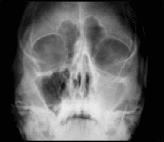
Pre-operative occipitomental view showing haziness in left maxillary sinus
Fig. 13.
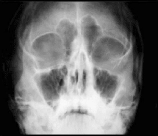
Post-operative occipitomental view at 3 months follow up showing clear maxillary sinus on both sides
Saleh et al. [12] supported the use of septal cartilage specially for larger defects and in their study failure was seen in 1 case out of 13 cases, in which closure was done under tension due to availability of less tissue on palatal side after radiation following tumour excision. But in our study failure was noticed in two out of 20 cases and in both the failed cases defect was larger than 10 mm2. Both patients developed post operative infection leading to extrusion of graft.
Conclusion
We found that the autogenous auricular cartilage graft is an effective sealing material in oro-antral fistula closure. Regardless of the chosen technique for closure of OAF, sinus infection must be treated with adequate nasal drainage, supported with use of appropriate antibiotics in addition to topical and/or systemic decongestants.
We recommend the use of auricular cartilage graft for the defect size ≤10 mm2 in which future dental implant placement is sought as it allows easy sinus lifting procedure, or in patients with multiple failed attempt for closure.
References
- 1.Sokler K, Vuksan V, Lauc T. Treatment of oroantral fistula. Acta Stomatol Croat. 2002;36(1):135–140. [Google Scholar]
- 2.Borgonovo AE, Berardinelli FV, Favale M, Maiorana C. Surgical options in oroantral fistula treatment. Open Dent J. 2012;6:94–98. doi: 10.2174/1874210601206010094. [DOI] [PMC free article] [PubMed] [Google Scholar]
- 3.Von Wowern N. Oro-antral communications and displacements of roots into the maxillary sinus: a followup of 231 cases. J Oral Surg. 1971;29(9):622–627. [PubMed] [Google Scholar]
- 4.Punwutikorn J, Waikakul A, Pairuchvej V. Clinically significant oroantral communications—a study of incidence and site. Int J Oral Maxillofac Surg. 1994;23:19–21. doi: 10.1016/S0901-5027(05)80320-0. [DOI] [PubMed] [Google Scholar]
- 5.Guven O. A clinical study on oroantral fistulae. J Craniomaxillofac Surg. 1998;26:267–271. doi: 10.1016/S1010-5182(98)80024-3. [DOI] [PubMed] [Google Scholar]
- 6.Lin PT, Bukachaevsky R, Blake M. Management of odontogenic sinusitis with persistent oro-antral fistula. Ear Nose Throat J. 1991;70:488–490. [PubMed] [Google Scholar]
- 7.Hanazawa Y, Itoh K, Mabashi T, Sato K. Closure of oroantral communications using a pedicled buccal fat pad graft. J Oral Maxillofac Surg. 1995;53(7):771–776. doi: 10.1016/0278-2391(95)90329-1. [DOI] [PubMed] [Google Scholar]
- 8.Poeschl PW, Baumann A, Russmueller G, Poeschl E, Klug C, et al. Closure of oroantral communications with Bichat’s buccal fat pad. J Oral Maxillofac Surg. 2009;67:1460–1466. doi: 10.1016/j.joms.2009.03.049. [DOI] [PubMed] [Google Scholar]
- 9.Lee JJ, Kok SH, Chang HH, Yang PJ, Hahn LJ, et al. Repair of oroantral communications in the third molar region by random palatal flap. Int J Oral Maxillofac Surg. 2002;31:677–680. doi: 10.1054/ijom.2001.0209. [DOI] [PubMed] [Google Scholar]
- 10.Kitagawa Y, Sano K, Nakamura M, Ogasawara T. Use of third molar transplantation for closure of the oroantral communication after tooth extraction: a report of 2 cases. Oral Surg Oral Med Oral Path Oral Radiol Endod. 2003;95:409–415. doi: 10.1067/moe.2003.122. [DOI] [PubMed] [Google Scholar]
- 11.Ogunsalu C. A new surgical management for oro-antral communication. The resorbable guided tissue regeneration membrane—bone substitute sandwich technique. West Indian Med J. 2005;54(4):261–263. doi: 10.1590/s0043-31442005000400011. [DOI] [PubMed] [Google Scholar]
- 12.Saleh EA, Issa IA. Closure of large oroantral fistulas using septal cartilage. Otolaryngol Head Neck Surg. 2013;148(6):1048–1050. doi: 10.1177/0194599813482091. [DOI] [PubMed] [Google Scholar]
- 13.Berger A. Oroantral openings and their surgical corrections. Arch Otolaryngol. 1939;130:400–402. doi: 10.1001/archotol.1939.00650060434007. [DOI] [Google Scholar]
- 14.Martensson G. Operative method in fistulas to the maxillary sinus. Acta Otolaryngol. 1957;48:253. doi: 10.3109/00016485709124378. [DOI] [PubMed] [Google Scholar]
- 15.Edgerton M, Zoviekian T. Reconstruction of major defects of the palate. Plast Reconstr Surg. 1956;17:105–107. doi: 10.1097/00006534-195602000-00002. [DOI] [PubMed] [Google Scholar]
- 16.Guerrero-Santos J, Altamirano JT. The use of lingual flaps in repair of fistulas of the hard palate. Plast Reconstr Surg. 1966;38:123–128. doi: 10.1097/00006534-196608000-00007. [DOI] [PubMed] [Google Scholar]
- 17.Isler SC, Demircan S, Cansiz E. Closure of oroantral fistula using auricular cartilage: a new method to repair an oroantral fistula. Br J Oral Maxillofac Surg. 2011;49:86–87. doi: 10.1016/j.bjoms.2011.03.262. [DOI] [PubMed] [Google Scholar]


