

Abstract
Introduction
In patients with habitual dislocation of the temporomandibular joint, anterior dislocation readily occurs with mouth opening in the normal range.
Methods
This short paper describes our novel procedure, modified zygomatic arch osteotomy reported by LeClerc et al., with oblique osteotomy to create a V-shaped notch at the tip for more accurate locking.
Conclusion
The technique obtained favorable results without the recurrence of TMJ dislocation.
Keywords: Habitual dislocation, Temporomandibular joint, Oblique osteotomy
In patients with habitual dislocation of the temporomandibular joint (TMJ), anterior dislocation readily occurs with mouth opening in the normal range. Once dislocation occurs, not only pain at the joint area, but also anxiety about the development of dislocation, is marked, and excessive salivation often interferes with the diet and conversation.
As invasive treatment for habitual TMJ dislocation, various methods have been reported. In 1943, LeClerc [1] reported a surgical procedure in which the zygomatic arch was vertically osteotomized at a site anterior to the articular eminence and moved medio-inferiorly, forming a block anterior to the eminence. Subsequently, to elevate the articular eminence, Lindemann et al. [2] split the posterior part of the eminence, and pushed the eminence fragment antero-inferiorly. Dingman et al. [3] split the inferior part of the articular eminence and inserted a small bone graft. In addition, Randzio and Fischer-Brandies et al. [4] split the anterior part of the eminence, pushed down the split part, and inserted an apatite block into the space.
The LeClerc procedure [1] is a classical method, and the position of the moved zygomatic arch is difficult to maintain. Therefore, Gosserez et al. reported osteotomy at a site anterior to the articular eminence obliquely in the posterior direction from the lower margin of the zygomatic arch towards its upper margin for more accurate fixation. In another study, to prevent return of the moved zygomatic arch, a wedge-shaped bone graft was inserted into the temporal bone side of the osteotomy end [5]. However, this method requires the collection of bone. A modified LeClerc’s operation involving a V-shaped notch made at the tip of the osteotomized segment was first reported by Akioka et al. [6].
We modified the original LeClerc procedure to set oblique osteotomy at the zygomatic arch and a V-shaped notch at the tip so as not to produce detachment or luxation, and performed this novel method. This method allowed proper fixation without bone grafting or the use of foreign materials such as fixation wire. In our department, this novel procedure led to a favorable result in three patients. The technique obtained favorable results without the recurrence of TMJ dislocation 7 years after the operation. In the future, the accumulation of cases treated using this procedure will be necessary to evaluate it comprehensively.
Technique
Under general anesthesia, a temporal approach to the right TMJ was used [7]. Detachment was advanced on the temporal fascia and reached the site immediately above the zygomatic arch. Incisions were made in the temporal fascia and zygomatic arch periosteum, and the zygomatic arch was exposed. Oblique osteotomy was performed at a site anterior to the articular eminence. To prevent return of the moved zygomatic arch, a 2-mm V-shaped notch was created at the anterior–superior margin of the osteotomized zygomatic arch (Fig. 1a). The portion was pushed laterally once, loosening the temporo-zygomatic suture, and turned down while retaining the suture. After the operation, the zygomatic arch was fixed (Fig. 1b). Bone grafting, wire fixation, or drain placement was not performed. Postoperative radiography and 3D CT showed anterior movement of the TMJ from the mandibular fossa, but confirmed locking by the osteotomized zygomatic arch (Fig. 2).
Fig. 1.

Osteotomy with our modified LeClerc procedure. a Oblique osteotomy line. b Return was prevented by creating a 2-mm V-shaped notch at the antero-superior margin of the osteotomized zygomatic arch
Fig. 2.
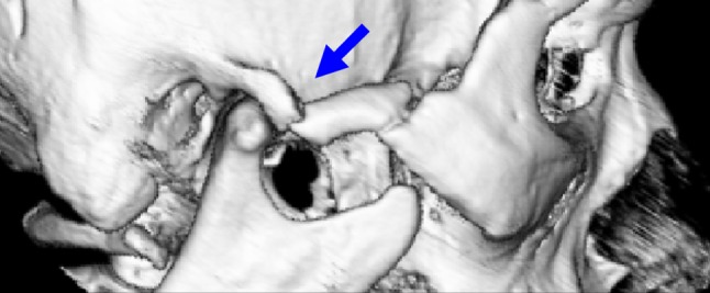
3D-CT image showing TMJ dislocation has not recurred, and fixation without detachment or luxation of the osteotomized zygomatic arch was observed (blue arrow)
Compliance with Ethical Standards
Ethical Approval
All procedures performed in studies involving human participants were in accordance with the ethical standards of the institutional and/or national research committee and with the 1964 Helsinki declaration and its later amendments or comparable ethical standards.
Informed Consent
Informed consent was obtained from all individual participants included in the study.
References
- 1.LeClerc G, Girard G. Un nouveau procede de butee dans le traitement chirurgical de la luxation reidivante de la machoire inferieure. Mem Acad Chir. 1943;69:457–459. [Google Scholar]
- 2.Lindemann A. Die chirurgische Behandlung der Erkrankungen des Kiefergelenks. Z Stomat. 1925;23:395. [Google Scholar]
- 3.Dingman RO, Dingman DL, Lawrence RA. Surgical correction of lesions of the temporomandibular joints. Plast Reconstr Surg. 1975;55(3):335–340. [PubMed] [Google Scholar]
- 4.Randzio J, Fischer-Brandies E. Augmentation of the articular tubercle in treatment of chronic recurrent temporomandibular joint luxations. A preliminary case report. Oral Surg Oral Med Oral Pathol. 1986;61(1):19–22. doi: 10.1016/0030-4220(86)90196-9. [DOI] [PubMed] [Google Scholar]
- 5.Sailer HF, Antonini N. Ergenisse der LeClerc-operation bei habitueller Kiefergelenk und Diskusluxation. Fortsch Kiefer Gesichtschir Bd. 1980;25:43–45. [PubMed] [Google Scholar]
- 6.Akioka J, Kusumoto K, Sasaki F, Ogawa Y. A case of habitual luxation of the temporomandibular joint treated by a modified LeClerc’s operation: addition of a “V-shape” bony notch to get a bony attachment. Jpn J Plast Surg. 1997;40(11):1107–1112. [Google Scholar]
- 7.Obwegeser HL. Temporal approach to the TMJ, the orbit, and the retromaxillary-infracranial region. Head Neck Surg. 1985;7(3):185–199. doi: 10.1002/hed.2890070302. [DOI] [PubMed] [Google Scholar]