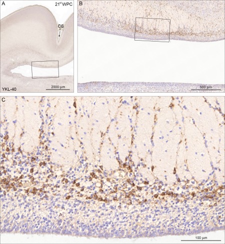
Figure 3.

YKL‐40 immunoreactivity in a coronal section of visual cortex from a 21st wpc fetus (CRL: 200 mm). A low power overview is shown in (A) where the calcarine sulcus (CS) is indicated with an arrow. The boxed area is shown in higher magnification in (B) which provides an overview of the distribution of the YKL‐40 positive cells in the inner and outer subventricular zones separated by the inner fibrous layer with migrating positive cells. The boxed area in (B) is shown in higher magnification in (C) where it is obvious that the YKL‐40‐immunoreactive population is not present in the ventricular zone whereas immunostained cells occupy a substantial part of the inner subventricular zone of the visual cortex. The many unstained cells might belong to groups of other progenitor cells, interneurons or microglia. Scale bars: A: 2,000 µm; B: 500 µm; C: 100 µm. [Color figure can be viewed in the online issue, which is available at wileyonlinelibrary.com.]
