Abstract
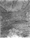
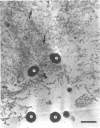
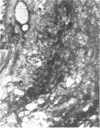
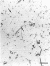
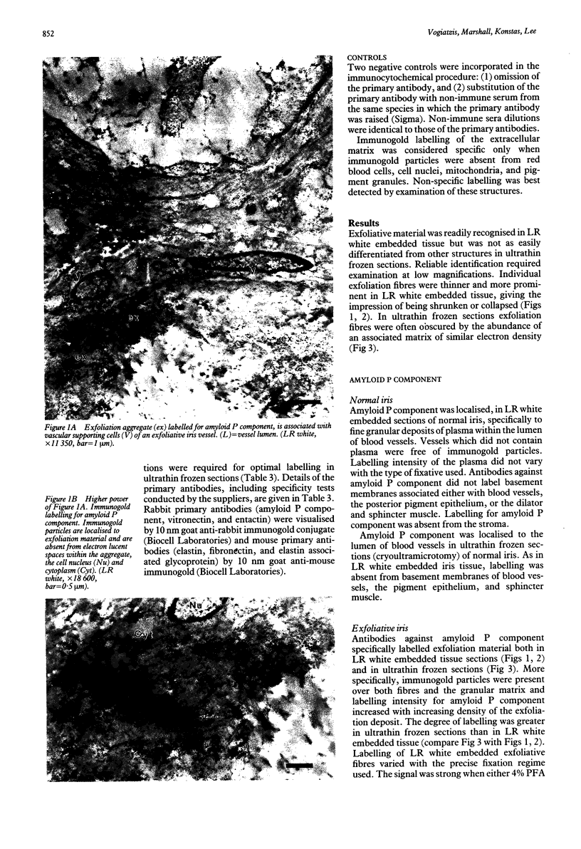
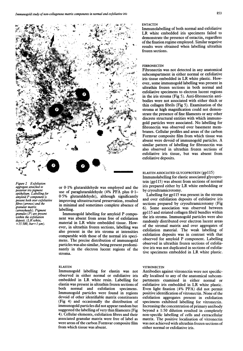
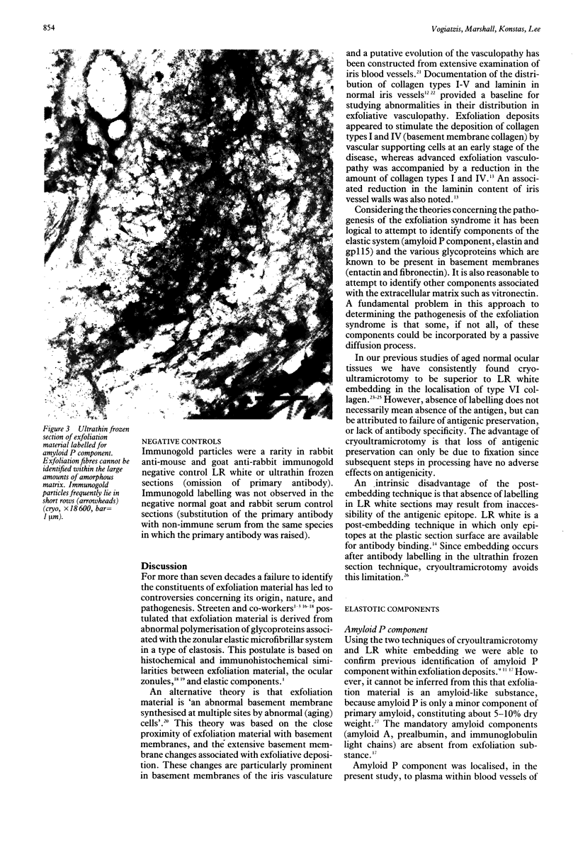
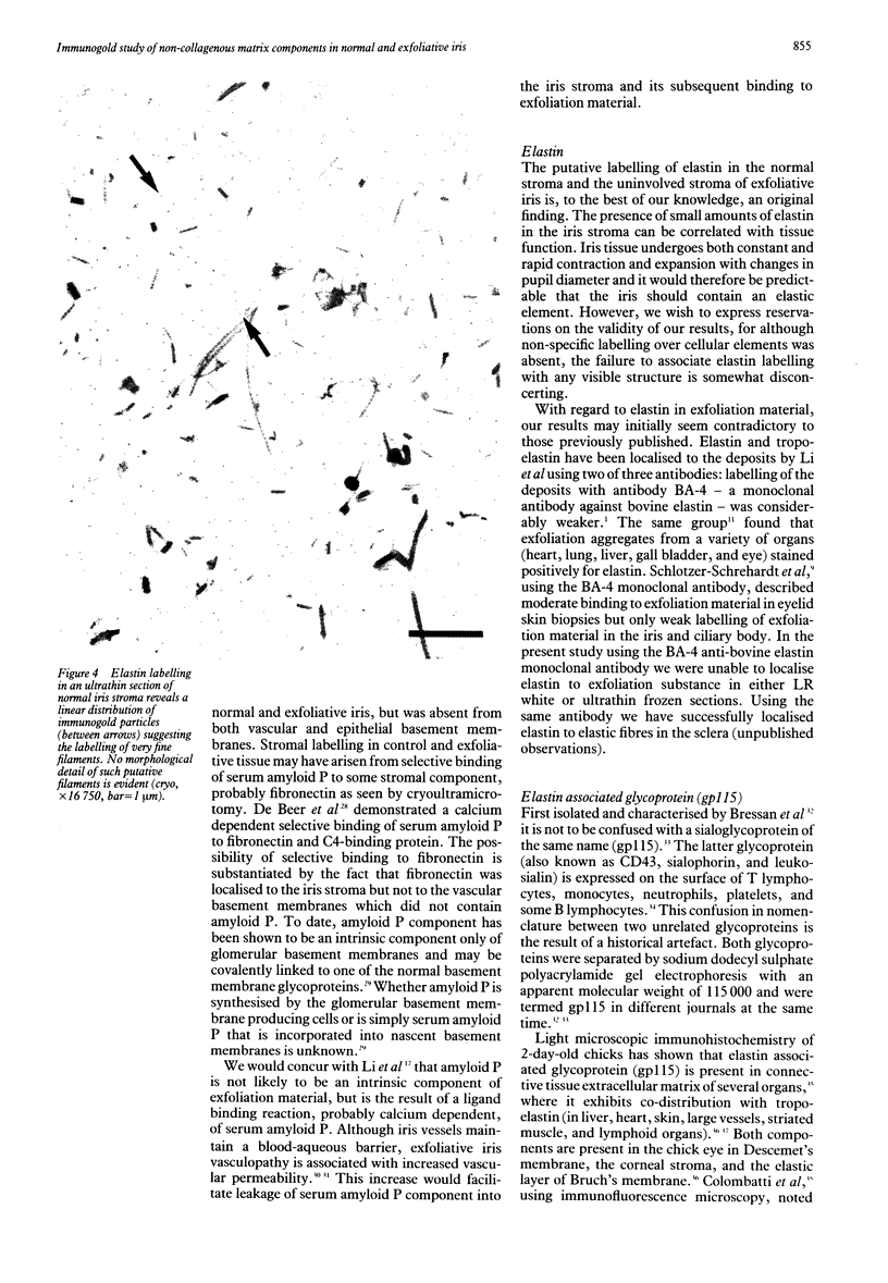
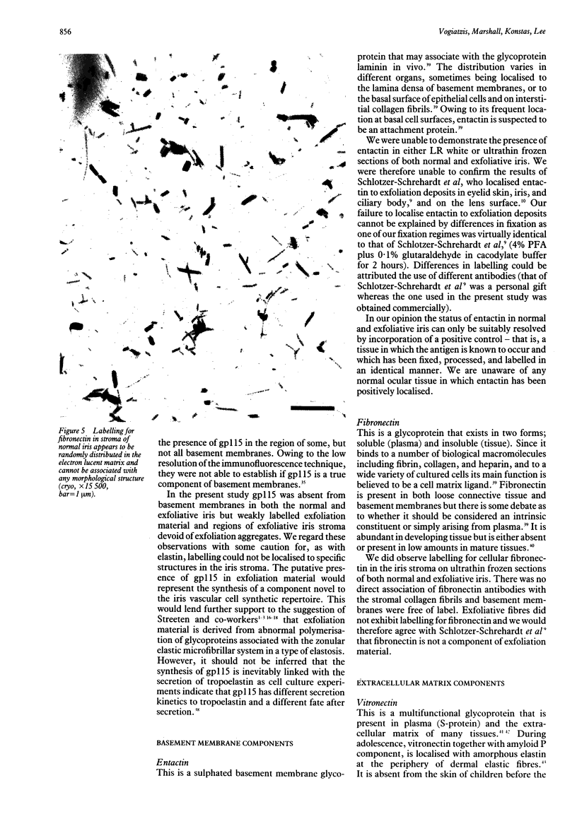
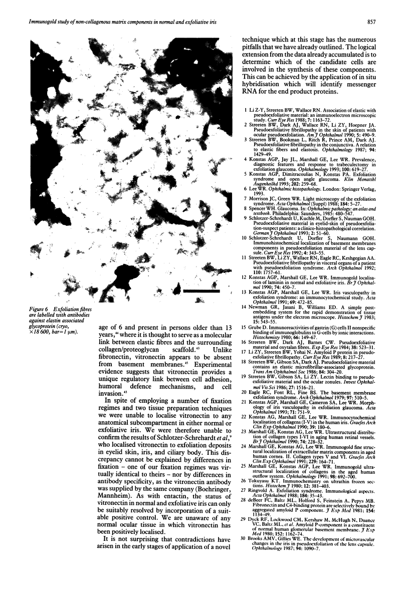
The present investigation was undertaken to determine if some of the components of exfoliation material in iris tissue were unique to exfoliation or were part of normal iris architecture. Eleven normal iris specimens and 10 exfoliative iris specimens were processed for cryoultramicrotomy and London resin white embedding. Immunogold electron microscopy was used to investigate the fine structural distribution of amyloid P component, elastin, entactin, fibronectin, gp115, and vitronectin in normal iris and their association with exfoliation material. Exfoliation material was positive for amyloid P component and possibly gp115, neither of which were present in normal iris tissue. Elastin and fibronectin were present in the normal iris stroma but were not associated with exfoliation material. The distribution of amyloid P component in the vessel lumen and wall led to the conclusion that amyloid P is a serum contaminant. The presence of gp115 in exfoliation material represents the synthesis of a component novel to the iris vascular cell synthetic repertoire.
Full text
PDF
Images in this article
Selected References
These references are in PubMed. This may not be the complete list of references from this article.
- Abrahamson D. R. Recent studies on the structure and pathology of basement membranes. J Pathol. 1986 Aug;149(4):257–278. doi: 10.1002/path.1711490402. [DOI] [PubMed] [Google Scholar]
- Barnes D. W., Silnutzer J. Isolation of human serum spreading factor. J Biol Chem. 1983 Oct 25;258(20):12548–12552. [PubMed] [Google Scholar]
- Bressan G. M., Castellani I., Colombatti A., Volpin D. Isolation and characterization of a 115,000-dalton matrix-associated glycoprotein from chick aorta. J Biol Chem. 1983 Nov 10;258(21):13262–13267. [PubMed] [Google Scholar]
- Brooks A. M., Gillies W. E. The development of microneovascular changes in the iris in pseudoexfoliation of the lens capsule. Ophthalmology. 1987 Sep;94(9):1090–1097. doi: 10.1016/s0161-6420(87)33329-9. [DOI] [PubMed] [Google Scholar]
- Colombatti A., Bonaldo P., Volpin D., Bressan G. M. The elastin associated glycoprotein gp115. Synthesis and secretion by chick cells in culture. J Biol Chem. 1988 Nov 25;263(33):17534–17540. [PubMed] [Google Scholar]
- Colombatti A., Bressan G. M., Castellani I., Volpin D. Glycoprotein 115, a glycoprotein isolated from chick blood vessels, is widely distributed in connective tissue. J Cell Biol. 1985 Jan;100(1):18–26. doi: 10.1083/jcb.100.1.18. [DOI] [PMC free article] [PubMed] [Google Scholar]
- Colombatti A., Poletti A., Bressan G. M., Carbone A., Volpin D. Widespread codistribution of glycoprotein gp 115 and elastin in chick eye and other tissues. Coll Relat Res. 1987 Sep;7(4):259–275. doi: 10.1016/s0174-173x(87)80032-8. [DOI] [PubMed] [Google Scholar]
- Colombatti A., Poletti A., Carbone A., Volpin D., Bressan G. M. Extracellular matrix of lymphoid tissues in the chick. J Histochem Cytochem. 1989 May;37(5):757–763. doi: 10.1177/37.5.2703709. [DOI] [PubMed] [Google Scholar]
- Dahlbäck K., Löfberg H., Alumets J., Dahlbäck B. Immunohistochemical demonstration of age-related deposition of vitronectin (S-protein of complement) and terminal complement complex on dermal elastic fibers. J Invest Dermatol. 1989 May;92(5):727–733. doi: 10.1111/1523-1747.ep12721619. [DOI] [PubMed] [Google Scholar]
- Dyck R. F., Lockwood C. M., Kershaw M., McHugh N., Duance V. C., Baltz M. L., Pepys M. B. Amyloid P-component is a constituent of normal human glomerular basement membrane. J Exp Med. 1980 Nov 1;152(5):1162–1174. doi: 10.1084/jem.152.5.1162. [DOI] [PMC free article] [PubMed] [Google Scholar]
- Eagle R. C., Jr, Font R. L., Fine B. S. The basement membrane exfoliation syndrome. Arch Ophthalmol. 1979 Mar;97(3):510–515. doi: 10.1001/archopht.1979.01020010254014. [DOI] [PubMed] [Google Scholar]
- Falk R. J., Podack E., Dalmasso A. P., Jennette J. C. Localization of S protein and its relationship to the membrane attack complex of complement in renal tissue. Am J Pathol. 1987 Apr;127(1):182–190. [PMC free article] [PubMed] [Google Scholar]
- Grube D. Immunoreactivities of gastrin (G-) cells. II. Non-specific binding of immunoglobulins to G-cells by ionic interactions. Histochemistry. 1980;66(2):149–167. doi: 10.1007/BF00494642. [DOI] [PubMed] [Google Scholar]
- Hayman E. G., Pierschbacher M. D., Ohgren Y., Ruoslahti E. Serum spreading factor (vitronectin) is present at the cell surface and in tissues. Proc Natl Acad Sci U S A. 1983 Jul;80(13):4003–4007. doi: 10.1073/pnas.80.13.4003. [DOI] [PMC free article] [PubMed] [Google Scholar]
- Konstas A. G., Dimitracoulias N., Konstas P. A. Exfoliationssyndrom und Offenwinkelglaukom. Originaltitel: Exfoliation Syndrome and Open Angle Glaucoma. Klin Monbl Augenheilkd. 1993 Apr;202(4):259–268. doi: 10.1055/s-2008-1045591. [DOI] [PubMed] [Google Scholar]
- Konstas A. G., Jay J. L., Marshall G. E., Lee W. R. Prevalence, diagnostic features, and response to trabeculectomy in exfoliation glaucoma. Ophthalmology. 1993 May;100(5):619–627. doi: 10.1016/s0161-6420(93)31596-4. [DOI] [PubMed] [Google Scholar]
- Konstas A. G., Marshall G. E., Cameron S. A., Lee W. R. Morphology of iris vasculopathy in exfoliation glaucoma. Acta Ophthalmol (Copenh) 1993 Dec;71(6):751–759. doi: 10.1111/j.1755-3768.1993.tb08595.x. [DOI] [PubMed] [Google Scholar]
- Konstas A. G., Marshall G. E., Lee W. R. Immunocytochemical localisation of collagens (I-V) in the human iris. Graefes Arch Clin Exp Ophthalmol. 1990;228(2):180–186. doi: 10.1007/BF00935730. [DOI] [PubMed] [Google Scholar]
- Konstas A. G., Marshall G. E., Lee W. R. Immunogold localisation of laminin in normal and exfoliative iris. Br J Ophthalmol. 1990 Aug;74(8):450–457. doi: 10.1136/bjo.74.8.450. [DOI] [PMC free article] [PubMed] [Google Scholar]
- Konstas A. G., Marshall G. E., Lee W. R. Iris vasculopathy in exfoliation syndrome. An immunocytochemical study. Acta Ophthalmol (Copenh) 1991 Aug;69(4):472–483. doi: 10.1111/j.1755-3768.1991.tb02025.x. [DOI] [PubMed] [Google Scholar]
- Li Z. Y., Streeten B. W., Wallace R. N. Association of elastin with pseudoexfoliative material: an immunoelectron microscopic study. Curr Eye Res. 1988 Dec;7(12):1163–1172. doi: 10.3109/02713688809033220. [DOI] [PubMed] [Google Scholar]
- Li Z. Y., Streeten B. W., Yohai N. Amyloid P protein in pseudoexfoliative fibrillopathy. Curr Eye Res. 1989 Feb;8(2):217–227. doi: 10.3109/02713688908995194. [DOI] [PubMed] [Google Scholar]
- Liakka K. A., Autio-Harmainen H. I. Distribution of the extracellular matrix proteins tenascin, fibronectin, and vitronectin in fetal, infant, and adult human spleens. J Histochem Cytochem. 1992 Aug;40(8):1203–1210. doi: 10.1177/40.8.1377736. [DOI] [PubMed] [Google Scholar]
- Marshall G. E., Konstas A. G., Lee W. R. Immunogold fine structural localization of extracellular matrix components in aged human cornea. II. Collagen types V and VI. Graefes Arch Clin Exp Ophthalmol. 1991;229(2):164–171. doi: 10.1007/BF00170551. [DOI] [PubMed] [Google Scholar]
- Marshall G. E., Konstas A. G., Lee W. R. Immunogold ultrastructural localization of collagens in the aged human outflow system. Ophthalmology. 1991 May;98(5):692–700. doi: 10.1016/s0161-6420(91)32232-2. [DOI] [PubMed] [Google Scholar]
- Marshall G. E., Konstas A. G., Lee W. R. Ultrastructural distribution of collagen types I-VI in aging human retinal vessels. Br J Ophthalmol. 1990 Apr;74(4):228–232. doi: 10.1136/bjo.74.4.228. [DOI] [PMC free article] [PubMed] [Google Scholar]
- Morrison J. C., Green W. R. Light microscopy of the exfoliation syndrome. Acta Ophthalmol Suppl. 1988;184:5–27. doi: 10.1111/j.1755-3768.1988.tb02624.x. [DOI] [PubMed] [Google Scholar]
- Newman G. R., Jasani B., Williams E. D. A simple post-embedding system for the rapid demonstration of tissue antigens under the electron microscope. Histochem J. 1983 Jun;15(6):543–555. doi: 10.1007/BF01954145. [DOI] [PubMed] [Google Scholar]
- Preissner K. T. Structure and biological role of vitronectin. Annu Rev Cell Biol. 1991;7:275–310. doi: 10.1146/annurev.cb.07.110191.001423. [DOI] [PubMed] [Google Scholar]
- Ringvold A. Exfoliation syndrome immunological aspects. Acta Ophthalmol Suppl. 1988;184:35–43. doi: 10.1111/j.1755-3768.1988.tb02626.x. [DOI] [PubMed] [Google Scholar]
- Rosenstein Y., Park J. K., Hahn W. C., Rosen F. S., Bierer B. E., Burakoff S. J. CD43, a molecule defective in Wiskott-Aldrich syndrome, binds ICAM-1. Nature. 1991 Nov 21;354(6350):233–235. doi: 10.1038/354233a0. [DOI] [PubMed] [Google Scholar]
- Schlötzer-Schredhardt U., Küchle M., Dörfler S., Naumann G. O. Pseudoexfoliative material in the eyelid skin of pseudoexfoliation-suspect patients: a clinico-histopathological correlation. Ger J Ophthalmol. 1993 Feb;2(1):51–60. [PubMed] [Google Scholar]
- Schlötzer-Schrehardt U., Dörfler S., Naumann G. O. Immunohistochemical localization of basement membrane components in pseudoexfoliation material of the lens capsule. Curr Eye Res. 1992 Apr;11(4):343–355. doi: 10.3109/02713689209001788. [DOI] [PubMed] [Google Scholar]
- Spring F. A., Anstee D. J. Lectin-binding components of normal granulocytes and leukaemic myeloid cells. Biochem J. 1983 Sep 1;213(3):661–670. doi: 10.1042/bj2130661. [DOI] [PMC free article] [PubMed] [Google Scholar]
- Streeten B. W., Dark A. J., Barnes C. W. Pseudoexfoliative material and oxytalan fibers. Exp Eye Res. 1984 May;38(5):523–531. doi: 10.1016/0014-4835(84)90130-1. [DOI] [PubMed] [Google Scholar]
- Streeten B. W., Dark A. J., Wallace R. N., Li Z. Y., Hoepner J. A. Pseudoexfoliative fibrillopathy in the skin of patients with ocular pseudoexfoliation. Am J Ophthalmol. 1990 Nov 15;110(5):490–499. doi: 10.1016/s0002-9394(14)77871-7. [DOI] [PubMed] [Google Scholar]
- Streeten B. W., Gibson S. A., Dark A. J. Pseudoexfoliative material contains an elastic microfibrillar-associated glycoprotein. Trans Am Ophthalmol Soc. 1986;84:304–320. [PMC free article] [PubMed] [Google Scholar]
- Streeten B. W., Gibson S. A., Li Z. Y. Lectin binding to pseudoexfoliative material and the ocular zonules. Invest Ophthalmol Vis Sci. 1986 Oct;27(10):1516–1521. [PubMed] [Google Scholar]
- Streeten B. W., Li Z. Y., Wallace R. N., Eagle R. C., Jr, Keshgegian A. A. Pseudoexfoliative fibrillopathy in visceral organs of a patient with pseudoexfoliation syndrome. Arch Ophthalmol. 1992 Dec;110(12):1757–1762. doi: 10.1001/archopht.1992.01080240097039. [DOI] [PubMed] [Google Scholar]
- Tokuyasu K. T. Immunochemistry on ultrathin frozen sections. Histochem J. 1980 Jul;12(4):381–403. doi: 10.1007/BF01011956. [DOI] [PubMed] [Google Scholar]
- Vannas A. Fluorescein angiography of the vessels of the iris in pseudoexfoliation of the lens capsule, capsular glaucoma and some other forms of glaucoma. Acta Ophthalmol Suppl. 1969;105:1–75. [PubMed] [Google Scholar]
- de Beer F. C., Baltz M. L., Holford S., Feinstein A., Pepys M. B. Fibronectin and C4-binding protein are selectively bound by aggregated amyloid P component. J Exp Med. 1981 Oct 1;154(4):1134–1139. doi: 10.1084/jem.154.4.1134. [DOI] [PMC free article] [PubMed] [Google Scholar]


