

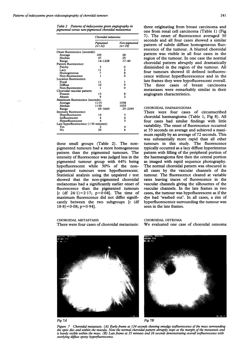
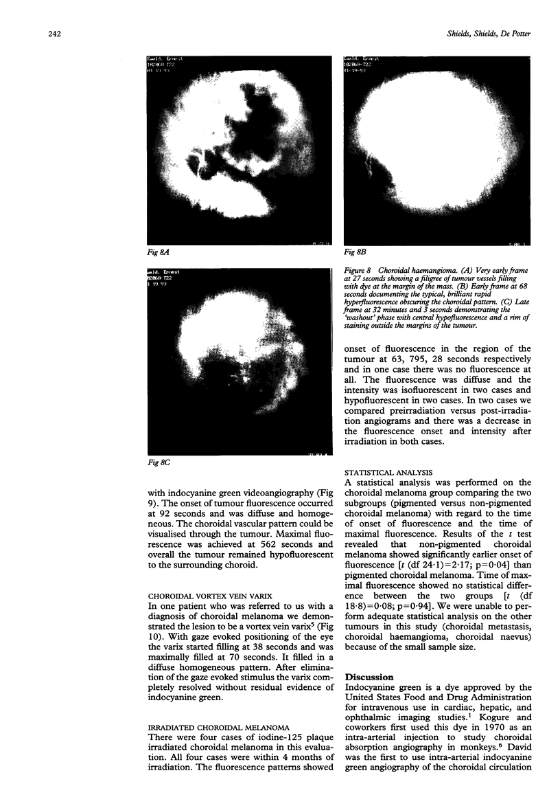
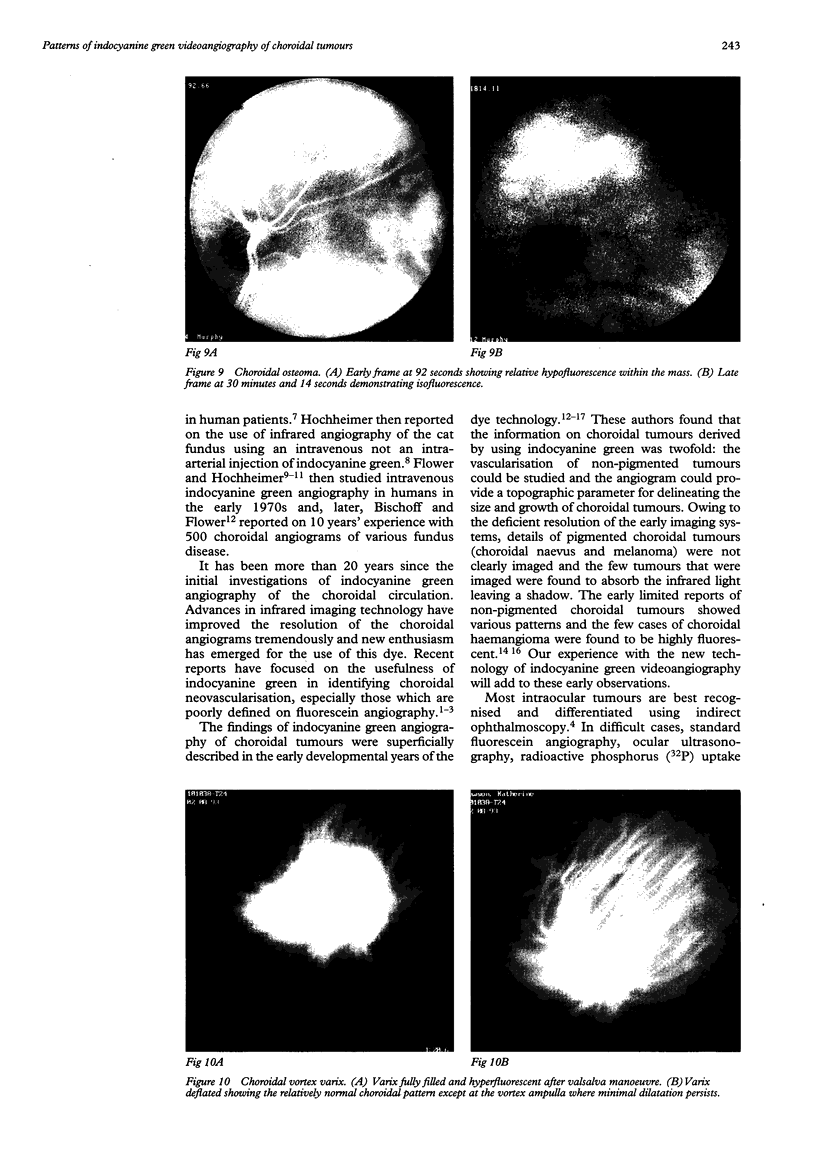
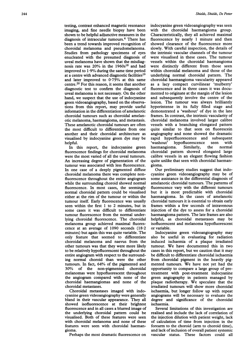
Abstract
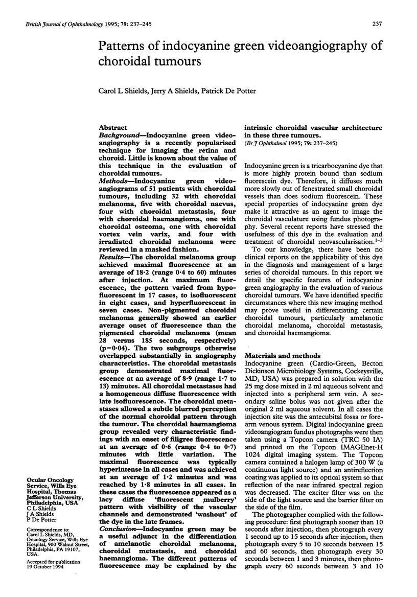
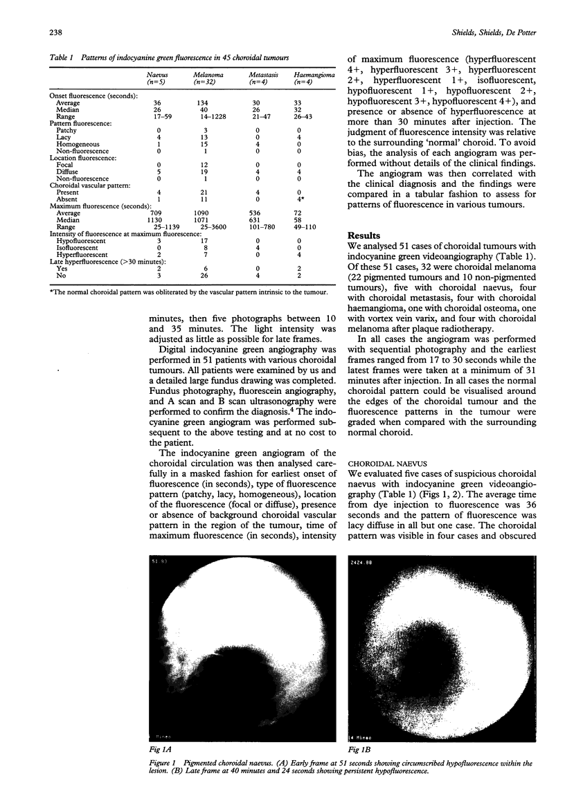
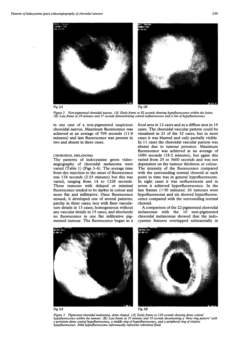
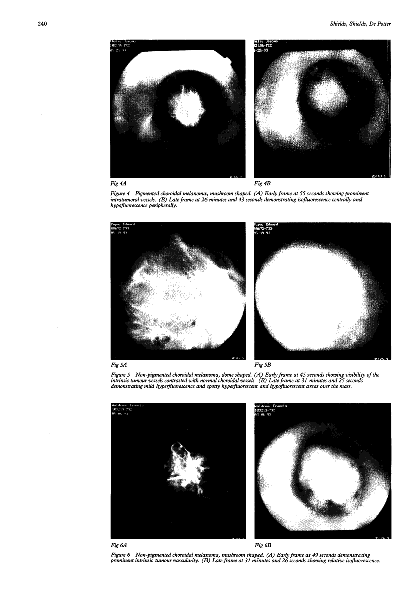
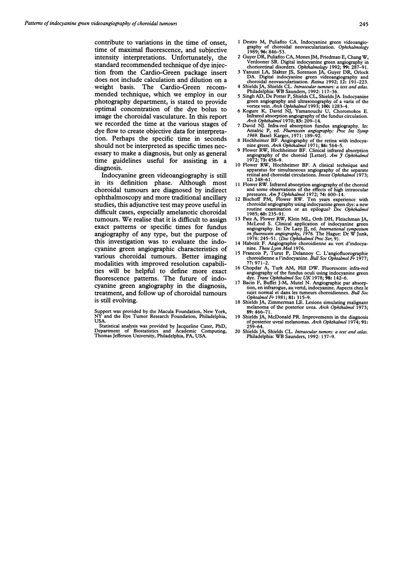
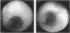
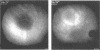
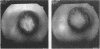
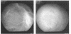
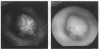
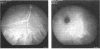
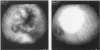
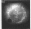
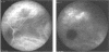
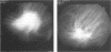
BACKGROUND--Indocyanine green video-angiography is a recently popularised technique for imaging the retina and choroid. Little is known about the value of this technique in the evaluation of choroidal tumours. METHODS--Indocyanine green video-angiograms of 51 patients with choroidal tumours, including 32 with choroidal melanoma, five with choroidal naevus, four with choroidal metastasis, four with choroidal haemangioma, one with choroidal osteoma, one with choroidal vortex vein varix, and four with irradiated choroidal melanoma were reviewed in a masked fashion. RESULTS--The choroidal melanoma group achieved maximal fluorescence at an average of 18.2 (range 0.4 to 60) minutes after injection. At maximum fluorescence, the pattern varied from hypofluorescent in 17 cases, to isofluorescent in eight cases, and hyperfluorescent in seven cases. Non-pigmented choroidal melanoma generally showed an earlier average onset of fluorescence than the pigmented choroidal melanoma (mean 28 versus 185 seconds, respectively) (p = 0.04). The two subgroups otherwise overlapped substantially in angiography characteristics. The choroidal metastasis group demonstrated maximal fluorescence at an average of 8.9 (range 1.7 to 13) minutes. All choroidal metastases had a homogeneous diffuse fluorescence with late isofluorescence. The choroidal metastases allowed a subtle blurred perception of the normal choroidal pattern through the tumour. The choroidal haemangioma group revealed very characteristic findings with an onset of filigree fluorescence at an average of 0.6 (range 0.4 to 0.7) minutes with little variation. The maximal fluorescence was typically hyperintense in all cases and was achieved at an average of 1.2 minutes and was reached by 1.8 minutes in all cases. In these cases the fluorescence appeared as a lacy diffuse 'fluorescent mulberry' pattern with visibility of the vascular channels and demonstrated 'washout' of the dye in the late frames. CONCLUSION--Indocyanine green may be a useful adjunct in the differentiation of amelanotic choroidal melanoma, choroidal metastasis, and choroidal haemangioma. The different patterns of fluorescence may be explained by the intrinsic choroidal vascular architecture in these three tumours.
Full text
PDF
Images in this article
Selected References
These references are in PubMed. This may not be the complete list of references from this article.
- Bacin F., Buffet J. M., Mutel N. Angiographie par absorption, en infrarouge, au vert d'indocyanine. Aspects chez le sujet normal et dans les tumeurs choroïdiennes. Bull Soc Ophtalmol Fr. 1981 Mar;81(3):315–319. [PubMed] [Google Scholar]
- Bischoff P. M., Flower R. W. Ten years experience with choroidal angiography using indocyanine green dye: a new routine examination or an epilogue? Doc Ophthalmol. 1985 Sep 30;60(3):235–291. doi: 10.1007/BF00157827. [DOI] [PubMed] [Google Scholar]
- Chopdar A., Turk A. M., Hill D. W. Fluorescent infra-red angiography of the fundus oculi using indocyanine green dye. Trans Ophthalmol Soc U K. 1978 Apr;98(1):142–146. [PubMed] [Google Scholar]
- Destro M., Puliafito C. A. Indocyanine green videoangiography of choroidal neovascularization. Ophthalmology. 1989 Jun;96(6):846–853. doi: 10.1016/s0161-6420(89)32826-0. [DOI] [PubMed] [Google Scholar]
- Flower R. W., Hochheimer B. F. A clinical technique and apparatus for simultaneous angiography of the separate retinal and choroidal circulations. Invest Ophthalmol. 1973 Apr;12(4):248–261. [PubMed] [Google Scholar]
- Flower R. W., Hochheimer B. F. Clinical infrared absorption angiography of the choroid. Am J Ophthalmol. 1972 Mar;73(3):458–459. doi: 10.1016/0002-9394(72)90079-7. [DOI] [PubMed] [Google Scholar]
- Flower R. W. Infrared absorption angiography of the choroid and some observations on the effects of high intraocular pressures. Am J Ophthalmol. 1972 Oct;74(4):600–614. doi: 10.1016/0002-9394(72)90819-7. [DOI] [PubMed] [Google Scholar]
- Francois P., Turut P., Delannoy C. L'angiofluorographie choroïdienne a l'indocyanine. Bull Soc Ophtalmol Fr. 1977 Nov-Dec;77(11-12):971–972. [PubMed] [Google Scholar]
- Guyer D. R., Puliafito C. A., Monés J. M., Friedman E., Chang W., Verdooner S. R. Digital indocyanine-green angiography in chorioretinal disorders. Ophthalmology. 1992 Feb;99(2):287–291. doi: 10.1016/s0161-6420(92)31981-5. [DOI] [PubMed] [Google Scholar]
- Hochheimer B. F. Angiography of the retina with indocyanine green. Arch Ophthalmol. 1971 Nov;86(5):564–565. doi: 10.1001/archopht.1971.01000010566014. [DOI] [PubMed] [Google Scholar]
- Kogure K., David N. J., Yamanouchi U., Choromokos E. Infrared absorption angiography of the fundus circulation. Arch Ophthalmol. 1970 Feb;83(2):209–214. doi: 10.1001/archopht.1970.00990030211015. [DOI] [PubMed] [Google Scholar]
- Shields J. A. Lesions simulating malignant melanoma of the posterior uvea. Arch Ophthalmol. 1973 Jun;89(6):466–471. doi: 10.1001/archopht.1973.01000040468004. [DOI] [PubMed] [Google Scholar]
- Shields J. A., McDonald P. R. Improvements in the diagnosis of posterior uveal melanomas. Arch Ophthalmol. 1974 Apr;91(4):259–264. [PubMed] [Google Scholar]
- Singh A. D., De Potter P., Shields C. L., Shields J. A. Indocyanine green angiography and ultrasonography of a varix of vortex vein. Arch Ophthalmol. 1993 Sep;111(9):1283–1284. doi: 10.1001/archopht.1993.01090090135031. [DOI] [PubMed] [Google Scholar]
- Yannuzzi L. A., Slakter J. S., Sorenson J. A., Guyer D. R., Orlock D. A. Digital indocyanine green videoangiography and choroidal neovascularization. Retina. 1992;12(3):191–223. [PubMed] [Google Scholar]