

Abstract
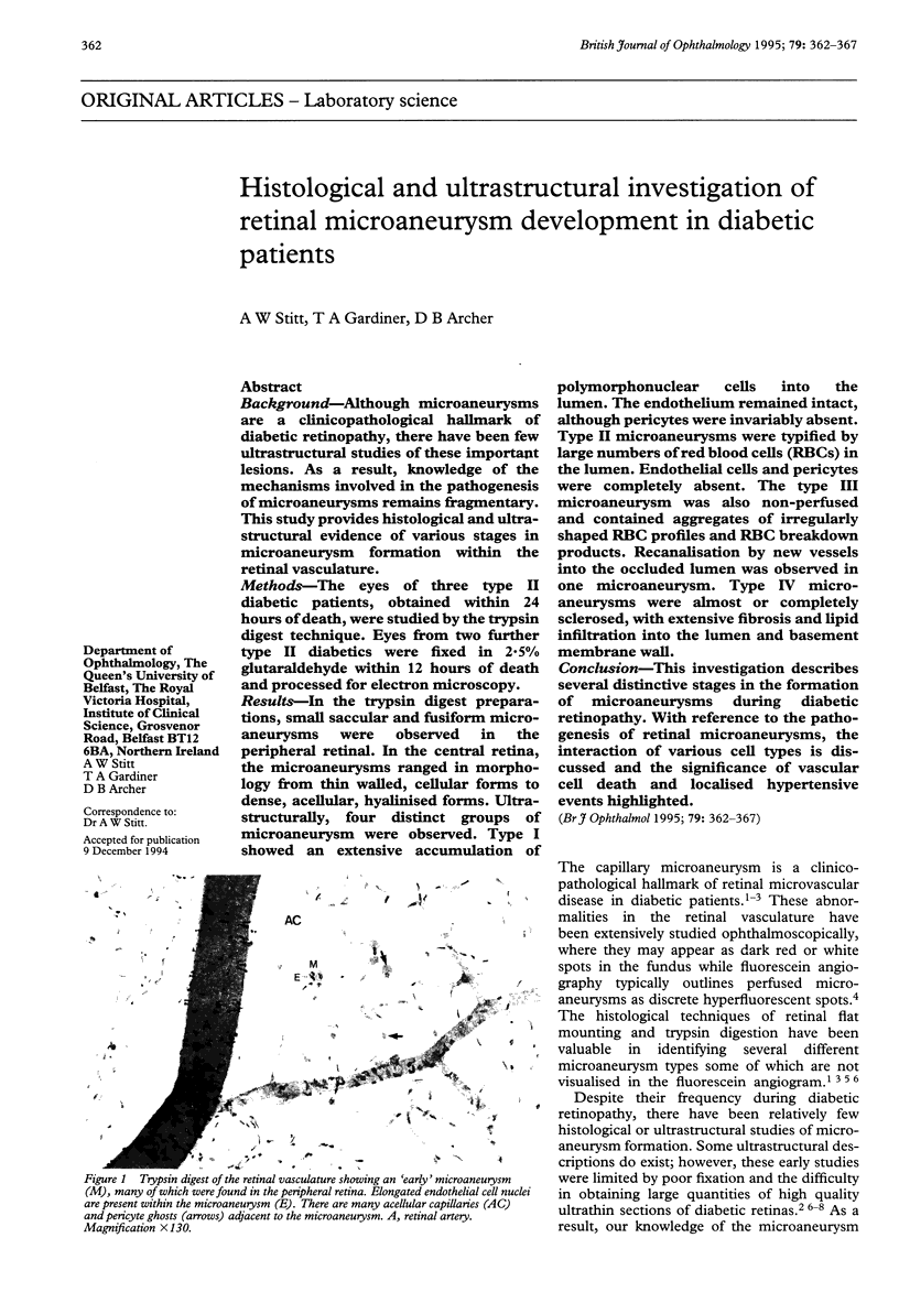
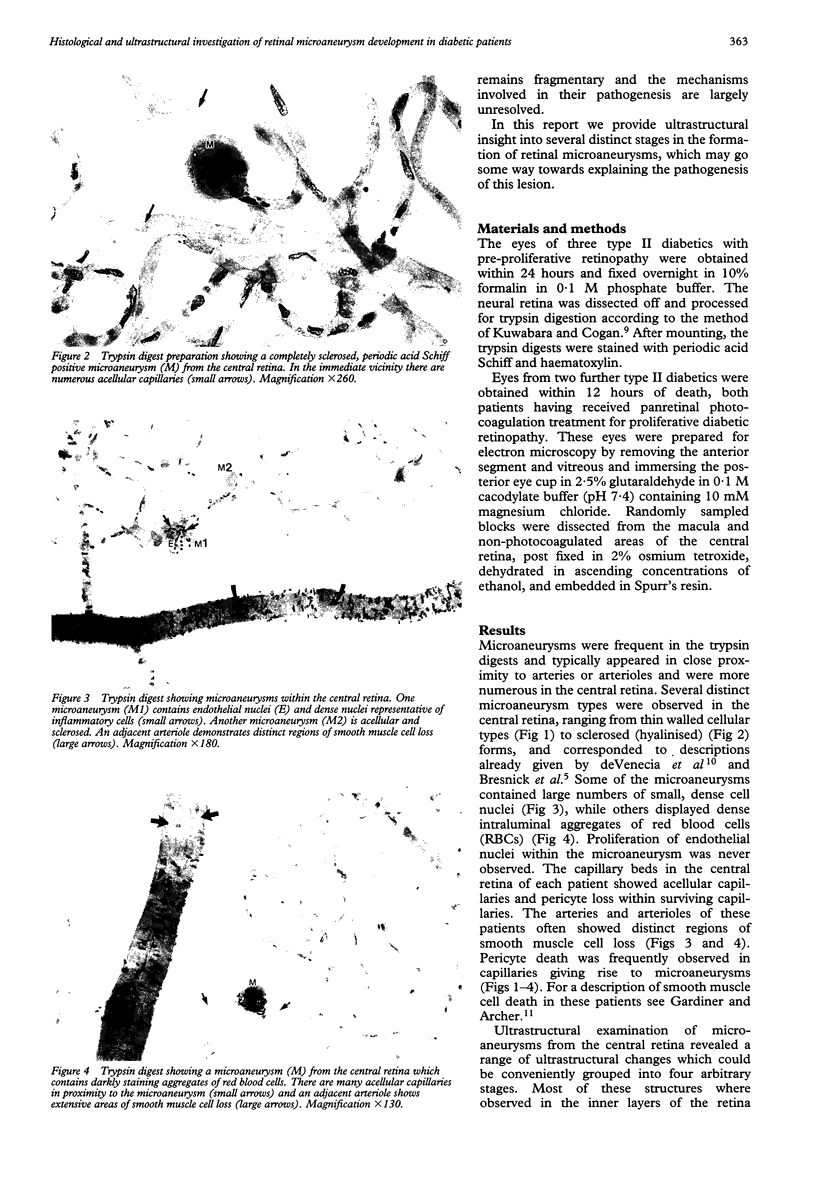
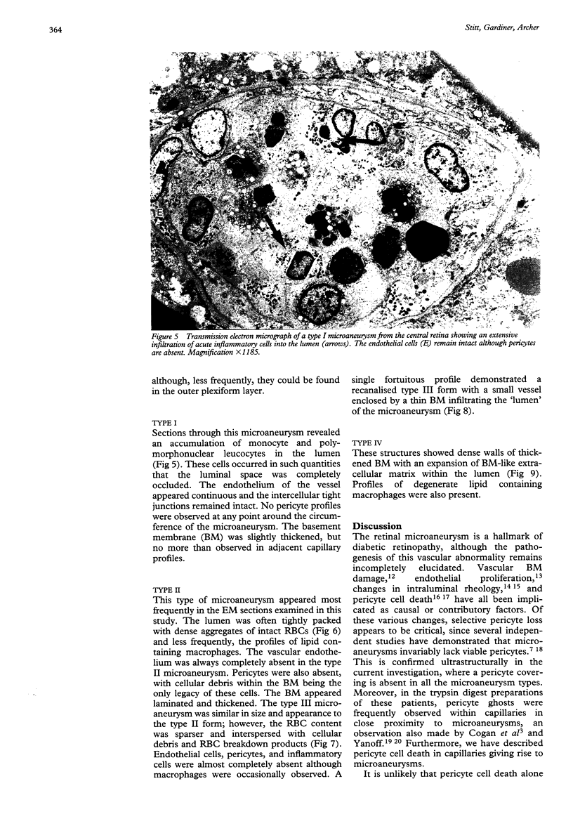
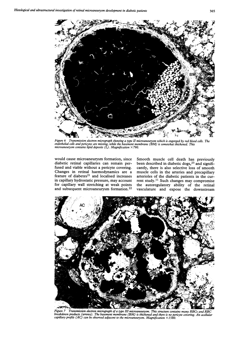
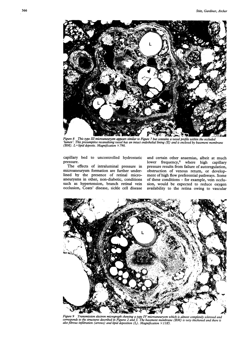
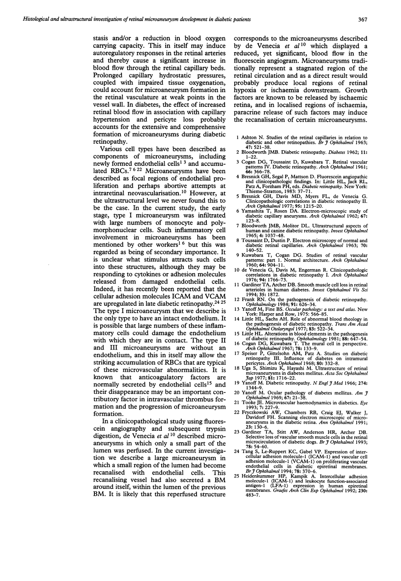
BACKGROUND--Although microaneurysms are a clinicopathological hallmark of diabetic retinopathy, there have been few ultrastructural studies of these important lesions. As a result, knowledge of the mechanisms involved in the pathogenesis of microaneurysms remains fragmentary. This study provides histological and ultrastructural evidence of various stages in microaneurysm formation within the retinal vasculature. METHODS--The eyes of three type II diabetic patients, obtained within 24 hours of death, were studied by the trypsin digest technique. Eyes from two further type II diabetics were fixed in 2.5% glutaraldehyde within 12 hours of death and processed for electron microscopy. RESULTS--In the trypsin digest preparations, small saccular and fusiform microaneurysms were observed in the peripheral retinal. In the central retina, the microaneurysms ranged in morphology from thin walled, cellular forms to dense, acellular, hyalinised forms. Ultrastructurally, four distinct groups of microaneurysm were observed. Type I showed an extensive accumulation of polymorphonuclear cells into the lumen. The endothelium remained intact, although pericytes were invariably absent. Type II microaneurysms were typified by large numbers of red blood cells (RBCs) in the lumen. Endothelial cells and pericytes were completely absent. The type III microaneurysm was also non-perfused and contained aggregates of irregularly shaped RBC profiles and RBC breakdown products. Recanalisation by new vessels into the occluded lumen was observed in one microaneurysm. Type IV microaneurysms were almost or completely sclerosed, with extensive fibrosis and lipid infiltration into the lumen and basement membrane wall. CONCLUSION--This investigation describes several distinctive stages in the formation of microaneurysms during diabetic retinopathy. With reference to the pathogenesis of retinal microaneurysms, the interaction of various cell types is discussed and the significance of vascular cell death and localised hypertensive events highlighted.
Full text
PDF
Images in this article
Selected References
These references are in PubMed. This may not be the complete list of references from this article.
- ASHTON N. STUDIES OF THE RETINAL CAPILLARIES IN RELATION TO DIABETIC AND OTHER RETINOPATHIES. Br J Ophthalmol. 1963 Sep;47:521–538. doi: 10.1136/bjo.47.9.521. [DOI] [PMC free article] [PubMed] [Google Scholar]
- BLOODWORTH J. M., Jr Diabetic retinopathy. Diabetes. 1962 Jan-Feb;11:1–22. [PubMed] [Google Scholar]
- Bloodworth J. M., Jr, Molitor D. L. Ultrastructural aspects of human and canine diabetic retinopathy. Invest Ophthalmol. 1965 Dec;4(6):1037–1048. [PubMed] [Google Scholar]
- Bresnick G. H., Davis M. D., Myers F. L., de Venecia G. Clinicopathologic correlations in diabetic retinopathy. II. Clinical and histologic appearances of retinal capillary microaneurysms. Arch Ophthalmol. 1977 Jul;95(7):1215–1220. doi: 10.1001/archopht.1977.04450070113010. [DOI] [PubMed] [Google Scholar]
- COGAN D. G., TOUSSAINT D., KUWABARA T. Retinal vascular patterns. IV. Diabetic retinopathy. Arch Ophthalmol. 1961 Sep;66:366–378. doi: 10.1001/archopht.1961.00960010368014. [DOI] [PubMed] [Google Scholar]
- Cogan D. G., Kuwabara T. The mural cell in perspective. Arch Ophthalmol. 1967 Aug;78(2):133–139. doi: 10.1001/archopht.1967.00980030135005. [DOI] [PubMed] [Google Scholar]
- De Venecia G., Davis M., Engerman R. Clinicopathologic correlations in diabetic retinopathy. I. Histology and fluorescein angiography of microaneurysms. Arch Ophthalmol. 1976 Oct;94(10):1766–1773. doi: 10.1001/archopht.1976.03910040540013. [DOI] [PubMed] [Google Scholar]
- Frank R. N. On the pathogenesis of diabetic retinopathy. Ophthalmology. 1984 Jun;91(6):626–634. doi: 10.1016/s0161-6420(84)34258-0. [DOI] [PubMed] [Google Scholar]
- Fryczkowski A. W., Chambers R. B., Craig E. J., Walker J., Davidorf F. H. Scanning electron microscopic study of microaneurysms in the diabetic retina. Ann Ophthalmol. 1991 Apr;23(4):130–136. [PubMed] [Google Scholar]
- Gardiner T. A., Stitt A. W., Anderson H. R., Archer D. B. Selective loss of vascular smooth muscle cells in the retinal microcirculation of diabetic dogs. Br J Ophthalmol. 1994 Jan;78(1):54–60. doi: 10.1136/bjo.78.1.54. [DOI] [PMC free article] [PubMed] [Google Scholar]
- Heidenkummer H. P., Kampik A. Intercellular adhesion molecule-1 (ICAM-1) and leukocyte function-associated antigen-1 (LFA-1) expression in human epiretinal membranes. Graefes Arch Clin Exp Ophthalmol. 1992;230(5):483–487. doi: 10.1007/BF00175938. [DOI] [PubMed] [Google Scholar]
- JAMPEL R. S., TITONE C. Congenital paradoxical gustatory-lacrimal reflex and lateral rectus paralysis. Case report. Arch Ophthalmol. 1962 Feb;67:123–126. doi: 10.1001/archopht.1962.00960020125004. [DOI] [PubMed] [Google Scholar]
- KUWABARA T., COGAN D. G. Studies of retinal vascular patterns. I. Normal architecture. Arch Ophthalmol. 1960 Dec;64:904–911. doi: 10.1001/archopht.1960.01840010906012. [DOI] [PubMed] [Google Scholar]
- Little H. L. Alterations in blood elements in the pathogenesis of diabetic retinopathy. Ophthalmology. 1981 Jul;88(7):647–654. doi: 10.1016/s0161-6420(81)34971-9. [DOI] [PubMed] [Google Scholar]
- Speiser P., Gittelsohn A. M., Patz A. Studies on diabetic retinopathy. 3. Influence of diabetes on intramural pericytes. Arch Ophthalmol. 1968 Sep;80(3):332–337. doi: 10.1001/archopht.1968.00980050334007. [DOI] [PubMed] [Google Scholar]
- Tang S., Le-Ruppert K. C., Gabel V. P. Expression of intercellular adhesion molecule-1 (ICAM-1) and vascular cell adhesion molecule-1 (VCAM-1) on proliferating vascular endothelial cells in diabetic epiretinal membranes. Br J Ophthalmol. 1994 May;78(5):370–376. doi: 10.1136/bjo.78.5.370. [DOI] [PMC free article] [PubMed] [Google Scholar]
- Tooke J. E. Microvascular haemodynamics in diabetes. Eye (Lond) 1993;7(Pt 2):227–229. doi: 10.1038/eye.1993.54. [DOI] [PubMed] [Google Scholar]
- Uga S., Shimizu K., Hayashi M. [Ultrastructure or retinal microaneurysm in diabetes mellitus (author's transl)]. Nippon Ganka Gakkai Zasshi. 1977 Nov 10;81(11):1716–1722. [PubMed] [Google Scholar]
- Yanoff M. Ocular pathology of diabetes mellitus. Am J Ophthalmol. 1969 Jan;67(1):21–38. doi: 10.1016/0002-9394(69)90004-x. [DOI] [PubMed] [Google Scholar]