The nucleus is the center of vital activity and is the brain of any cell. This stands true in case of physiological and pathological cells. Nucleus is the most active and prominent cellular organelle which shows many forms, has varied shapes and sizes, depicting the activity of the cell. Morphology of the nuclear shape and presentation can get altered because of changes in nuclear lamina or it can also be because of the forces exerted by the cytoplasm.[1] Change in nuclear shape might affect the function of a cell, though it is not very clear. Two hypothesis are given for this: The first states that nuclear shape change might give it a beneficial plasticity whereas the second states that the changed shape results in chromatin reorganization which might affect gene expression.[1]
Common forms of nucleus physiologically, are open, closed and semi-open.[1] A variety of nuclear shapes, sizes and presentation are associated with numerous pathologies. The cleaved nucleus, owl eye nucleus, Orphan Annie nucleus and buckled nucleus are a few to name. An interesting and uncommon change in the nucleus is nuclear vacuoles and vacuolization.
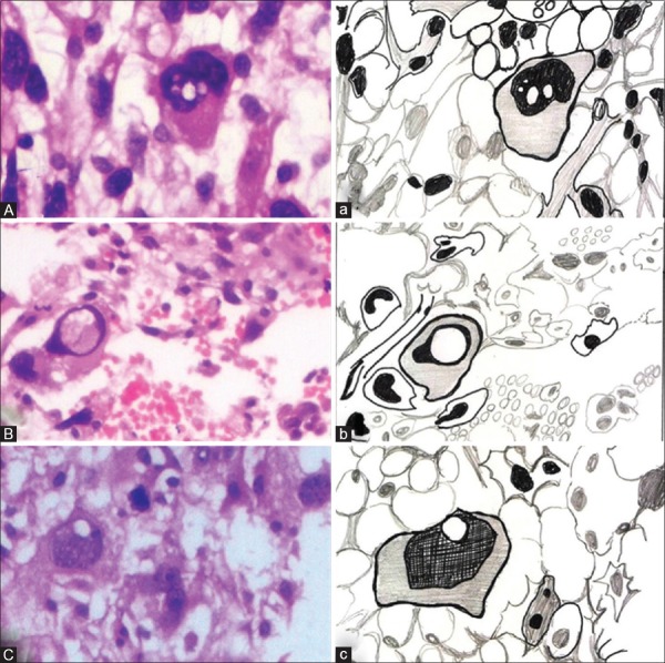
This is the morphological change in the nucleus, detectable by light microscopy on routine hematoxylin and eosin (H&E) stained slides. It is spoken in relation with a few morphological cells such as hepatocyte nuclei,[2] in the neoplastic cells of acute lymphoblastic leukemia,[3] and Lochkern cells associated with pathologies of adipocytes.[4] These changes may be associated with deranged cellular function. Nuclear vacuolization- Lockhern change is said to be seen classically in normal adipocytes and lipogenic tumors.[5,6] Different opinions are given on this nuclear change, a few speak of cells with slightly hypochromic nucleus with intranuclear vacuoles[6] whereas the others speak of nucleus which can show hyperchromatic or pleomorphic nucleus and has vacuoles in it.[7] Lochkern (German: Loch: Hole, kern: Nucleus)[7,8] cells may be present in normal adipocytes or in the neoplastic proliferation of the same. The presentation of Lochkern cells is of three types as given by Winckler.[9]
Lochkern cells-nucleus with one or multiple holes [Figure 1 A and a]
Ringkern-nucleus with a big central hole producing a ring-like an appearance [Figure 1 B and b]
Kerbenkern or Eingekerbterkern-notched nucleus. [Figure 1 C and c]
Figure 1.
(A) Photomicrograph of a neoplastic cell showing lochkern nucleus (H&E stain, ×400), (a) hand-drawn illustration showing Lochkern nucleus. (B) Photomicrograph of a neoplastic cell showing Ringkern nucleus (H&E stain, ×400), (b) hand-drawn illustration showing Ringkern nucleus. (C) Photomicrograph of a neoplastic cell showing Kerbkern nucleus (H&E stain, ×400), (c) hand-drawn illustration showing Kerbkern nucleus
The cause of these nuclear vacuolization was investigated by many authors and pathologists including Unna, Sack, Winkler, Rable and Plaut. There are two schools of thoughts for the nuclear vacuolization. The first as proposed by Unna, Sack and Winkler who believed that Lochkern change was due to true hole formation in the nucleus.[8,10] The second school of thought was as proposed by Rable and Plaut that subsequently got confirmed by the ultra-structural work of Ghadially.[8,10] They proposed that Lochkern change was due to nuclear invagination by cytoplasm. Both, true lipoid inclusions and pseudo-inclusions could occur within the nucleus.
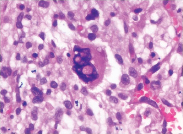
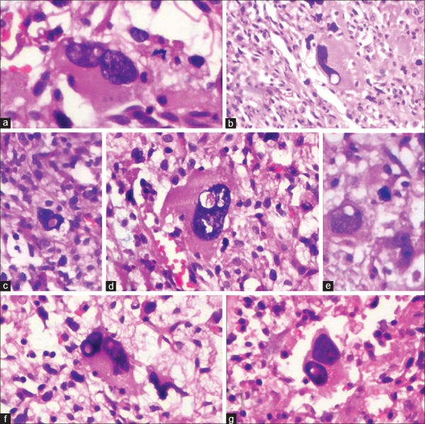
In the present case, large giant neoplastic cells of a high-grade pleomorphic liposarcoma showed the presence of nuclear vacuolization. These cells were large, mono/multinucleated with pronounced pleomorphism in the cell and nucleus. In the pleomorphic hyperchromatic nucleus various patterns of Lochkern, Ringkern and Kerbenkern patterns [Figure 2] were clearly seen. Nuclear vacuoles were seen in the nucleus as single/multiple punched out areas. This observation was seen in single large nucleus and multinucleated cells also. In some instances, the nucleus showed multiple punched out areas giving the nucleus a “Swiss cheese pattern” of appearance [Figure 3]. What these changes represent might be elusive, but it might be associated with lipid inclusions as the cells are part of sarcoma associated with adipose tissue-liposarcoma. Whether it leads to nuclear derangement is questionable, although morphologically, the cells are showing signs of derangement by the way of exhibiting extensive pleomorphism in nucleus and cell, along with the formation of mono/multinucleated giant cells.
Figure 2.
(a-g) Photomicrographs showing plethora of nuclei exhibiting Lochkern, Ringkern, Kerbkern patterns (H&E, stain, [a] ×400, [b] ×100, [c] ×200, [d-f] ×400)
Figure 3.

Photomicrograph showing Lockhern nucleus exhibiting swiss cheese pattern in the nucleus (H&E stain, ×200)
Financial support and sponsorship
Nil.
Conflicts of interest
There are no conflicts of interest.
Acknowledgment
The author would like to acknowledge Dr. Shruti Singh, Dr. Sameer Kumar V, Dr. Shiny Bopaiah (Postgraduate students, Department of Oral Pathology, Krishnadevaraya College of Dental Sciences, Bengaluru) for their contribution while preparing this manuscript.
REFERENCES
- 1.Webster M, Witkin KL, Cohen-Fix O. Sizing up the nucleus: Nuclear shape, size and nuclear-envelope assembly. J Cell Sci. 2009;122(Pt 10):1477–86. doi: 10.1242/jcs.037333. [DOI] [PMC free article] [PubMed] [Google Scholar]
- 2.Aravinthan A, Verma S, Coleman N, Davies S, Allison M, Alexander G. Vacuolation in hepatocyte nuclei is a marker of senescence. J Clin Pathol. 2012;65:557–60. doi: 10.1136/jclinpath-2011-200641. [DOI] [PubMed] [Google Scholar]
- 3.Ganick DJ, Finlay JL. Acute lymphoblastic leukemia with Burkitt cell morphology and cytoplasmic immunoglobulin. Blood. 1980;56:311–4. [PubMed] [Google Scholar]
- 4.Gnepp DR. Diagnostic Surgical Pathology of the Head and Neck. 2nd ed. Philadelphia: Elsevier Health Sciences; 2009. [Google Scholar]
- 5.Lindeburg MR. Diagnostic Pathology: Soft Tissue Tumors. 2nd ed. Philadelphia: Amirsys, Elsevier Health Sciences; 2015. [Google Scholar]
- 6.Hisaoka M. Lipoblast: Morphologic features and diagnostic value. J UOEH. 2014;36:115–21. doi: 10.7888/juoeh.36.115. [DOI] [PubMed] [Google Scholar]
- 7.Yuge S, Hayashi T, Kinoshita H, Toriyama E, Kinoshita N, Abe K, et al. Subconjunctival orbital fat herniation mimicking lipomatous tumors. Acta Med Nagasaki. 2011;56:19–22. [Google Scholar]
- 8.Resnik KS, Kutzner H. Original observation to rediscovery: Nuclear findings in adipocytes as example. Am J Dermatopathol. 2004;26:493–8. doi: 10.1097/00000372-200412000-00009. [DOI] [PubMed] [Google Scholar]
- 9.Winckler G. Characteristics of the vacuolar nucleus of the fat cell in man. Z Anat Entwicklungsgesch. 1960;122:241–6. [PubMed] [Google Scholar]
- 10.Ghadially FN. 1st ed. Vol. 1. London: Buterworths; 1988. Nucleus: Ultrastructural Pathology of Cell and Matrix. A Text and Atlas of Physiological and Pathological Alterations in Fine Structure in Cellular and Extracellular Component; pp. 1–180. [Google Scholar]


