Abstract
Background:
Various clinical and histological factors have helped in predicting the survival of patients with oral squamous cell carcinoma (OSCC). However, there has been a need for more specialized diagnostic and prognostic factors to avoid subjective variation among opinion. Thus, fractal dimension (FD) can be used as an index of the morphological changes that the epithelial cells undergo during their transformation into neoplastic cell. In oral cancer study, nuclear FD (NFD) can be used as a quantitative index to discriminate between normal, dysplastic and neoplastic oral mucosa.
Aim:
To use nuclear fractal geometry to compare the morphometric complexity in the normal, epithelial dysplasia and OSCC cases and to verify the difference among the various histological grades of dysplasia and OSCC. It was fulfilled by estimating the FDs of the nuclear surface.
Materials and Methods:
Histopathologically diagnosed cases of epithelial dysplasia and OSCC were taken from the archives. Photomicrographs were captured with the help of Lawrence and Mayo research microscope. The images were then subjected to image analysis using the Image J software with FracLac plugin java 1.6 to obtain FDs. FD of ten selected nuclei was calculated using the box-counting algorithm.
Statistical Analysis:
was done using descriptive analysis, ANOVA and Tukey's honest significant difference post hoc tests with STATAIC-13 software.
Results and Conclusion:
NFD can provide valuable information to discriminate between normal mucosa, dysplasia and carcinoma objectively without subjective discrimination.
Keywords: Epithelial dysplasia, fractal geometry, nuclear fractal dimension, squamous cell carcinoma
INTRODUCTION
Oral squamous cell carcinoma (OSCC) is the most common type of oral malignancy. Despite progress in therapeutic approaches, the 5-year survival rate for oral cancer has not improved significantly over the past several decades, and it remains about 50%, making this disease a serious public health problem. Although tumor-node-metastasis clinical staging system has proved to be a useful prognostic tool, the biological behavior of individual tumors remains however unpredictable. The research has focused on discovering improvised biologic marker and other factors related to the morphology of the neoplastic cells and tissues, which can be studied through computer-aided image analysis. Image analysis methods are divided into two categories: conventional methods, which usually focus on the size of the nuclei and more modern and accurate methods, fractal dimension (FD) that assess the complexity.[1]
Fractal geometry is a new development in mathematics, established by Benoit Mandelbrot in 1970. It aids the accurate study of the structural properties of natural objects, including histopathological specimens. The assessment and quantification of the degree of complexity and irregularity of these objects give measurements called “FDs.” An FD is a ratio providing a statistical index of complexity comparing how the detail in a pattern (strictly speaking, a fractal pattern) changes with the scale at which it is measured.[2] The dimension is simply the exponent of the number of self-similar pieces with magnification factor N into which the figure may be broken. For a biological or natural object to be called as a fractal, it should have a high level of organization, shape irregularities, functional morphological and temporal auto-similarities, scale invariance. Broccoli is the best natural fractal object.
Fractal geometry has been applied to various fields of medicine such as cardiovascular system, neurobiology, pathology and molecular biology. The heart rate of healthy individuals in different time scale shows essentially similar pattern. This pattern is self-similar and fractal. In various diseases of heart, the heart rate variability loses its complex fractal pattern. Hence, it may be possible to predict impending arrhythmia.[3] The fractal concept has been applied to measure the infiltrative margin of the malignant tumor, to assess the tumor angiogenesis and also to measure the irregular distribution of collagen in tissue.[3]
Few studies have used fractal analysis to distinguish the normal mucosa/cells from dysplastic and carcinomatous tissue. Very few studies have been done pertaining to oral mucosa, and ours is the first such study to assess FD in various histological grades of epithelial dysplasia and OSCC.
The aim of our present study was to use nuclear fractal geometry to compare the morphometric complexity in the normal, epithelial dysplasia and OSCC cases; and to verify the difference among different grades of epithelial dysplasia and OSCC.
MATERIALS AND METHODS
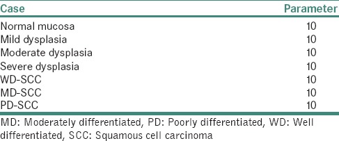
Histological H&E-stained sections of normal mucosa, different histological grades of epithelial dysplasia and squamous cell carcinoma were retrieved from the archives. The sample size was set at 70 as given by the statistician. The detailed sample distribution is given in Table 1.
Table 1.
Sample distribution for the present study
These tissue specimens were chosen with confirmed histopathologic diagnosis from buccal mucosa to standardize the results in both study and control groups as the FDs of different zones of oral mucosa vary considerably.
The histological pictures were captured at ×400 magnifications with the help of Lawrence and Mayo research microscope, USA, using TS view. The obtained images were then subjected to image analysis using Image J software with FracLac plugin java 1.6 (public domain - National Institutes of Health) to obtain FDs. The images were then converted into binary 8 bit image by the image analysis [Figure 1]. Later, using free hand tool selection, five nuclei are selected from each field and two fields per slide were taken into account. Thus, ten nuclei per case were studied.
Figure 1.
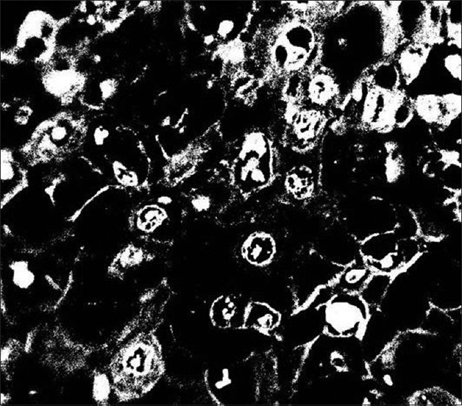
8 bit binary image of Broccoli as natural fractal obtained by the Image J software
Image analysis was performed using the fractal software to quantify FD by the box-counting method. To standardize our results and to avoid complications of multiple scans, we kept the box count from 2 to 12 only. Output was generated in the graph form with the slope of the regression line generated from the software. The slope [Figure 2] generated by the software gives the logarithmic value of the measure of FD.
Figure 2.
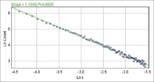
Slope generated as the result from the Image J software
A single fractal value was obtained for each nucleus, and an average FD of all ten nuclei was obtained for each slide. FD value thus obtained for each case was suitably tabulated and subjected to statistical analysis using descriptive analysis, ANOVA and Tukey's honest significant difference (HSD) post hoc tests using the STATAIC-13 software(StataCorp LP, Texas, USA). The P value was set at 0.005.
RESULTS
Descriptive analysis was used to tabulate the mean, standard deviation and standard error [Table 2]. It can be observed that there is a progressive increase in mean from normal mucosa to poorly differentiated SCC, with statistically significant difference between the groups (P ≤ 0.001) as shown in [Figure 3].
Table 2.
Descriptive analysis of the result obtained

Figure 3.
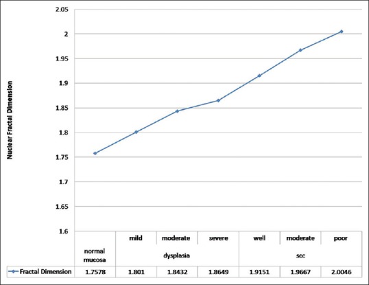
Mean fractal dimension of normal mucosa, Oral epithelial dysplasia and OSCC
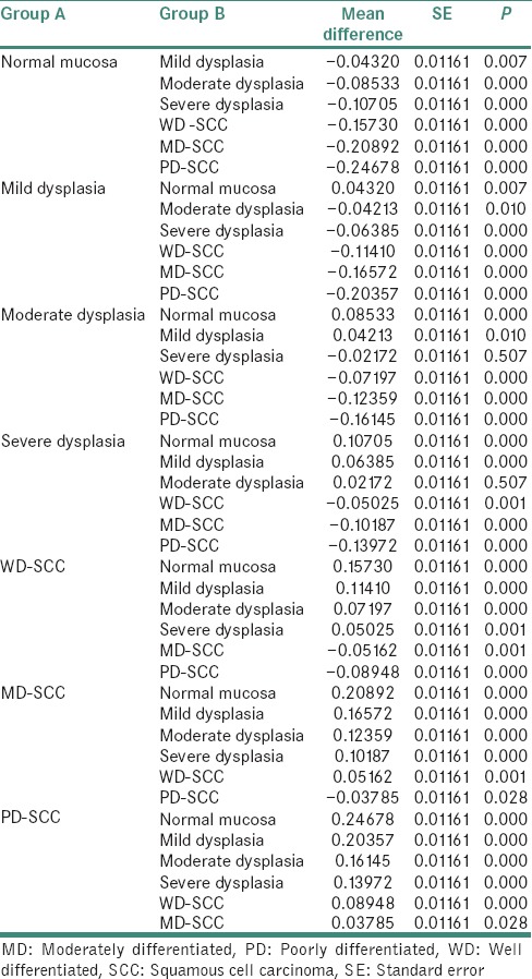
After applying ANOVA analysis, we found statistically significant difference with P < 0.001 between the groups. Similarly, Tukey's HSD post hoc tests for multiple comparisons indicated statistical difference within Group A except for the difference between normal mucosa and mild dysplasia (P = 0.007). The statistical difference between mild to moderate dysplasia (P = 0.010), moderate dysplasia and severe dysplasia (P = 0.507) are as shown in Table 3.
Table 3.
Results of statistical difference between group Tukey's honest significant difference post hoc tests for multiple comparisons
DISCUSSION
Many natural objects, including most objects studied in pathology, have complex structural characteristics and the complexity of their structures, for example, the degree of branching of vessels or the irregularity of a tumor boundary, remains at a constant level over a wide range of magnifications. These structures also have patterns that repeat themselves at different magnifications, a property known as scaling self-similarity. This has important implications for the measurement of parameters such as length and area since Euclidean measurements of these may be invalid. The fractal system of geometry overcomes the limitations of the Euclidean geometry for such objects, and the measurement of the FD gives an index of their space-filling properties.[4]
To be called fractals, biological and/or natural objects must fulfill a certain number of theoretic and methodological criteria including a high level of organization, shape irregularity, functional morphological and temporal auto-similarity, scale invariance, iterative pathways and a noninteger peculiar FD.[5] The assessment and quantification of the degree of complexity and irregularity of these objects gives measurements called “FDs.” The application of the fractal principle is very valuable for measuring dimensional properties and spatial parameters of irregular biological structures, for understanding the architectural/morphological organization of living tissues and organs and for achieving an objective comparison among complex morphogenetic changes occurring through the development of physiological, pathologic and neoplastic process.[5]
Fractal geometry can be used to assess epithelial connective tissue interface (ECTI) and nuclear FD (NFD). The structural organization of ECTI is useful in diagnostic histopathology to distinguish between normal mucosa and OSCC and appears to be crucial in tumor invasion and metastasis.[6,7] Moreover, NFD has been proved to be an independent prognostic factor of survival in oral, cervical and prostate cancer patients.[8,9]
Sedivy et al.[8] in 1998 studied fractal analysis to quantify structural irregularities of atypical nuclei associated with dysplastic lesions of the uterine cervix. A box-counting algorithm was used to determine the FD of atypical nuclei in the dysplastic cervical epithelium. They found that the FDs of the nuclei increased as the degree of dysplasia increased from 1.02 to 1.32. In our present study, we had compared normal oral mucosa, epithelial dysplasia and OSCC. Although the values obtained in our study do not coincide with other studies, we have obtained an increasing mean FD from normal mucosa (1.7578), epithelial dysplasia (1.836) to squamous cell carcinoma (1.9567).
Bauer and Mackenzie 1999[10] utilized FD of perimeter surface of cell section as a new observable factor to characterize cells of cancerous and healthy origin. They performed this distinction between patients with hairy-cell lymphocytic leukemia and normal blood lymphocytes. They observed that none of the healthy cells have a fractal value higher than 1.28. Similar studies were performed by Noroozinia et al.[11] in 2011 on the nuclear boundary of cancer cells in urinary smears. They utilized 41 positive urine cytology samples and 33 negative samples to pinpoint objective method for the differences between malignant and nonmalignant epithelial cells in urine cytology. They selected a cutoff value of 1.732 to discriminate the malignant and nonmalignant epithelial cells.
Among the few studies, some have evaluated NFD on oral tissue and all have noted a significant change in NFD between normal mucosa and OSCC. Some studies have also observed statistically significant difference between the groups of OSCC.
Goutzanis et al.[12] studied FDs on histological sections from 48 OSCCs as well as from 17 nonmalignant mucosa specimens which were stained with H and E and Feulgen for nuclear complexity evaluation. They observed that in normal epithelium and in well-differentiated neoplasms, FD values were low, while in less differentiated tumors they were high. Patients with lower NFD values presented with significantly better survival than patients with larger FD values. These results are in agreement with our study. In this present study, we observed a significant change in value with NFD of 1.9151, 1.9667 and 2.0046 for well-differentiated SCC, moderately differentiated SCC and poorly differentiated SCC, respectively.
Mincione et al.[13] evaluated NFD values in different stages of OSCC and the correlations with clinicopathological variables and patient survival were investigated. Histological sections from OSCC and control nonneoplastic mucosa specimens were stained with H and E for pathological analysis and with Feulgen for nuclear evaluation. FD in OSCC groups versus controls revealed statistically significant differences (P < 0.001). In addition, a progressive increase of FD from stage I and II lesions and stage III and IV lesions was observed, with statistically significant differences (P = 0.003). Moreover, different degrees of tumor differentiation showed a significant difference in the average NFD values (P = 0.001). A relationship between FD and patients’ survival was also detected with lower FD values associated with longer survival time and higher FD values with shorter survival time (P = 0.034). These data showed that FD significantly increased during OSCC progression. Even in our study, we have observed statistically significant difference between various grades of OSCC with P value being < 0.005.
Yinti et al.[14] evaluated the NFD in OSCC using computer-aided image analysis. Histological sections of 14 selected cases of OSCC and six samples of normal buccal mucosa (as control) were stained with H and E and Feulgen stain for histopathological examination and evaluation of nuclear complexity, respectively. They observed that the NFD increased progressively toward worst tumor staging as compared to the normal buccal mucosa.
CONCLUSION
Our study has shown a reliable method to distinguish between normal mucosa, dysplasia and carcinoma. We have obtained a mean NFD of 1.7578 for normal mucosa, 1.8363 for epithelial dysplasia and 1.9621 for squamous cell carcinoma. This indicates a substantial increase in mean signifying the further use of fractal geometry for histopathological analysis. As fractal analysis technique is noninvasive and cost-effective, it can be used in a developing country.
With growing literature on FD, interest has been cultivated to use fractal dimension as a diagnostic and prognostic tool for epithelial dysplasia and OSCC.
Financial support and sponsorship
Nil.
Conflicts of interest
There are no conflicts of interest.
REFERENCES
- 1.Silverman S., Jr Demographics and occurrence of oral and pharyngeal cancers. The outcomes, the trends, the challenge. J Am Dent Assoc. 2001;132(Suppl):7S–11S. doi: 10.14219/jada.archive.2001.0382. [DOI] [PubMed] [Google Scholar]
- 2.Losa GA, Nonnenmacher TF. Self-similarity and fractal irregularity in pathologic tissues. Mod Pathol. 1996;9:174–82. [PubMed] [Google Scholar]
- 3.Losa GA. Fractals and their contribution to biology and medicine. Medicographia. 2012;34:365–74. [Google Scholar]
- 4.Metze K. Fractal dimension of chromatin: Potential molecular diagnostic applications for cancer prognosis. Expert Rev Mol Diagn. 2013;13:719–35. doi: 10.1586/14737159.2013.828889. [DOI] [PMC free article] [PubMed] [Google Scholar]
- 5.Mandelbrot BB. The Fractal Geometry of Nature. San Francisco, CA: WH Freeman and Co; 1982. [Google Scholar]
- 6.Upadhyaya N, Khandekar S, Dive A, Mishra R, Moharil R. A study of morphometrical differences between normal mucosa, dysplasia; squamous cell carcinoma and pseudoepitheliomatous hyperplasia of the oral mucosa. J Pharm Biol Sci. 2013;5:66–70. [Google Scholar]
- 7.Khandekar S, Dive A, Mishra R, Upadhyaya N. Study of epithelial connective tissue interface using fractal geometry: A pilot study. Journlal of evolution of medical and dental science. 2013;2:41–5. [Google Scholar]
- 8.Sedivy R, Windischberger C, Svozil K, Moser E, Breitenecker G. Fractal analysis: An objective method for identifying atypical nuclei in dysplastic lesions of the cervix uteri. Gynecol Oncol. 1999;75:78–83. doi: 10.1006/gyno.1999.5516. [DOI] [PubMed] [Google Scholar]
- 9.Kademani D, Bell RB, Bagheri S, Holmgren E, Dierks E, Potter B, et al. Prognostic factors in intraoral squamous cell carcinoma: The influence of histologic grade. J Oral Maxillofac Surg. 2005;63:1599–605. doi: 10.1016/j.joms.2005.07.011. [DOI] [PubMed] [Google Scholar]
- 10.Bauer W, Mackenzie CD. Cancer Detection via Determination of Fractal Cell Dimension: In Michigan State University (US) 1999 Jan;:p9. Report no.: Patt-sol/9506003; MSUCL-980. [Google Scholar]
- 11.Noroozinia F, Behjati G, Shahabi S. Fractal study on nuclear boundary of cancer cells in urinary smears. Iran J Pathol. 2011;6:63–7. [Google Scholar]
- 12.Goutzanis L, Papadogeorgakis N, Pavlopoulos PM, Katti K, Petsinis V, Plochoras I, et al. Nuclear fractal dimension as a prognostic factor in oral squamous cell carcinoma. Oral Oncol. 2008;44:345–53. doi: 10.1016/j.oraloncology.2007.04.005. [DOI] [PubMed] [Google Scholar]
- 13.Mincione G, Di Nicola M, Di Marcantonio MC, Muraro R, Piattelli A, Rubini C, et al. Nuclear fractal dimension in oral squamous cell carcinoma: A novel method for the evaluation of grading, staging, and survival. J Oral Pathol Med. 2015;44:680–4. doi: 10.1111/jop.12280. [DOI] [PubMed] [Google Scholar]
- 14.Yinti SR, Srikant N, Boaz K, Lewis AJ, Ashokkumar PJ, Kapila SN. Nuclear fractal dimensions as a tool for prognostication of oral squamous cell carcinoma. J Clin Diagn Res. 2015;9:EC21–5. doi: 10.7860/JCDR/2015/12931.6837. [DOI] [PMC free article] [PubMed] [Google Scholar]


