

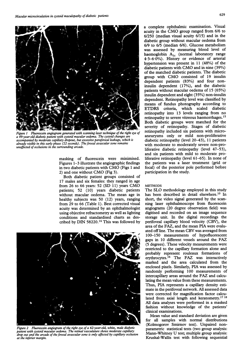
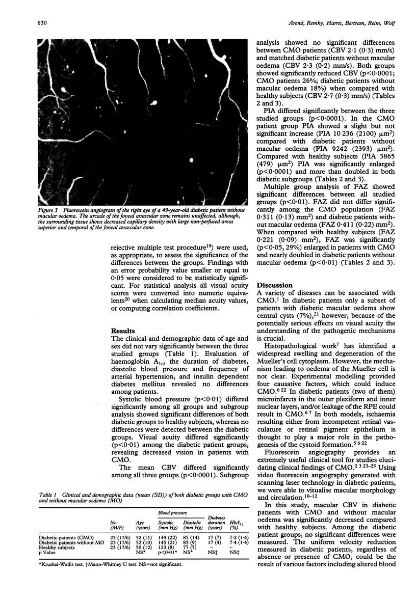
Abstract
BACKGROUND--In patients with diabetic macular oedema and central cysts ischaemia of the retina appears to be an important contributing factor in the pathogenesis of cysts. This study was performed to further elucidate the role of the inner retinal microcirculation in diabetic cystoid macular oedema (CMO). METHODS--Video fluorescein angiography allows visualisation of the macular microvasculature and measurements of the capillary blood velocity (CBV), foveal avascular zone (FAZ), and perifoveal intercapillary area (PIA, characterising capillary density). RESULTS--Twenty three diabetic subjects with CMO, matched diabetic patients without macular oedema (n = 23), and healthy subjects (n = 23) were included. CBV, PIA, and FAZ did not differ significantly among diabetic groups regardless of presence of cystoid changes. CBV was significantly reduced (p < 0.0001) and PIA was more than doubled in both diabetic groups (p < 0.0001) when compared with healthy subjects. Furthermore, FAZ showed a nearly doubled size in diabetic patients without macular oedema (p < 0.01) and a less pronounced enlargement (by 29%) in diabetics with CMO (p < 0.05). CONCLUSION--The results indicate that the retinal microcirculation in diabetic patients is markedly altered when compared with healthy subjects, regardless of CMO presence. In CMO patients the microcirculatory changes are similar to those of diabetic patients without macular oedema. Thus inner retinal perfusion does not contribute to tissue ischaemia leading to cystoid formations in diabetic maculopathy.
Full text
PDF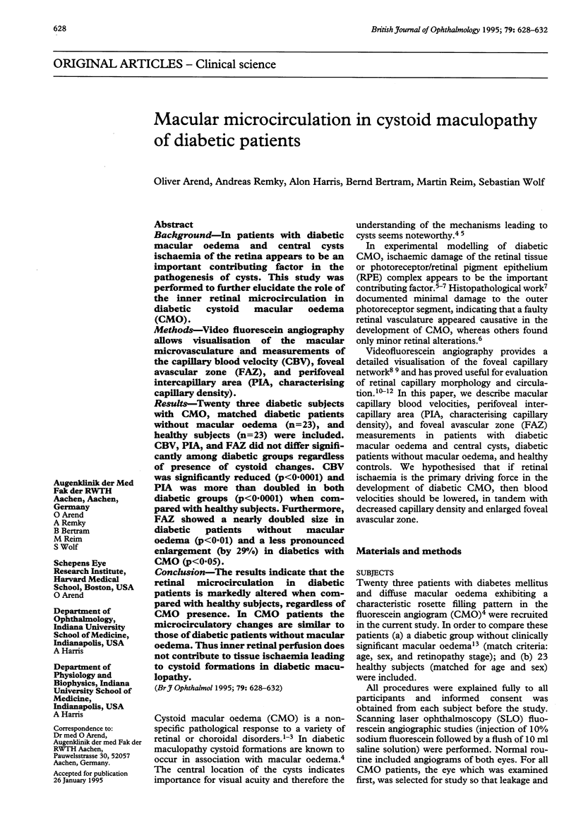
Images in this article
Selected References
These references are in PubMed. This may not be the complete list of references from this article.
- ASHTON N. STUDIES OF THE RETINAL CAPILLARIES IN RELATION TO DIABETIC AND OTHER RETINOPATHIES. Br J Ophthalmol. 1963 Sep;47:521–538. doi: 10.1136/bjo.47.9.521. [DOI] [PMC free article] [PubMed] [Google Scholar]
- Arend O., Harris A., Wolf S. Capillary blood flow velocity measurements in cystoid macular edema with the scanning laser ophthalmoscope. Am J Ophthalmol. 1994 Jun 15;117(6):819–820. doi: 10.1016/s0002-9394(14)70339-3. [DOI] [PubMed] [Google Scholar]
- Arend O., Wolf S., Jung F., Bertram B., Pöstgens H., Toonen H., Reim M. Retinal microcirculation in patients with diabetes mellitus: dynamic and morphological analysis of perifoveal capillary network. Br J Ophthalmol. 1991 Sep;75(9):514–518. doi: 10.1136/bjo.75.9.514. [DOI] [PMC free article] [PubMed] [Google Scholar]
- Arend O., Wolf S., Remky A., Sponsel W. E., Harris A., Bertram B., Reim M. Perifoveal microcirculation with non-insulin-dependent diabetes mellitus. Graefes Arch Clin Exp Ophthalmol. 1994 Apr;232(4):225–231. doi: 10.1007/BF00184010. [DOI] [PubMed] [Google Scholar]
- Ashton N. Vascular basement membrane changes in diabetic retinopathy. Montgomery lecture, 1973. Br J Ophthalmol. 1974 Apr;58(4):344–366. doi: 10.1136/bjo.58.4.344. [DOI] [PMC free article] [PubMed] [Google Scholar]
- Bellhorn R. W. Analysis of animal models of macular edema. Surv Ophthalmol. 1984 May;28 (Suppl):520–524. doi: 10.1016/0039-6257(84)90235-2. [DOI] [PubMed] [Google Scholar]
- Bennett A. G., Rudnicka A. R., Edgar D. F. Improvements on Littmann's method of determining the size of retinal features by fundus photography. Graefes Arch Clin Exp Ophthalmol. 1994 Jun;232(6):361–367. doi: 10.1007/BF00175988. [DOI] [PubMed] [Google Scholar]
- Bresnick G. H., Condit R., Syrjala S., Palta M., Groo A., Korth K. Abnormalities of the foveal avascular zone in diabetic retinopathy. Arch Ophthalmol. 1984 Sep;102(9):1286–1293. doi: 10.1001/archopht.1984.01040031036019. [DOI] [PubMed] [Google Scholar]
- Bresnick G. H. Diabetic macular edema. A review. Ophthalmology. 1986 Jul;93(7):989–997. doi: 10.1016/s0161-6420(86)33650-9. [DOI] [PubMed] [Google Scholar]
- Coscas G., Gaudric A. Natural course of nonaphakic cystoid macular edema. Surv Ophthalmol. 1984 May;28 (Suppl):471–484. doi: 10.1016/0039-6257(84)90229-7. [DOI] [PubMed] [Google Scholar]
- Fine B. S., Brucker A. J. Macular edema and cystoid macular edema. Am J Ophthalmol. 1981 Oct;92(4):466–481. doi: 10.1016/0002-9394(81)90638-3. [DOI] [PubMed] [Google Scholar]
- Gass J. D., Anderson D. R., Davis E. B. A clinical, fluorescein angiographic, and electron microscopic correlation of cystoid macular edema. Am J Ophthalmol. 1985 Jul 15;100(1):82–86. doi: 10.1016/s0002-9394(14)74988-8. [DOI] [PubMed] [Google Scholar]
- Gass J. D., Norton E. W. Cystoid macular edema and papilledema following cataract extraction. A fluorescein fundoscopic and angiographic study. Arch Ophthalmol. 1966 Nov;76(5):646–661. doi: 10.1001/archopht.1966.03850010648005. [DOI] [PubMed] [Google Scholar]
- Hamilton A. M., Kohner E. M., Rosen D., Bird A. C., Dollery C. T. Experimental retinal branch vein occlusion in rhesus monkeys. I. Clinical appearances. Br J Ophthalmol. 1979 Jun;63(6):377–387. doi: 10.1136/bjo.63.6.377. [DOI] [PMC free article] [PubMed] [Google Scholar]
- Hartmann E. Sehschärfebestimmung. Klin Monbl Augenheilkd. 1987 Jul;191(1):62–68. doi: 10.1055/s-2008-1050468. [DOI] [PubMed] [Google Scholar]
- Klein R., Klein B. E., Moss S. E., Davis M. D., DeMets D. L. The Wisconsin epidemiologic study of diabetic retinopathy. IV. Diabetic macular edema. Ophthalmology. 1984 Dec;91(12):1464–1474. doi: 10.1016/s0161-6420(84)34102-1. [DOI] [PubMed] [Google Scholar]
- Littmann H. Zur Bestimmung der wahren Grösse eines Objektes auf dem Hintergrund eines lebenden Auges. Klin Monbl Augenheilkd. 1988 Jan;192(1):66–67. doi: 10.1055/s-2008-1050076. [DOI] [PubMed] [Google Scholar]
- Mansour A. M., Schachat A., Bodiford G., Haymond R. Foveal avascular zone in diabetes mellitus. Retina. 1993;13(2):125–128. doi: 10.1097/00006982-199313020-00006. [DOI] [PubMed] [Google Scholar]
- Ohnishi Y., Fujisawa K., Ishibashi T., Kojima H. Capillary blood flow velocity measurements in cystoid macular edema with the scanning laser ophthalmoscope. Am J Ophthalmol. 1994 Jan 15;117(1):24–29. doi: 10.1016/s0002-9394(14)73011-9. [DOI] [PubMed] [Google Scholar]
- Patz A. Cystoid maculopathy in diabetics. Arch Ophthalmol. 1976 May;94(5):761–768. doi: 10.1001/archopht.1976.03910030369004. [DOI] [PubMed] [Google Scholar]
- Schröder S., Palinski W., Schmid-Schönbein G. W. Activated monocytes and granulocytes, capillary nonperfusion, and neovascularization in diabetic retinopathy. Am J Pathol. 1991 Jul;139(1):81–100. [PMC free article] [PubMed] [Google Scholar]
- Smith R. T., Lee C. M., Charles H. C., Farber M., Cunha-Vaz J. G. Quantification of diabetic macular edema. Arch Ophthalmol. 1987 Feb;105(2):218–222. doi: 10.1001/archopht.1987.01060020072032. [DOI] [PubMed] [Google Scholar]
- Tso M. O. Animal modeling of cystoid macular edema. Surv Ophthalmol. 1984 May;28 (Suppl):512–519. doi: 10.1016/0039-6257(84)90234-0. [DOI] [PubMed] [Google Scholar]
- Tso M. O. Pathology of cystoid macular edema. Ophthalmology. 1982 Aug;89(8):902–915. doi: 10.1016/s0161-6420(82)34698-9. [DOI] [PubMed] [Google Scholar]
- Wolf S., Arend O., Toonen H., Bertram B., Jung F., Reim M. Retinal capillary blood flow measurement with a scanning laser ophthalmoscope. Preliminary results. Ophthalmology. 1991 Jun;98(6):996–1000. doi: 10.1016/s0161-6420(91)32192-4. [DOI] [PubMed] [Google Scholar]
- Wolf S., Toonen H., Arend O., Jung F., Kaupp A., Kiesewetter H., Meyer-Ebrecht D., Reim M. Zur Quantifizierung der retinalen Kapillardurchblutung mit Hilfe des Scanning-Laser-Ophthalmoskops. Biomed Tech (Berl) 1990 Jun;35(6):131–134. [PubMed] [Google Scholar]
- Yanoff M., Fine B. S., Brucker A. J., Eagle R. C., Jr Pathology of human cystoid macular edema. Surv Ophthalmol. 1984 May;28 (Suppl):505–511. doi: 10.1016/0039-6257(84)90233-9. [DOI] [PubMed] [Google Scholar]