

Abstract
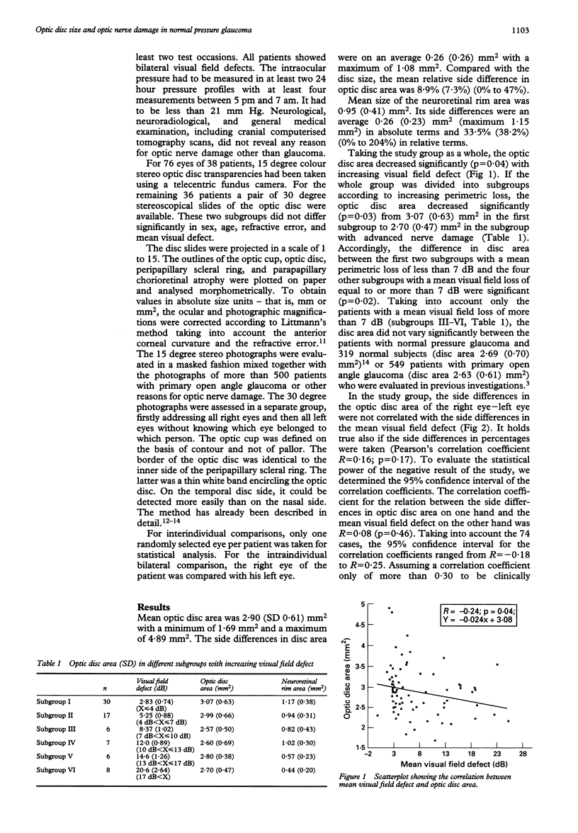
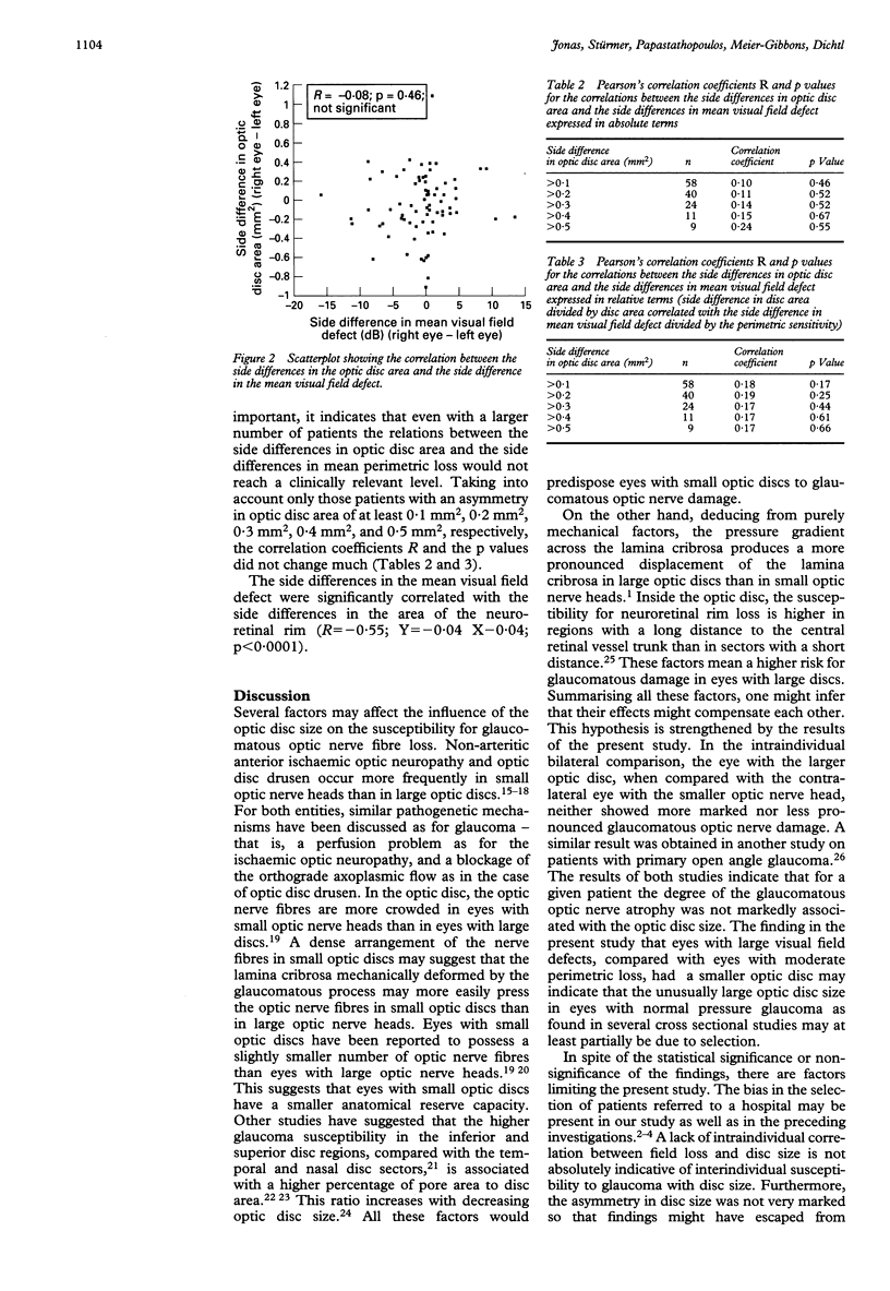
BACKGROUND--Recent reports indicate that eyes with normal pressure glaucoma have larger optic discs than eyes with primary open angle glaucoma or normal eyes. This study was performed to find whether, in normal pressure glaucoma, a large disc is associated with more optic nerve damage than a small disc. METHODS--Colour optic disc photographs of 74 patients with normal pressure glaucoma were assessed morphometrically. RESULTS--Taking the study group as a whole, the optic disc size decreased significantly (p = 0.04) with increasing visual field defect. In an intraindividual bilateral comparison, the side differences in the disc area of the right minus the left eye of the same individual were not significantly correlated with the side differences in the mean visual field defect. CONCLUSIONS--The results indicate that the eye with the larger optic disc, when compared with the contralateral eye with the smaller optic nerve head, showed neither a significantly more marked nor less pronounced glaucomatous optic nerve damage. It suggests that for a given patient the degree of glaucomatous optic nerve atrophy was not markedly associated with the optic disc size. The finding that patients with large visual field defects had smaller discs than patients with moderate perimetric loss may indicate that the results of previous cross sectional studies reporting on an unusually large disc size in normal pressure glaucoma may be due partially to selection.
Full text
PDF
Selected References
These references are in PubMed. This may not be the complete list of references from this article.
- Airaksinen P. J., Drance S. M., Schulzer M. Neuroretinal rim area in early glaucoma. Am J Ophthalmol. 1985 Jan 15;99(1):1–4. doi: 10.1016/s0002-9394(14)75856-8. [DOI] [PubMed] [Google Scholar]
- Beck R. W., Savino P. J., Repka M. X., Schatz N. J., Sergott R. C. Optic disc structure in anterior ischemic optic neuropathy. Ophthalmology. 1984 Nov;91(11):1334–1337. doi: 10.1016/s0161-6420(84)34146-x. [DOI] [PubMed] [Google Scholar]
- Bengtsson B. The variation and covariation of cup and disc diameters. Acta Ophthalmol (Copenh) 1976 Dec;54(6):804–818. doi: 10.1111/j.1755-3768.1976.tb01801.x. [DOI] [PubMed] [Google Scholar]
- Betz P., Camps F., Collignon-Brach C., Weekers R. Photographie stéréoscopique et photogrammétrie de l'excavation physiologique de la papille. J Fr Ophtalmol. 1981;4(3):193–203. [PubMed] [Google Scholar]
- Burk R. O., Rohrschneider K., Noack H., Völcker H. E. Are large optic nerve heads susceptible to glaucomatous damage at normal intraocular pressure? A three-dimensional study by laser scanning tomography. Graefes Arch Clin Exp Ophthalmol. 1992;230(6):552–560. doi: 10.1007/BF00181778. [DOI] [PubMed] [Google Scholar]
- Caprioli J., Miller J. M. Optic disc rim area is related to disc size in normal subjects. Arch Ophthalmol. 1987 Dec;105(12):1683–1685. doi: 10.1001/archopht.1987.01060120081030. [DOI] [PubMed] [Google Scholar]
- Chi T., Ritch R., Stickler D., Pitman B., Tsai C., Hsieh F. Y. Racial differences in optic nerve head parameters. Arch Ophthalmol. 1989 Jun;107(6):836–839. doi: 10.1001/archopht.1989.01070010858029. [DOI] [PubMed] [Google Scholar]
- Drance S. M., Balazsi G. Die neuroretinale Randzone beim frühen Glaukom. Klin Monbl Augenheilkd. 1984 Apr;184(4):271–273. doi: 10.1055/s-2008-1054462. [DOI] [PubMed] [Google Scholar]
- Jonas J. B., Fernandez M. C., Naumann G. O. Glaucomatous optic nerve atrophy in small discs with low cup-to-disc ratios. Ophthalmology. 1990 Sep;97(9):1211–1215. doi: 10.1016/s0161-6420(90)32434-x. [DOI] [PubMed] [Google Scholar]
- Jonas J. B., Fernández M. C., Naumann G. O. Correlation of the optic disc size to glaucoma susceptibility. Ophthalmology. 1991 May;98(5):675–680. doi: 10.1016/s0161-6420(91)32234-6. [DOI] [PubMed] [Google Scholar]
- Jonas J. B., Fernández M. C. Shape of the neuroretinal rim and position of the central retinal vessels in glaucoma. Br J Ophthalmol. 1994 Feb;78(2):99–102. doi: 10.1136/bjo.78.2.99. [DOI] [PMC free article] [PubMed] [Google Scholar]
- Jonas J. B., Gusek G. C., Guggenmoos-Holzmann I., Naumann G. O. Optic nerve head drusen associated with abnormally small optic discs. Int Ophthalmol. 1987 Dec;11(2):79–82. doi: 10.1007/BF00136734. [DOI] [PubMed] [Google Scholar]
- Jonas J. B., Gusek G. C., Naumann G. O. Anterior ischemic optic neuropathy: nonarteritic form in small and giant cell arteritis in normal sized optic discs. Int Ophthalmol. 1988;12(2):119–125. doi: 10.1007/BF00137137. [DOI] [PubMed] [Google Scholar]
- Jonas J. B., Gusek G. C., Naumann G. O. Optic disk morphometry in high myopia. Graefes Arch Clin Exp Ophthalmol. 1988;226(6):587–590. doi: 10.1007/BF02169209. [DOI] [PubMed] [Google Scholar]
- Jonas J. B., Mardin C. Y., Schlötzer-Schrehardt U., Naumann G. O. Morphometry of the human lamina cribrosa surface. Invest Ophthalmol Vis Sci. 1991 Feb;32(2):401–405. [PubMed] [Google Scholar]
- Jonas J. B., Schmidt A. M., Müller-Bergh J. A., Schlötzer-Schrehardt U. M., Naumann G. O. Human optic nerve fiber count and optic disc size. Invest Ophthalmol Vis Sci. 1992 May;33(6):2012–2018. [PubMed] [Google Scholar]
- Jonas J. B. Size of glaucomatous optic discs. Ger J Ophthalmol. 1992;1(1):41–44. [PubMed] [Google Scholar]
- Jonas J. B., Xu L. Parapapillary chorioretinal atrophy in normal-pressure glaucoma. Am J Ophthalmol. 1993 Apr 15;115(4):501–505. doi: 10.1016/s0002-9394(14)74453-8. [DOI] [PubMed] [Google Scholar]
- Quigley H. A., Addicks E. M., Green W. R., Maumenee A. E. Optic nerve damage in human glaucoma. II. The site of injury and susceptibility to damage. Arch Ophthalmol. 1981 Apr;99(4):635–649. doi: 10.1001/archopht.1981.03930010635009. [DOI] [PubMed] [Google Scholar]
- Quigley H. A., Addicks E. M. Regional differences in the structure of the lamina cribrosa and their relation to glaucomatous optic nerve damage. Arch Ophthalmol. 1981 Jan;99(1):137–143. doi: 10.1001/archopht.1981.03930010139020. [DOI] [PubMed] [Google Scholar]
- Quigley H. A., Coleman A. L., Dorman-Pease M. E. Larger optic nerve heads have more nerve fibers in normal monkey eyes. Arch Ophthalmol. 1991 Oct;109(10):1441–1443. doi: 10.1001/archopht.1991.01080100121056. [DOI] [PubMed] [Google Scholar]
- Radius R. L. Regional specificity in anatomy at the lamina cribrosa. Arch Ophthalmol. 1981 Mar;99(3):478–480. doi: 10.1001/archopht.1981.03930010480020. [DOI] [PubMed] [Google Scholar]
- Spencer W. H. Drusen of the optic disk and aberrant axoplasmic transport. The XXXIV Edward Jackson memorial lecture. Am J Ophthalmol. 1978 Jan;85(1):1–12. doi: 10.1016/s0002-9394(14)76658-9. [DOI] [PubMed] [Google Scholar]
- Tuulonen A., Airaksinen P. J. Optic disc size in exfoliative, primary open angle, and low-tension glaucoma. Arch Ophthalmol. 1992 Feb;110(2):211–213. doi: 10.1001/archopht.1992.01080140067029. [DOI] [PubMed] [Google Scholar]