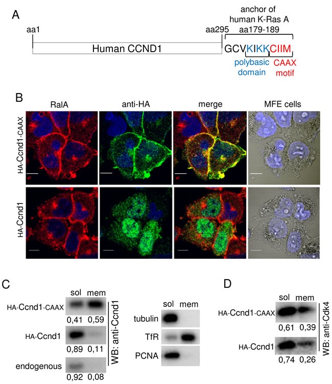
Figure 2. Ccnd1-CAAX is localized in cell membranes of tumor cells.

A. Schematic representation of C-terminal fusion of the anchor domain of human K-Ras with Ccnd1 protein. B. MFE cells infected with HA-Ccnd1 or HA-Ccnd1-CAAX or an empty vector were fixed in 4% paraformaldehyde and permeabilized with 0.2% triton-100X. Images were acquired by confocal microscopy (10μm bar). Nuclei were stained with Hoescht (blue). The antibodies used were anti-HA (rat monoclonal 3F10, green) and anti-RalA (mouse monoclonal, red). C. Ishikawa cells infected with HA-Ccnd1 or HA-Ccnd1-CAAX were submitted to subcellular fractionation (see “Materials and Methods”). Fractions were analyzed by immunoblotting to detect Ccnd1 in soluble and membrane fractions. Quantification of Ccnd1 levels are shown at the bottom of the panel. Tubulin as a cytosol marker, Transferrin Receptor as a membrane marker and PCNA as a nucleoplasm marker were used to control fractionation. D. Cdk4 distribution in soluble and membrane fractions from the experiment in C. Quantification of Cdk4 levels are shown at the bottom of the panel.
