
Figure 5. UHRF1 mRNA and protein expression after si-UHRF1 transfection and effects of UHRF1 silencing in BC cell lines.
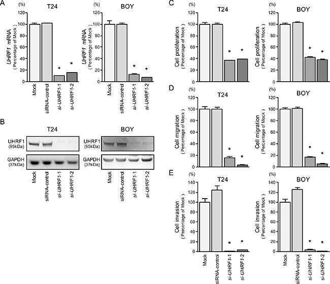
(A) UHRF1 mRNA expression was evaluated by qRT-PCR in T24 and BOY 72 hours after transfection with si-UHRF1-1 and si-UHRF1-2. GUSB was used as an internal control. (B) UHRF1 protein expression was evaluated by Western blot analysis in T24 and BOY 72 - 96 hours after transfection with miR-145-5p or miR-145-3p. GAPDH was used as a loading control. (C) Cell proliferation was determined with the XTT assays 72 hours after transfection with 10 nM si-UHRF1-1 or si-UHRF1-2. *P < 0.0001. (D) Cell migration activity was determined by wound-healing assays. *P < 0.0001. (E) Cell invasion activity was determined using Matrigel invasion assays. *P < 0.0001.