Abstract
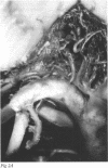
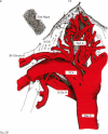
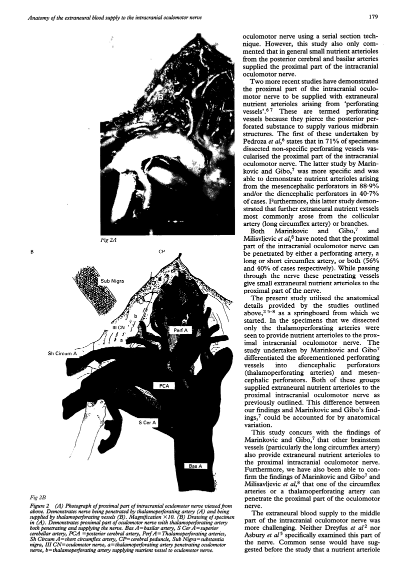
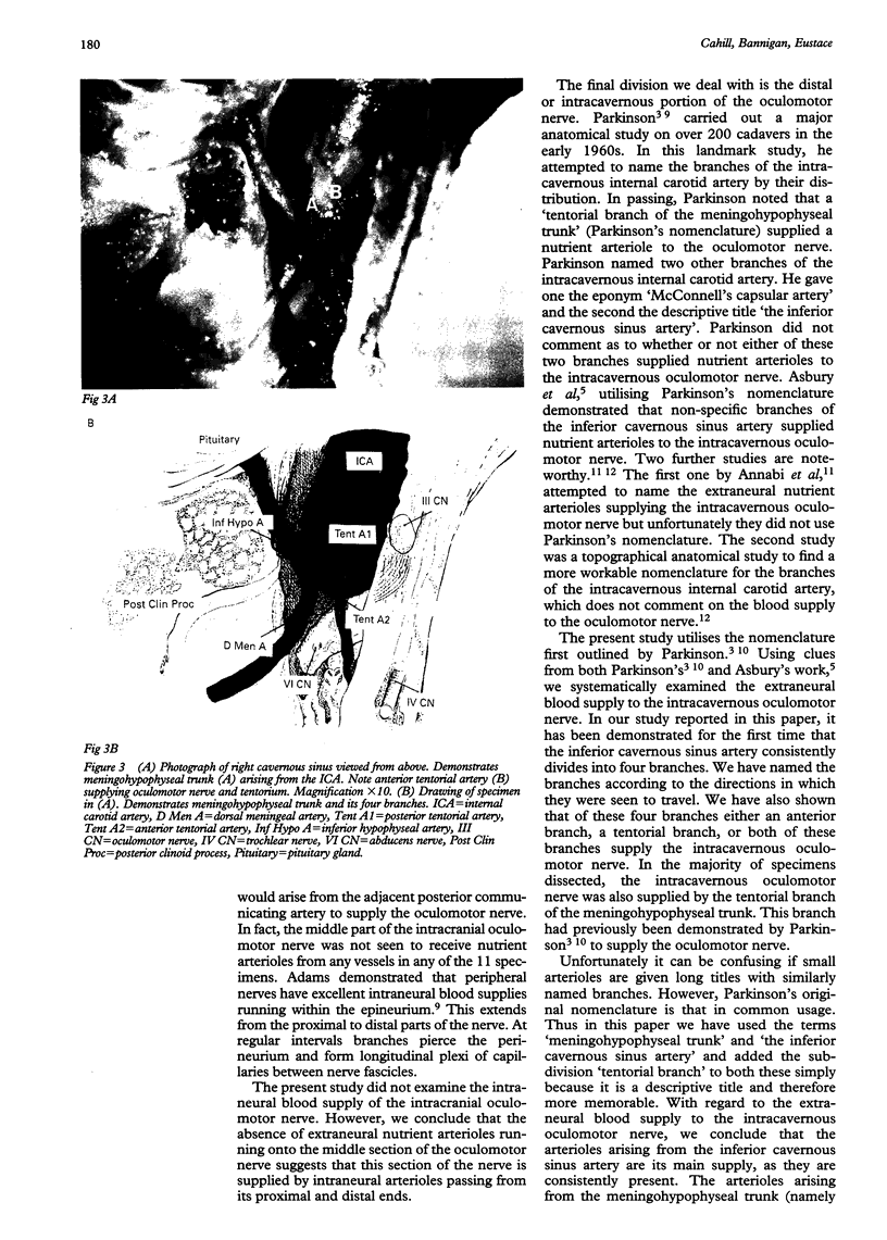
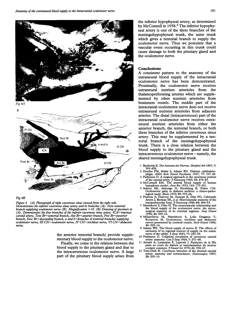
AIMS: An anatomical study was undertaken to determine the extraneural blood supply to the intracranial oculomotor nerve. METHODS: Human tissue blocks containing brainstem, cranial nerves II-VI, body of sphenoid, and associated cavernous sinuses were obtained, injected with contrast material, and dissected using a stereoscopic microscope. RESULTS: Eleven oculomotor nerves were dissected, the intracranial part being divided into proximal, middle, and distal (intracavernous) parts. The proximal part of the intracranial oculomotor nerve received extraneural nutrient arterioles from thalamoperforating arteries in all specimens and in six nerves this blood supply was supplemented by branches from other brainstem vessels. Four nerves were seen to be penetrated by branches of brainstem vessels and these penetrating arteries also supplied nutrient arterioles. The middle part of the intracranial oculomotor nerve did not receive nutrient arterioles from adjacent arteries. The distal part of the intracranial oculomotor nerve received nutrient arterioles from the inferior cavernous sinus artery in all 11 nerves and in seven nerves this was supplemented by a tentorial artery arising from the meningohypophyseal trunk. The inferior hypophyseal artery arose from the meningohypophyseal trunk in all 11 cavernous sinuses dissected. CONCLUSION: This study shows a constant pattern to the blood supply of the intracranial oculomotor nerve. It also highlights the close relation between the blood supplies to the intracavernous oculomotor nerve and the pituitary gland.
Full text
PDF
Images in this article
Selected References
These references are in PubMed. This may not be the complete list of references from this article.
- Adams W. E. The blood supply of nerves: II. The effects of exclusion of its regional sources of supply on the sciatic nerve of the rabbit. J Anat. 1943 Apr;77(Pt 3):243–250.3. doi: 10.1093/oxfordjournals.bmb.a070234. [DOI] [PMC free article] [PubMed] [Google Scholar]
- Annabi A., Lasjaunias P., Lapresle J. Paralysies de la IIIe paire au cours du diabete et vascularisation du moteur oculaire commun. J Neurol Sci. 1979 May;41(3):359–367. doi: 10.1016/0022-510x(79)90095-9. [DOI] [PubMed] [Google Scholar]
- Asbury A. K., Aldredge H., Hershberg R., Fisher C. M. Oculomotor palsy in diabetes mellitus: a clinico-pathological study. Brain. 1970;93(3):555–566. doi: 10.1093/brain/93.3.555. [DOI] [PubMed] [Google Scholar]
- DREYFUS P. M., HAKIM S., ADAMS R. D. Diabetic ophthalmoplegia; report of case, with postmortem study and comments on vascular supply of human oculomotor nerve. AMA Arch Neurol Psychiatry. 1957 Apr;77(4):337–349. [PubMed] [Google Scholar]
- Friedlander W. J. Who was 'the father of bromide treatment of epilepsy'? Arch Neurol. 1986 May;43(5):505–507. doi: 10.1001/archneur.1986.00520050077027. [DOI] [PubMed] [Google Scholar]
- MCCONNELL E. M. The arterial blood supply of the human hypophysis cerebri. Anat Rec. 1953 Feb;115(2):175–203. doi: 10.1002/ar.1091150204. [DOI] [PubMed] [Google Scholar]
- Marinković S., Gibo H. The neurovascular relationships and the blood supply of the oculomotor nerve: the microsurgical anatomy of its cisternal segment. Surg Neurol. 1994 Dec;42(6):505–516. doi: 10.1016/0090-3019(94)90081-7. [DOI] [PubMed] [Google Scholar]
- PARKINSON D. COLLATERAL CIRCULATION OF CAVERNOUS CAROTID ARTERY: ANATOMY. Can J Surg. 1964 Jul;7:251–268. [PubMed] [Google Scholar]
- Parkinson D. A surgical approach to the cavernous portion of the carotid artery. Anatomical studies and case report. J Neurosurg. 1965 Nov;23(5):474–483. doi: 10.3171/jns.1965.23.5.0474. [DOI] [PubMed] [Google Scholar]
- Pedroza A., Dujovny M., Ausman J. I., Diaz F. G., Cabezudo Artero J., Berman S. K., Mirchandani H. G., Umansky F. Microvascular anatomy of the interpeduncular fossa. J Neurosurg. 1986 Mar;64(3):484–493. doi: 10.3171/jns.1986.64.3.0484. [DOI] [PubMed] [Google Scholar]
- Tran-Dinh H. Cavernous branches of the internal carotid artery: anatomy and nomenclature. Neurosurgery. 1987 Feb;20(2):205–210. doi: 10.1227/00006123-198702000-00001. [DOI] [PubMed] [Google Scholar]


