Abstract
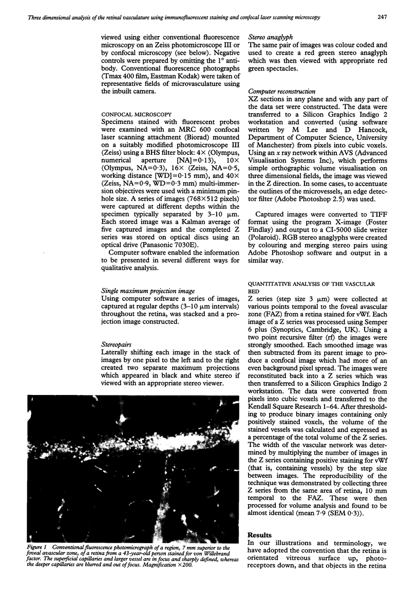
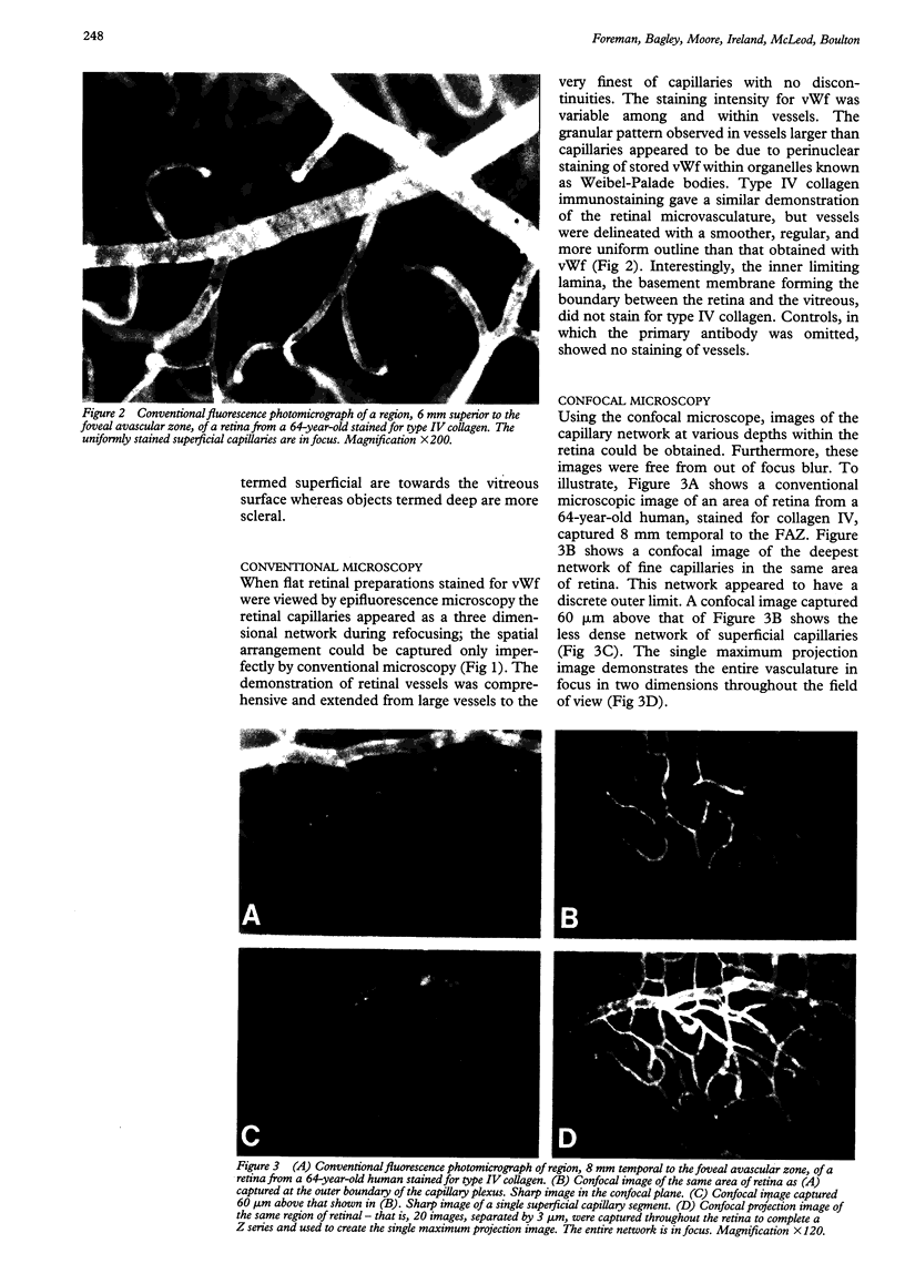
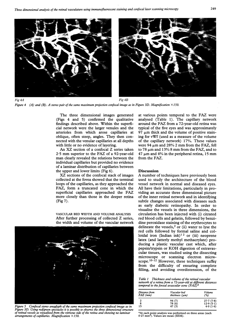
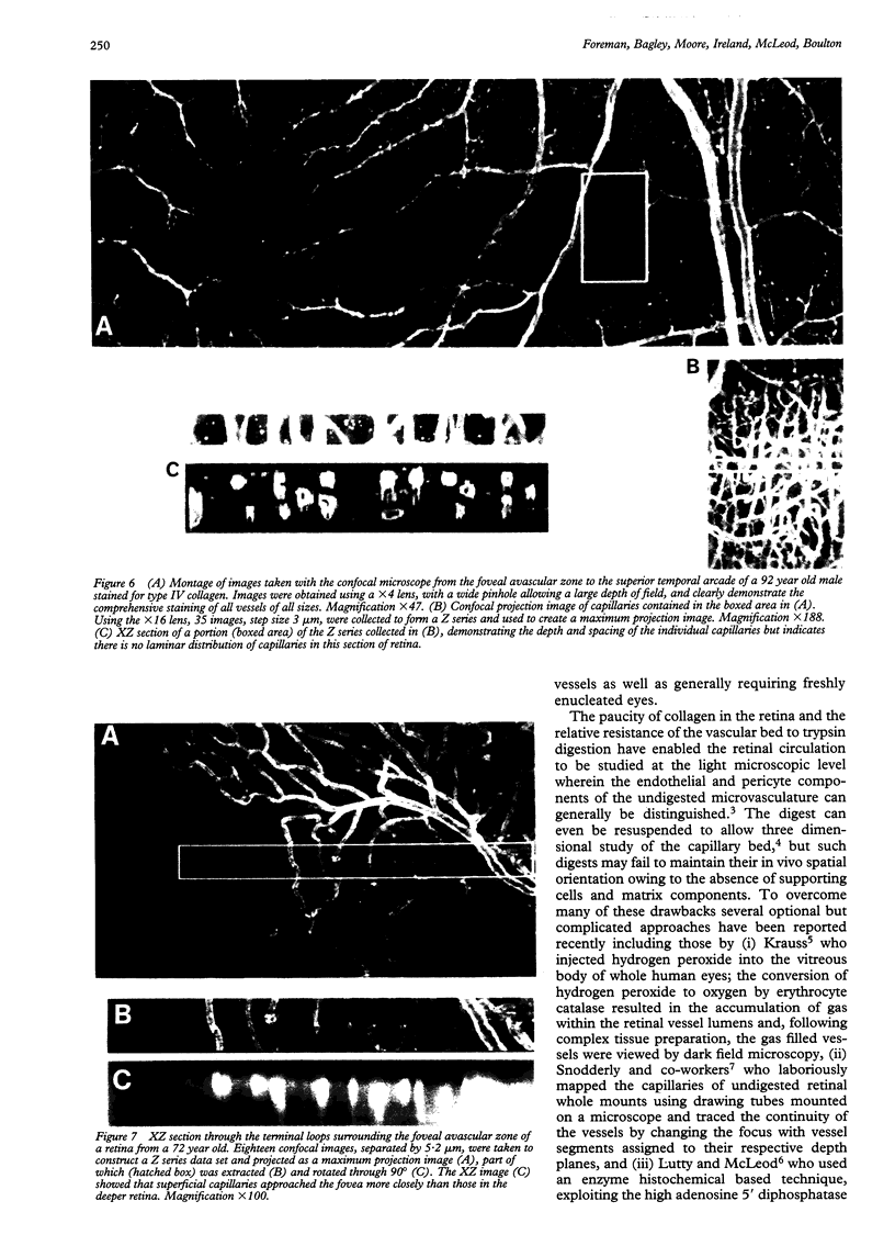
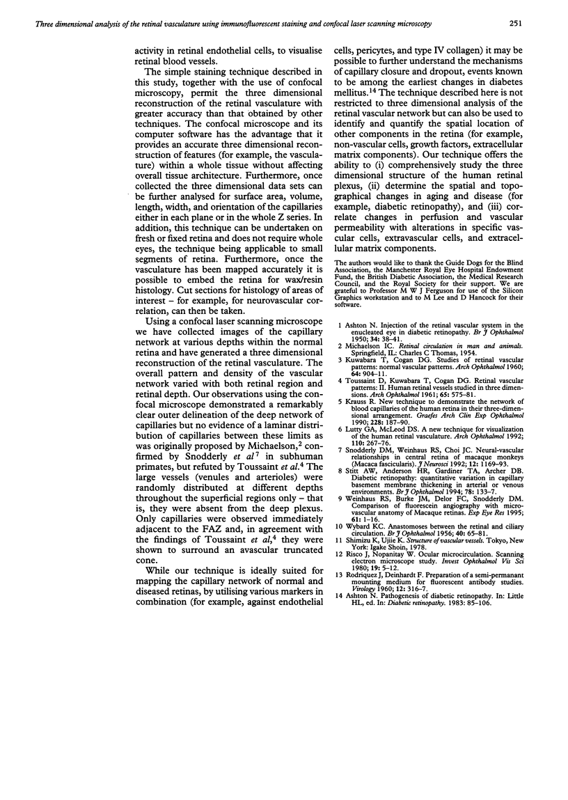
AIM: To undertake a qualitative and quantitative analysis in three dimensions of the human retinal vasculature. METHOD: Fixed and excised whole retinas were permeabilised and subjected to immunofluorescent staining for blood vessel components followed by confocal laser scanning microscopy. Single projection and stereoimages were constructed using computer software. XZ sections through the retina were constructed and the vasculature analysed using appropriate software. RESULTS: Immunofluorescent staining with no discontinuities was present in vessels of all sizes, the confocal images of the capillary network being free of out of focus blur at all depths. Quantitative analysis of XZ sections confirmed the qualitative impression of sharp delineation of the deep retinal capillary plexus, an absence of laminar arrangement of capillaries within the inner retina, and a truncated cone of capillaries around the foveal avascular zone (FAZ) wherein the superficial capillaries approached the FAZ more closely than those in the deeper retina. CONCLUSION: Immunofluorescent staining of the retina and confocal laser scanning microscopy were shown to be useful in analysing accurate three dimensional reconstructions of the normal retinal vasculature without affecting overall tissue architecture.
Full text
PDF
Images in this article
Selected References
These references are in PubMed. This may not be the complete list of references from this article.
- ASHTON N. Injection of the retinal vascular system in the enucleated eye in diabetic retinopathy. Br J Ophthalmol. 1950 Jan;34(1):38-41, illust. doi: 10.1136/bjo.34.1.38. [DOI] [PMC free article] [PubMed] [Google Scholar]
- KUWABARA T., COGAN D. G. Studies of retinal vascular patterns. I. Normal architecture. Arch Ophthalmol. 1960 Dec;64:904–911. doi: 10.1001/archopht.1960.01840010906012. [DOI] [PubMed] [Google Scholar]
- Krauss R. New technique to demonstrate the network of blood capillaries of the human retina in their three-dimensional arrangement. Graefes Arch Clin Exp Ophthalmol. 1990;228(2):187–190. doi: 10.1007/BF00935731. [DOI] [PubMed] [Google Scholar]
- Lutty G. A., McLeod D. S. A new technique for visualization of the human retinal vasculature. Arch Ophthalmol. 1992 Feb;110(2):267–276. doi: 10.1001/archopht.1992.01080140123039. [DOI] [PubMed] [Google Scholar]
- RODRIGUEZ J., DEINHARDT F. Preparation of a semipermanent mounting medium for fluorescent antibody studies. Virology. 1960 Oct;12:316–317. doi: 10.1016/0042-6822(60)90205-1. [DOI] [PubMed] [Google Scholar]
- Risco J. M., Nopanitaya W. Ocular microcirculation. Scanning electron microscopic study. Invest Ophthalmol Vis Sci. 1980 Jan;19(1):5–12. [PubMed] [Google Scholar]
- Snodderly D. M., Weinhaus R. S., Choi J. C. Neural-vascular relationships in central retina of macaque monkeys (Macaca fascicularis). J Neurosci. 1992 Apr;12(4):1169–1193. doi: 10.1523/JNEUROSCI.12-04-01169.1992. [DOI] [PMC free article] [PubMed] [Google Scholar]
- Stitt A. W., Anderson H. R., Gardiner T. A., Archer D. B. Diabetic retinopathy: quantitative variation in capillary basement membrane thickening in arterial or venous environments. Br J Ophthalmol. 1994 Feb;78(2):133–137. doi: 10.1136/bjo.78.2.133. [DOI] [PMC free article] [PubMed] [Google Scholar]
- TOUSSAINT D., KUWABARA T., COGAN D. G. Retinal vascular patterns. II. Human retinal vessels studied in three dimensions. Arch Ophthalmol. 1961 Apr;65:575–581. doi: 10.1001/archopht.1961.01840020577022. [DOI] [PubMed] [Google Scholar]
- WYBAR K. C. Anastomoses between the retinal and ciliary arterial circulations. Br J Ophthalmol. 1956 Feb;40(2):65–81. doi: 10.1136/bjo.40.2.65. [DOI] [PMC free article] [PubMed] [Google Scholar]
- Weinhaus R. S., Burke J. M., Delori F. C., Snodderly D. M. Comparison of fluorescein angiography with microvascular anatomy of macaque retinas. Exp Eye Res. 1995 Jul;61(1):1–16. doi: 10.1016/s0014-4835(95)80053-0. [DOI] [PubMed] [Google Scholar]


