Key Clinical Message
Mahjong, a game similar to bridge and chess in Western cultures, can cause reflex seizure. We report a case of Mahjong‐induced seizures with the first documentation of ictal electroencephalography (EEG) findings, which showed secondarily generalized partial seizure of the right parietal origin.
Keywords: Mahjong, reflex epilepsy, seizures, system epilepsy
Introduction
Mahjong, a game similar to bridge and chess in Western cultures, is popular in Asian countries. Traditional Mahjong tiles consist of 144 or 136 small rectangular blocks with faces showing various characters and designs. This game is usually played with four players. Players continue exchanging their tiles with other players while sorting and arranging their own tiles into desired spatial sequences. Thinking, memory, intuition, and decision‐making are continuously involved in this complicated game. Due to the high cognitive demands of Mahjong, it can induce a subtype of reflex seizures or cognition‐induced epilepsy; however, it is rarely reported 1, 2, 3. Therefore, no studies have shown ictal electroencephalography (EEG) findings of reflex seizures induced by Mahjong. Here, we report a case that clearly demonstrated the focal origin in the right parietal area.
Clinical Case
The patient was a right‐handed male and had been a regular Mahjong player since the age of 38 years. At the age of 62 years, he experienced his first attack while playing Mahjong, with loss of consciousness and incontinence. At the age of 64 years, he experienced his second tonic–clonic seizure lasted for 2 min while playing the computer version of Mahjong. Both attacks occurred without alcohol consumption or sleep deprivation and lasted for approximately 20 min, including postictal confusion. He tried to avoid playing Mahjong and, thus, had no attacks until he experienced habitual attacks while playing Mahjong at the age of 78 years. The patient frequently played Mahjong since the age of 81 years and experienced attacks whenever he played the game. The attacks usually occurred more than an hour after starting the game. In June 2014, at the age of 82 years, he was admitted to our hospital to examine the cause of attacks.
He had a history of right idiopathic facial paralysis, hypertension, dyslipidemia, diabetes, asymptomatic cerebral infarction, and angina. He had no family history of epilepsy. A physical examination did not reveal any neurological deficits except for a sequel of right facial nerve palsy. Magnetic resonance imaging (MRI) of the brain demonstrated punctate lesions as asymptomatic cerebral infarctions in the deep white matter of the right frontal and parietal lobe on fluid‐attenuated inversion recovery (FLAIR) scans, and magnetic resonance angiography revealed moderate stenosis in the right middle and posterior cerebral arteries and the right vertebral artery.
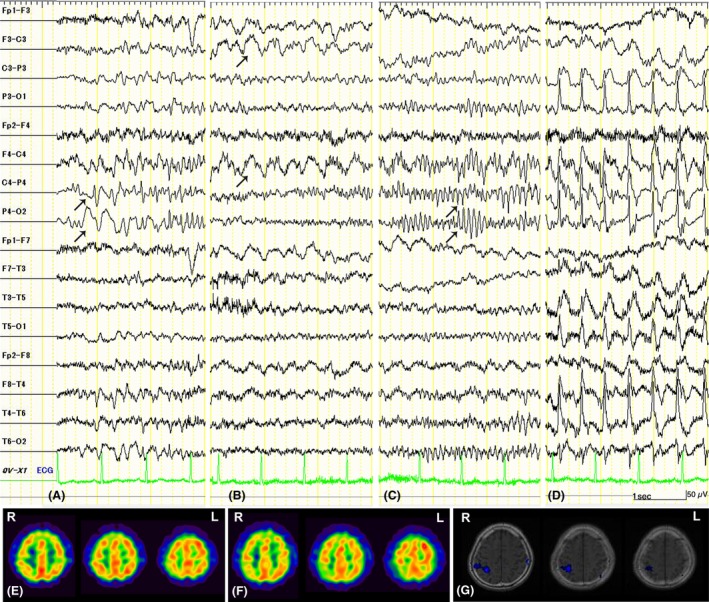
Single photon emission computed tomography (SPECT) was performed using 99 mTc‐ethyl cysteinate dimer (ECD) at a dose of 600 MBq in the interictal phase and postictal phase (Fig. 1E and F). Postictal SPECT was performed an hour after a bilateral tonic–clonic seizure that lasted for 30 sec followed by a very brief, postictal confusion induced by a computer version of Mahjong. The subtraction image of postictal SPECT from interictal SPECT coregistered with MRI 4 showed a significant difference in the right parietal area with the differential value greater than two standard deviations above the mean values of all voxels, suggesting the presence of postictal hypoperfusion in the right parietal area (Fig. 1G). Interictal EEG showed intermittent irregular, 4‐Hz slow waves over the right frontoparietal region. Ictal EEG was recorded with video‐EEG monitoring while playing the computer version of Mahjong (Fig. 1A–D). An informed consent was obtained from the current patient prior to the challenge test after providing him with the option to not play Mahjong during his EEG recording. Interictally, low‐voltage slow waves of 3–4 Hz occurred every 3–4 sec over the right parietal region initially while playing the game. Two hours after starting the game, he had a clinical seizure with loss of consciousness and head version to the left. EEG demonstrated irregular slow wave activity that began in the right parietal region (Fig. 1A) and spread to the bilateral frontal region (Fig. 1B). Subsequently, ictal, repetitive spike activities were localized in the right parietal region (Fig. 1C). Finally, bursts of spikes and slow waves spread from the right parietal region dominantly to the ipsilateral side with secondary generalized period (Fig. 1D).
Figure 1.

Ictal EEG performed with video‐EEG monitoring while playing the computer version of Mahjong. Slow wave activity (arrows) began in the right parietal region (A) and spread to the bilateral frontal region (B) at the clinical seizure onset. Subsequently, spike waves (arrows) were localized in the right parietal region during the ictal phase (C). Finally, bursts of spikes and slow waves spread from the right parietal region dominantly to the ipsilateral side with the secondarily generalized period (D). EEG was recorded with a bipolar montage with the high‐frequency filter set at 60 Hz. Vertical marker, 50 μv; horizontal marker, 1 sec. SPECT studies using 99 mTc‐ECD in the interictal phase (E) and postictal phase (F). Interictal SPECT showed no abnormal findings. Postictal SPECT showed hypoperfusion in the right parietal area by the subtraction image of postictal from interictal SPECT coregistered with MRI (G). R, right; L, left; EEG, electroencephalography; MRI, Magnetic resonance imaging; SPECT, Single photon emission computed tomography.
Eventually, the patient was diagnosed with Mahjong‐induced partial seizure (Mahjong epilepsy) with secondary generalized state of the right parietal lobe origin. He followed our advice to quit Mahjong and has since been in a seizure‐free state.
Discussion
Reflex seizures have traditionally been classified into “generalized” and “focal or partial.” Focal reflex seizures occur in patients with symptomatic or cryptogenic epilepsy following startle, eating, music, hot water, orgasm, or somatosensory stimuli. Generalized reflex seizures occur in patients with idiopathic generalized epilepsy following visual stimuli, nonverbal cognitive task (thinking and praxis), or verbal cognitive task (reading, talking, and writing).
Reflex seizures by nonverbal cognitive tasks have been considered to be focal seizures with quick secondary generalization 5. Recently, a new concept termed “system epilepsy” has been proposed with the hypothesis that some types of epilepsy reflect pathological expression (ictogenesis) of a specific brain system, which normally integrates activity that subserves normal physiological functions 6. Functional imaging studies indicate that praxis‐induced epilepsy is related to an underlying ictogenic mechanism that can cause hyperexcitability of the functional anatomical central nervous system network physiologically subserving visuomotor coordination 7. Other study suggests that reading epilepsy can arise from a hyperexcitable network of cortical regions where their cumulative effects result in greater reciprocal network propagation and electroclinical seizures 8. Furthermore, a previous study also showed that long‐term cognitive work induces mental fatigue, which may cause changes to the information flux at different brain cortical areas on EEG analysis 9.
In seizures induced by thinking during calculation or playing chess or similar games, focal abnormalities on EEG usually involve the right hemisphere over the frontal or parietal regions 5. Previously, 23 cases of “Mahjong‐induced epilepsy” were reported as a subtype of reflex seizures or cognition‐induced epilepsy 1, 2, 3. Clinically, Mahjong‐induced epilepsy manifests more frequently with generalized tonic–clonic seizures (78%) than partial‐onset seizure (22%) 1, although ictal EEG findings have not been reported so as to determine whether the attack is caused by primary generalized seizures or partial seizures with secondary generalization. In the present case, ictal epileptic discharges on the right parietal region spreading dominantly to the ipsilateral side corresponded to the partial seizures on the left side of the body. We assume that the seizures of the present patient result from hyperexcitability and cumulative effects of the network involved in spatial thought and praxis task in the right parietal lobe, but also from electrophysiological changes related to mental fatigue during continuous Mahjong playing, suggesting that Mahjong‐induced epilepsy is a form of system epilepsy. The current case showed an old infarction in the white matter but not in the cerebral cortex, which was less likely to lower the seizure threshold.
Focal seizure activity is known to be associated with increased cerebral blood flow (CBF) in the involved region of the cerebral cortex. The advantage of SPECT is that the tracer can be injected during the seizure (ictal SPECT) to identify the focus of seizures. To improve sensitivity and specificity, ictal SPECT images are typically compared with baseline SPECT images in the same patient without seizures (interictal SPECT). Another important factor to modify the sensitivity and specificity of ictal SPECT is the timing of tracer injection. Whether CBF change is identified in the cases of partial seizure depends on the generalization and the timing of tracer injection, namely, either (i) pregeneralization period (during the partial seizure phase prior to generalization), (ii) during the generalization period, or (iii) postictal period 10, 11. In the partial seizure without generalization, CBF often decreases postictally both within and around the seizure focus 10. In contrast, in the secondary generalized partial seizure, CBF tends to increase in multiple lobes ipsilateral to the seizure focus with relatively decreased CBF in the contralateral hemisphere 11. In the present case, postictal SPECT showed localized hypoperfusion in the parietal lobe of the right hemisphere, that is, the side of seizure focus. This finding contradicts the previous findings of partial seizures with generalization but is rather similar to those observed in partial seizures without generalization. Such a difference might be related to fairly short‐term seizure duration in the present case, but additional case series are needed to examine the correlation between postictal SPECT findings and ictal semiology.
In conclusion, we report a case of Mahjong‐induced seizures with the first documentation of ictal EEG findings, which showed secondarily generalized partial seizure of the right parietal origin.
Conflict of Interest
The authors have no conflicts of interest to declare.
References
- 1. Chang, R. S. , Cheung R. T., Ho S. L., and Mak W.. 2007. Mah‐jong‐induced seizures: case reports and review of twenty‐three patients. Hong Kong Med. J. 13:314–318. [PubMed] [Google Scholar]
- 2. Kwan, S. Y. , and Su M. S.. 2000. Mah‐jong epilepsy: a new reflex epilepsy. Zhonghua. Yi. Xue. Za. Zhi. 63:316–321. [PubMed] [Google Scholar]
- 3. Wan, C. L. , Lin T. K., Lu C. H., Chang C. S., Chen S. D., and Chuang Y. C.. 2005. Mah‐Jong‐induced epilepsy: a special reflex epilepsy in Chinese society. Seizure 14:19–22. [DOI] [PubMed] [Google Scholar]
- 4. O'Brien, T. J. , O'Connor M. K., Mullan B. P., Brinkmann B. H., Hanson D., Jack C. R., et al. 1998. Subtraction ictal SPET co‐registered to MRI in partial epilepsy: description and technical validation of the method with phantom and patient studies. Nucl. Med. Commun. 19:31–45. [DOI] [PubMed] [Google Scholar]
- 5. Italiano, D. , Ferlazzo E., Gasparini S., Spina E., Mondello S., Labate A., et al. 2014. Generalized versus partial reflex seizures: a review. Seizure 23:512–520. [DOI] [PubMed] [Google Scholar]
- 6. Avanzini, G. , Manganotti P., Meletti S., Moshe S. L., Panzica F., Wolf P., et al. 2012. The system epilepsies: a pathophysiological hypothesis. Epilepsia 53:771–778. [DOI] [PubMed] [Google Scholar]
- 7. Yacubian, E. M. , and Wolf P.. 2014. Praxis induction. Definition, relation to epilepsy syndromes, nosological and prognostic significance. A focused review. Seizure 23:247–251. [DOI] [PubMed] [Google Scholar]
- 8. Fumuro, T. , Matsumoto R., Shimotake A., Matsuhashi M., Inouchi M., Urayama S., et al. 2015. Network hyperexcitability in a patient with partial reading epilepsy: converging evidence from magnetoencephalography, diffusion tractography, and functional magnetic resonance imaging. Clin. Neurophysiol. 126:675–681. [DOI] [PubMed] [Google Scholar]
- 9. Liu, J. P. , Zhang C., and Zheng C. X.. 2010. Estimation of the cortical functional connectivity by directed transfer function during mental fatigue. Appl. Ergon. 42:114–121. [DOI] [PubMed] [Google Scholar]
- 10. Varghese, G. I. , Purcaro M. J., Motelow J. E., Enev M., McNally K. A., Levin A. R., et al. 2009. Clinical use of ictal SPECT in secondarily generalized tonic‐clonic seizures. Brain 132(Pt 8):2102–2113. [DOI] [PMC free article] [PubMed] [Google Scholar]
- 11. Blumenfeld, H. , Varghese G. I., Purcaro M. J., Motelow J. E., Enev M., McNally K. A., et al. 2009. Cortical and subcortical networks in human secondarily generalized tonic‐clonic seizures. Brain 132(Pt 4):999–1012. [DOI] [PMC free article] [PubMed] [Google Scholar]


