

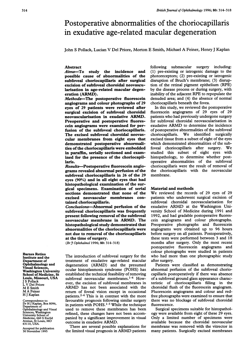
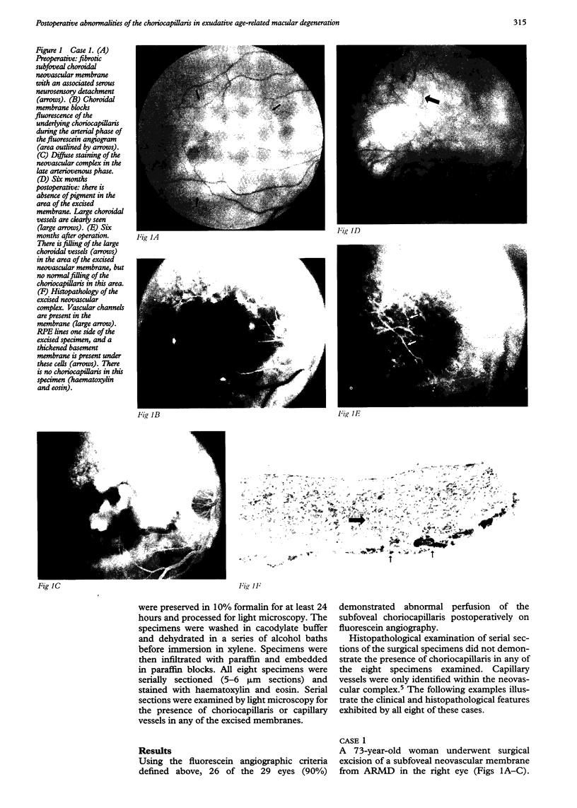
Abstract
AIMS: To study the incidence and possible cause of abnormalities of the subfoveal choriocapillaris after surgical excision of subfoveal choroidal neovascularisation in age-related macular degeneration (ARMD). METHODS: The postoperative fluorescein angiograms and colour photographs of 29 eyes of 29 patients were reviewed after surgical excision of subfoveal choroidal neovascularisation in exudative ARMD. Preoperative and postoperative fluorescein angiograms were examined for perfusion of the subfoveal choriocapillaris. The excised subfoveal choroidal neovascular membranes from eight eyes that demonstrated postoperative abnormalities of the choriocapillaris were embedded in paraffin, serially sectioned and examined for the presence of the choriocapillaris. RESULTS: Postoperative fluorescein angiograms revealed abnormal perfusion of the subfoveal choriocapillaris in 26 of the 29 eyes (90%) and in all eight eyes that had histopathological examination of the surgical specimens. Examination of serial sections demonstrated that none of the excised neovascular membranes contained choriocapillaris. CONCLUSIONS: Abnormal perfusion of the subfoveal choriocapillaris was frequently present following removal of the subfoveal neovascular membrane in ARMD. The histopathological study demonstrated that abnormalities of the choriocapillaris were not due to removal of the choriocapillaris at the time of surgery.
Full text
PDF
Images in this article
Selected References
These references are in PubMed. This may not be the complete list of references from this article.
- Adelberg D. A., Del Priore L. V., Kaplan H. J. Surgery for subfoveal membranes in myopia, angioid streaks, and other disorders. Retina. 1995;15(3):198–205. doi: 10.1097/00006982-199515030-00003. [DOI] [PubMed] [Google Scholar]
- Algvere P. V., Berglin L., Gouras P., Sheng Y. Transplantation of fetal retinal pigment epithelium in age-related macular degeneration with subfoveal neovascularization. Graefes Arch Clin Exp Ophthalmol. 1994 Dec;232(12):707–716. doi: 10.1007/BF00184273. [DOI] [PubMed] [Google Scholar]
- Berger A. S., Kaplan H. J. Clinical experience with the surgical removal of subfoveal neovascular membranes. Short-term postoperative results. Ophthalmology. 1992 Jun;99(6):969–976. doi: 10.1016/s0161-6420(92)31869-x. [DOI] [PubMed] [Google Scholar]
- Bird A. C. Bruch's membrane change with age. Br J Ophthalmol. 1992 Mar;76(3):166–168. doi: 10.1136/bjo.76.3.166. [DOI] [PMC free article] [PubMed] [Google Scholar]
- Del Priore L. V., Hornbeck R., Kaplan H. J., Jones Z., Valentino T. L., Mosinger-Ogilvie J., Swinn M. Débridement of the pig retinal pigment epithelium in vivo. Arch Ophthalmol. 1995 Jul;113(7):939–944. doi: 10.1001/archopht.1995.01100070113034. [DOI] [PubMed] [Google Scholar]
- FRIEDMAN E., SMITH T. R., KUWABARA T. Senile choroidal vascular patterns and drusen. Arch Ophthalmol. 1963 Feb;69:220–230. doi: 10.1001/archopht.1963.00960040226014. [DOI] [PubMed] [Google Scholar]
- Green W. R., Key S. N., 3rd Senile macular degeneration: a histopathologic study. Trans Am Ophthalmol Soc. 1977;75:180–254. [PMC free article] [PubMed] [Google Scholar]
- Henkind P., Gartner S. The relationship between retinal pigment epithelium and the choriocapillaris. Trans Ophthalmol Soc U K. 1983;103(Pt 4):444–447. [PubMed] [Google Scholar]
- Korte G. E., Reppucci V., Henkind P. RPE destruction causes choriocapillary atrophy. Invest Ophthalmol Vis Sci. 1984 Oct;25(10):1135–1145. [PubMed] [Google Scholar]
- Lambert H. M., Capone A., Jr, Aaberg T. M., Sternberg P., Jr, Mandell B. A., Lopez P. F. Surgical excision of subfoveal neovascular membranes in age-related macular degeneration. Am J Ophthalmol. 1992 Mar 15;113(3):257–262. doi: 10.1016/s0002-9394(14)71576-4. [DOI] [PubMed] [Google Scholar]
- Miller F. S., 3rd, Bunt-Milam A. H., Kalina R. E. Clinical-ultrastructural study of thioridazine retinopathy. Ophthalmology. 1982 Dec;89(12):1478–1488. doi: 10.1016/s0161-6420(82)34613-8. [DOI] [PubMed] [Google Scholar]
- Peyman G. A., Blinder K. J., Paris C. L., Alturki W., Nelson N. C., Jr, Desai U. A technique for retinal pigment epithelium transplantation for age-related macular degeneration secondary to extensive subfoveal scarring. Ophthalmic Surg. 1991 Feb;22(2):102–108. [PubMed] [Google Scholar]
- Reddy V. M., Zamora R. L., Kaplan H. J. Distribution of growth factors in subfoveal neovascular membranes in age-related macular degeneration and presumed ocular histoplasmosis syndrome. Am J Ophthalmol. 1995 Sep;120(3):291–301. doi: 10.1016/s0002-9394(14)72158-0. [DOI] [PubMed] [Google Scholar]
- Sarks S. H., Van Driel D., Maxwell L., Killingsworth M. Softening of drusen and subretinal neovascularization. Trans Ophthalmol Soc U K. 1980 Sep;100(3):414–422. [PubMed] [Google Scholar]
- Thomas M. A., Grand M. G., Williams D. F., Lee C. M., Pesin S. R., Lowe M. A. Surgical management of subfoveal choroidal neovascularization. Ophthalmology. 1992 Jun;99(6):952–976. doi: 10.1016/s0161-6420(92)31888-3. [DOI] [PubMed] [Google Scholar]
- Valentino T. L., Kaplan H. J., Del Priore L. V., Fang S. R., Berger A., Silverman M. S. Retinal pigment epithelial repopulation in monkeys after submacular surgery. Arch Ophthalmol. 1995 Jul;113(7):932–938. doi: 10.1001/archopht.1995.01100070106033. [DOI] [PubMed] [Google Scholar]
- Yang Q. R., Smets R. M., Neetens A., Vanden Berghe D. Human retinal pigment epithelial cells from different donors continuously produce a vascular endothelial cell-stimulating factor into serum-free medium. J Cell Sci. 1993 Jan;104(Pt 1):211–218. doi: 10.1242/jcs.104.1.211. [DOI] [PubMed] [Google Scholar]