

Abstract
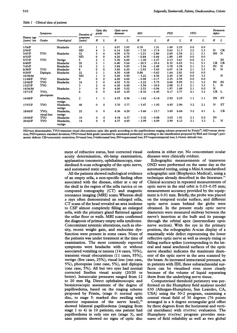
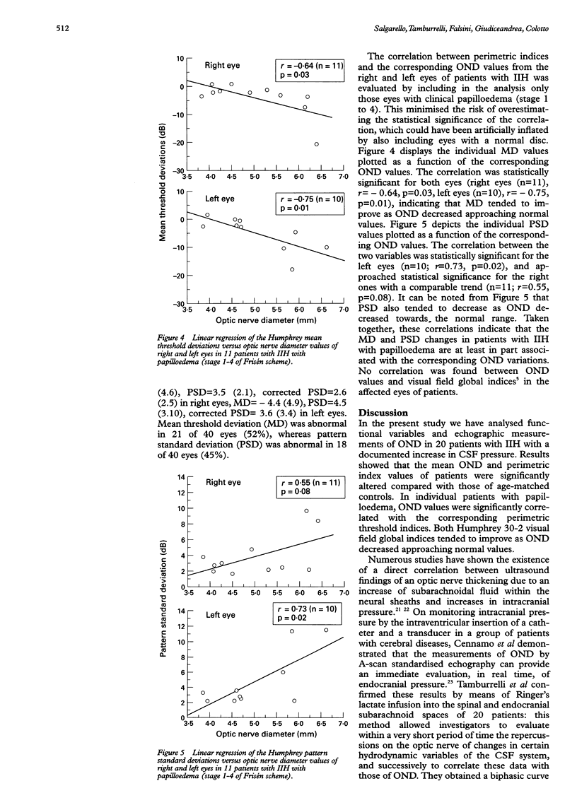
AIMS/BACKGROUND--Idiopathic intracranial hypertension (IIH) is a central nervous disorder characterised by abnormally increased cerebrospinal fluid (CSF) pressure leading to optic nerve compression. An indirect estimate of increased CSF pressure can be obtained by the ultrasonographic determination of optic nerve sheaths diameters. Computerised static perimetry is regarded as the method of choice for monitoring the course of the optic neuropathy in IIH. The aims were to compare the echographic optic nerve diameters (ONDs) and the perimetric thresholds of patients with IIH with those of age-matched controls, and to examine the correlation between these two variables in individual patients with papilloedema. METHODS--Standardised A-scan echography of the mid orbital optic nerve transverse diameters and automated threshold perimetry (Humphrey 30-2) were performed in 20 patients with IIH with variable degree of papilloedema (according to the Frisén scheme) and no concomitant ocular diseases. Echographic and perimetric results were compared with those obtained from 20 age-matched controls. RESULTS--When compared with controls, patients with IIH showed a significant increase in mean ONDs and significantly reduced mean perimetric sensitivities. In individual patients with papilloedema, the transverse ONDs correlated negatively with Humphrey mean deviation values and positively with pattern standard deviation values. CONCLUSION--These results indicate that OND changes in IIH are associated with perimetric threshold losses, and suggest that IIH functional deficits may be related to the degree of distension of optic nerve sheaths as a result of an increased CSF pressure.
Full text
PDF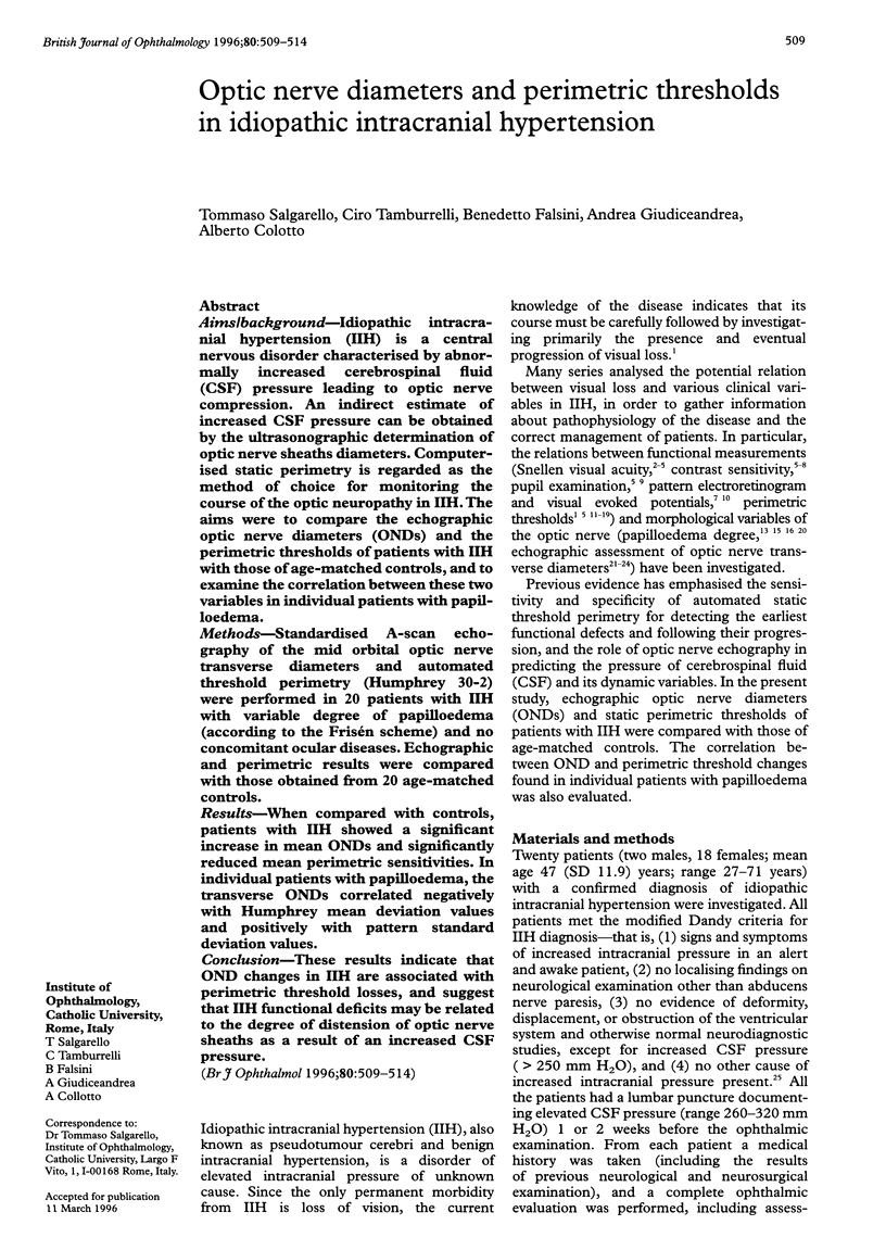
Selected References
These references are in PubMed. This may not be the complete list of references from this article.
- Bulens C., De Vries W. A., Van Crevel H. Benign intracranial hypertension. A retrospective and follow-up study. J Neurol Sci. 1979 Feb;40(2-3):147–157. doi: 10.1016/0022-510x(79)90200-4. [DOI] [PubMed] [Google Scholar]
- Bulens C., Meerwaldt J. D., Koudstaal P. J., Van der Wildt G. J. Spatial contrast sensitivity in benign intracranial hypertension. J Neurol Neurosurg Psychiatry. 1988 Oct;51(10):1323–1329. doi: 10.1136/jnnp.51.10.1323. [DOI] [PMC free article] [PubMed] [Google Scholar]
- Corbett J. J., Nerad J. A., Tse D. T., Anderson R. L. Results of optic nerve sheath fenestration for pseudotumor cerebri. The lateral orbitotomy approach. Arch Ophthalmol. 1988 Oct;106(10):1391–1397. doi: 10.1001/archopht.1988.01060140555022. [DOI] [PubMed] [Google Scholar]
- Corbett J. J., Savino P. J., Thompson H. S., Kansu T., Schatz N. J., Orr L. S., Hopson D. Visual loss in pseudotumor cerebri. Follow-up of 57 patients from five to 41 years and a profile of 14 patients with permanent severe visual loss. Arch Neurol. 1982 Aug;39(8):461–474. doi: 10.1001/archneur.1982.00510200003001. [DOI] [PubMed] [Google Scholar]
- Falsini B., Tamburrelli C., Porciatti V., Anile C., Porrello G., Mangiola N. Pattern electroretinograms and visual evoked potentials in idiopathic intracranial hypertension. Ophthalmologica. 1992;205(4):194–203. doi: 10.1159/000310341. [DOI] [PubMed] [Google Scholar]
- Foley K. M., Posner J. B. Does pseudotumor cerebri cause the empty sella syndrome? Neurology. 1975 Jun;25(6):565–569. doi: 10.1212/wnl.25.6.565. [DOI] [PubMed] [Google Scholar]
- Frisén L. Swelling of the optic nerve head: a staging scheme. J Neurol Neurosurg Psychiatry. 1982 Jan;45(1):13–18. doi: 10.1136/jnnp.45.1.13. [DOI] [PMC free article] [PubMed] [Google Scholar]
- Guidetti B., Giuffrè R., Gambacorta D. Follow-up study of 100 cases of pseudotumor cerebri. Acta Neurochir (Wien) 1968;18(4):259–267. doi: 10.1007/BF01405720. [DOI] [PubMed] [Google Scholar]
- Hayreh S. S. Optic disc edema in raised intracranial pressure. V. Pathogenesis. Arch Ophthalmol. 1977 Sep;95(9):1553–1565. doi: 10.1001/archopht.1977.04450090075006. [DOI] [PubMed] [Google Scholar]
- Holmes G. THE PROGNOSIS IN PAPILLOEDEMA. Br J Ophthalmol. 1937 Jul;21(7):337–342. doi: 10.1136/bjo.21.7.337. [DOI] [PMC free article] [PubMed] [Google Scholar]
- Kardon R. H., Haupert C. L., Thompson H. S. The relationship between static perimetry and the relative afferent pupillary defect. Am J Ophthalmol. 1993 Mar 15;115(3):351–356. doi: 10.1016/s0002-9394(14)73587-1. [DOI] [PubMed] [Google Scholar]
- Marcelis J., Silberstein S. D. Idiopathic intracranial hypertension without papilledema. Arch Neurol. 1991 Apr;48(4):392–399. doi: 10.1001/archneur.1991.00530160060014. [DOI] [PubMed] [Google Scholar]
- Orcutt J. C., Page N. G., Sanders M. D. Factors affecting visual loss in benign intracranial hypertension. Ophthalmology. 1984 Nov;91(11):1303–1312. doi: 10.1016/s0161-6420(84)34149-5. [DOI] [PubMed] [Google Scholar]
- Round R., Keane J. R. The minor symptoms of increased intracranial pressure: 101 patients with benign intracranial hypertension. Neurology. 1988 Sep;38(9):1461–1464. doi: 10.1212/wnl.38.9.1461. [DOI] [PubMed] [Google Scholar]
- Rush J. A. Pseudotumor cerebri: clinical profile and visual outcome in 63 patients. Mayo Clin Proc. 1980 Sep;55(9):541–546. [PubMed] [Google Scholar]
- Smith T. J., Baker R. S. Perimetric findings in pseudotumor cerebri using automated techniques. Ophthalmology. 1986 Jul;93(7):887–894. doi: 10.1016/s0161-6420(86)33645-5. [DOI] [PubMed] [Google Scholar]
- Sørensen P. S., Krogsaa B., Gjerris F. Clinical course and prognosis of pseudotumor cerebri. A prospective study of 24 patients. Acta Neurol Scand. 1988 Feb;77(2):164–172. doi: 10.1111/j.1600-0404.1988.tb05888.x. [DOI] [PubMed] [Google Scholar]
- Verplanck M., Kaufman D. I., Parsons T., Yedavally S., Kokinakis D. Electrophysiology versus psychophysics in the detection of visual loss in pseudotumor cerebri. Neurology. 1988 Nov;38(11):1789–1792. doi: 10.1212/wnl.38.11.1789. [DOI] [PubMed] [Google Scholar]
- Wall M. Contrast sensitivity testing in pseudotumor cerebri. Ophthalmology. 1986 Jan;93(1):4–7. doi: 10.1016/s0161-6420(86)33776-x. [DOI] [PubMed] [Google Scholar]
- Wall M., Conway M. D., House P. H., Allely R. Evaluation of sensitivity and specificity of spatial resolution and Humphrey automated perimetry in pseudotumor cerebri patients and normal subjects. Invest Ophthalmol Vis Sci. 1991 Dec;32(13):3306–3312. [PubMed] [Google Scholar]
- Wall M., George D. Idiopathic intracranial hypertension. A prospective study of 50 patients. Brain. 1991 Feb;114(Pt 1A):155–180. [PubMed] [Google Scholar]
- Wall M., George D. Visual loss in pseudotumor cerebri. Incidence and defects related to visual field strategy. Arch Neurol. 1987 Feb;44(2):170–175. doi: 10.1001/archneur.1987.00520140040015. [DOI] [PubMed] [Google Scholar]
- Wall M., Hart W. M., Jr, Burde R. M. Visual field defects in idiopathic intracranial hypertension (pseudotumor cerebri). Am J Ophthalmol. 1983 Nov;96(5):654–669. doi: 10.1016/s0002-9394(14)73425-7. [DOI] [PubMed] [Google Scholar]