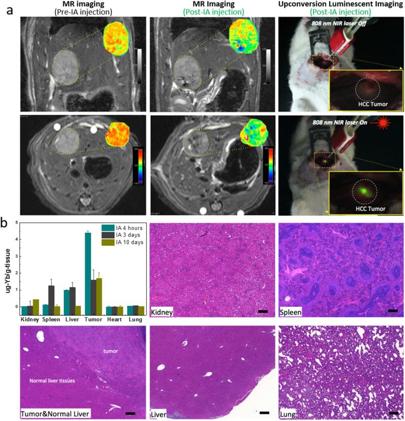
Figure 3.
(a) T2-weighted MR images acquired before and after catheter-directed hepatic IA anti-CD44-Nd-CSUCNP infusion and T2 maps (insets) of HCC tumor regions; (right) intraoperative upconversion luminescent imaging of anti-CD44-Nd-CSUCNP imaging reporters was also performed after IA transcatheter infusion. (b) Biodistribution of Nd-CSUCNPs following IA catheter-directed infusion (4 h, 3 days and 10 days follow-up intervals) and histological analysis of primary organs indicating no histologic changes in these rats 30 days after IA infusion of the anti-CD44-Nd-CSUCNP imaging reporters (scale bars=100 um). Retention of Nd-CSUCNPs in HCC tumor by IA infusion was still observed at 10 days post infusion.

