

Abstract
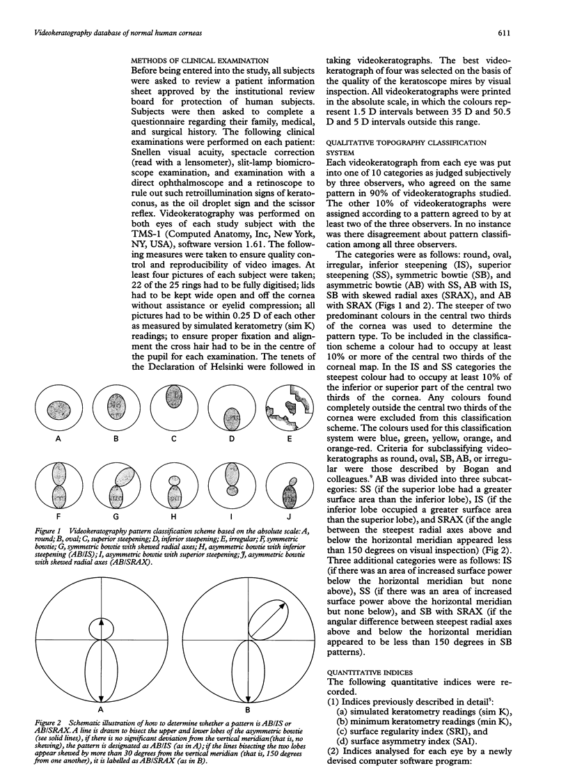
AIM: To form a database of videokeratography patterns and quantitative indices describing normal human corneas using the absolute scale. METHODS: Both eyes of 195 normal subjects were examined with a TMS-1 videokeratoscope. Videokeratographs were divided into 10 categories based on a classification scheme devised from the absolute scale and analysed with 10 quantitative indices devised to describe phenotypic features of keratoconus videokeratographs. Correlations were sought between videokeratograph patterns and quantitative indices. Additionally, data were analysed for differences in age, sex, and ethnicity. RESULTS: For symmetric videokeratography patterns, analysis in the absolute scale was similar to a previous study done in the normalised scale. In the asymmetric categories, analyses differed markedly. Using the absolute scale and our classification scheme more variation in normal videokeratography patterns could be appreciated. There was good correlation between quantitative indices and videokeratography patterns. Neither videokeratography patterns nor indices differed significantly between sex, ethnic groups, or age using two way analysis of variance. CONCLUSIONS: Pattern analysis of videokeratographs in the absolute scale using, a standard classification scheme, may be more useful in trying to determine whether a cornea is normal or represents subtle early disease than analysis in the normalised scale. Quantitative indices could remove the subjectivity from the decision making process thus facilitating universal reproducibility of videokeratography data interpretation.
Full text
PDF


Images in this article

Selected References
These references are in PubMed. This may not be the complete list of references from this article.
- Bogan S. J., Waring G. O., 3rd, Ibrahim O., Drews C., Curtis L. Classification of normal corneal topography based on computer-assisted videokeratography. Arch Ophthalmol. 1990 Jul;108(7):945–949. doi: 10.1001/archopht.1990.01070090047037. [DOI] [PubMed] [Google Scholar]
- Dingeldein S. A., Klyce S. D. Imaging of the cornea. Cornea. 1988;7(3):170–182. [PubMed] [Google Scholar]
- Dingeldein S. A., Klyce S. D. The topography of normal corneas. Arch Ophthalmol. 1989 Apr;107(4):512–518. doi: 10.1001/archopht.1989.01070010526024. [DOI] [PubMed] [Google Scholar]
- Edmund C. Location of the corneal apex and its influence on the stability of the central corneal curvature. A photokeratoscopy study. Am J Optom Physiol Opt. 1987 Nov;64(11):846–852. doi: 10.1097/00006324-198711000-00008. [DOI] [PubMed] [Google Scholar]
- Gormley D. J., Gersten M., Koplin R. S., Lubkin V. Corneal modeling. Cornea. 1988;7(1):30–35. [PubMed] [Google Scholar]
- Grandon S. C., Weber R. A. Radial keratotomy in patients with atypical inferior steepening. J Cataract Refract Surg. 1994 Jul;20(4):381–386. doi: 10.1016/s0886-3350(13)80171-x. [DOI] [PubMed] [Google Scholar]
- Hannush S. B., Crawford S. L., Waring G. O., 3rd, Gemmill M. C., Lynn M. J., Nizam A. Reproducibility of normal corneal power measurements with a keratometer, photokeratoscope, and video imaging system. Arch Ophthalmol. 1990 Apr;108(4):539–544. doi: 10.1001/archopht.1990.01070060087055. [DOI] [PubMed] [Google Scholar]
- Klein S. A., Mandell R. B. Shape and refractive powers in corneal topography. Invest Ophthalmol Vis Sci. 1995 Sep;36(10):2096–2109. [PubMed] [Google Scholar]
- Klyce S. D. Computer-assisted corneal topography. High-resolution graphic presentation and analysis of keratoscopy. Invest Ophthalmol Vis Sci. 1984 Dec;25(12):1426–1435. [PubMed] [Google Scholar]
- Mandell R. B., St Helen R. Position and curvature of the corneal apex. Am J Optom Arch Am Acad Optom. 1969 Jan;46(1):25–29. doi: 10.1097/00006324-196901000-00005. [DOI] [PubMed] [Google Scholar]
- Rabinowitz Y. S., Garbus J., McDonnell P. J. Computer-assisted corneal topography in family members of patients with keratoconus. Arch Ophthalmol. 1990 Mar;108(3):365–371. doi: 10.1001/archopht.1990.01070050063032. [DOI] [PubMed] [Google Scholar]
- Rabinowitz Y. S., McDonnell P. J. Computer-assisted corneal topography in keratoconus. Refract Corneal Surg. 1989 Nov-Dec;5(6):400–408. [PubMed] [Google Scholar]
- Roberts C. The accuracy of 'power' maps to display curvature data in corneal topography systems. Invest Ophthalmol Vis Sci. 1994 Aug;35(9):3525–3532. [PubMed] [Google Scholar]
- Tomlinson A., Schwartz C. The position of the corneal apex in the normal eye. Am J Optom Physiol Opt. 1979 Apr;56(4):236–240. doi: 10.1097/00006324-197904000-00004. [DOI] [PubMed] [Google Scholar]
- Wilson S. E., Klyce S. D. Quantitative descriptors of corneal topography. A clinical study. Arch Ophthalmol. 1991 Mar;109(3):349–353. doi: 10.1001/archopht.1991.01080030051037. [DOI] [PubMed] [Google Scholar]
- Wilson S. E., Verity S. M., Conger D. L. Accuracy and precision of the corneal analysis system and the topographic modeling system. Cornea. 1992 Jan;11(1):28–35. doi: 10.1097/00003226-199201000-00004. [DOI] [PubMed] [Google Scholar]