

Abstract
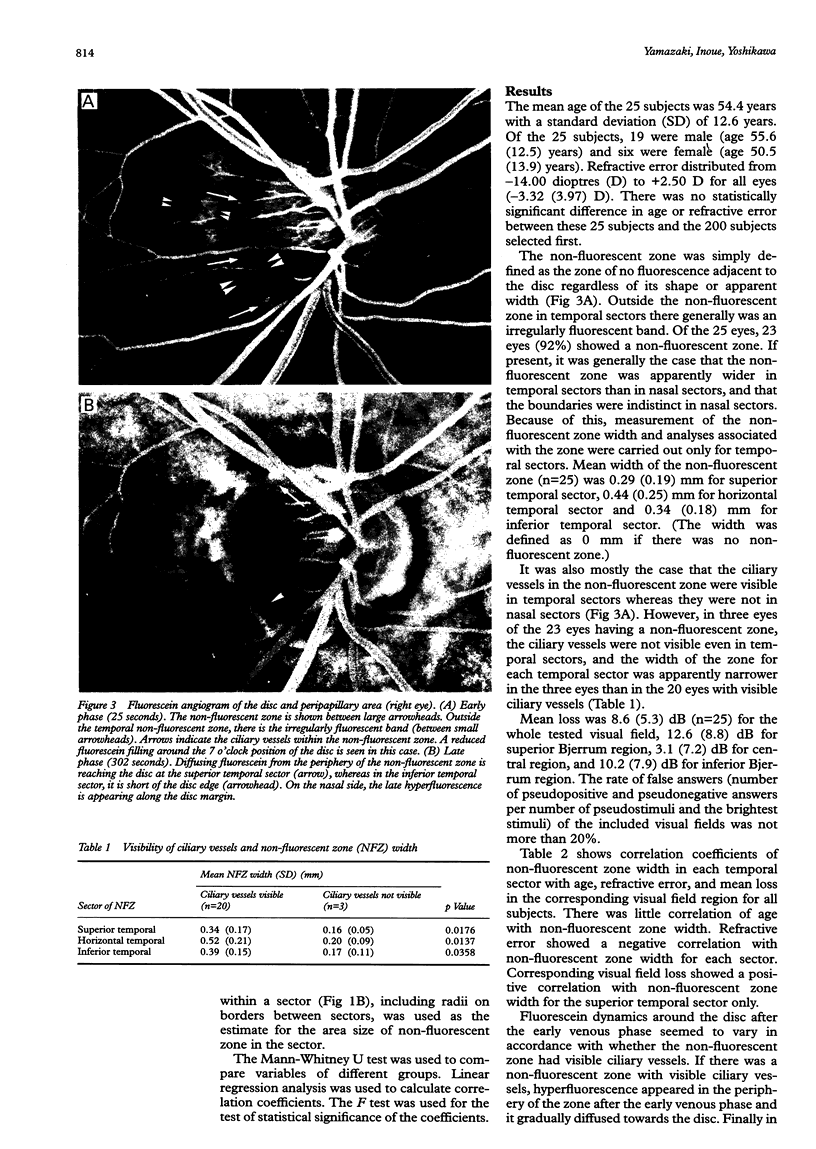
BACKGROUND: Detailed fluorescein angiographic findings in the disc circumference may be useful for evaluating the possible relation of the circumference to glaucomatous nerve damage. METHODS: Fluorescein angiograms of 25 eyes of 25 subjects with primary open angle glaucoma were observed after they had undertaken Octopus perimetry. Based on the retinotopic projection, disc sectors and corresponding visual field regions were set. RESULTS: Twenty three eyes (92%) showed a zone of no fluorescence around the disc (non-fluorescent zone). Of these, the zone width of the 20 eyes that had visible ciliary vessels within the zone was wider than that of the other three eyes, and showed fluorescein diffusion from the outer boundary of the zone towards the disc. The diffusion reached the disc if the zone was narrow. In those 20 eyes, a standardised difference in the zone width of inferior temporal sector minus superior temporal sector correlated with the difference in mean loss of corresponding visual field regions (r = 0.48, p = 0.0312). CONCLUSION: The visible ciliary vessels suggest the absence of the choriocapillaris in the non-fluorescent zone, the width of which correlated with the visual field defect and may affect the amount of the fluorescein diffusion to the disc. This suggests that the diffusion might be related to optic nerve damage in glaucoma.
Full text
PDF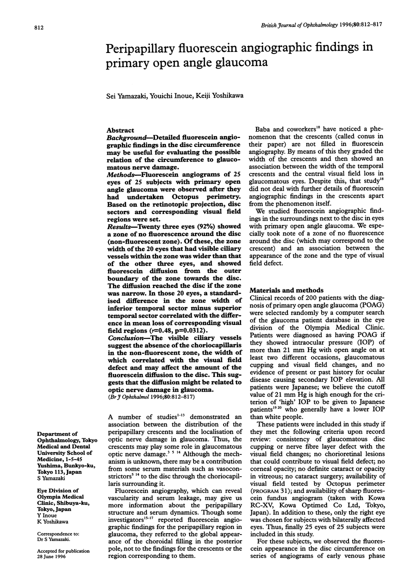


Images in this article
Selected References
These references are in PubMed. This may not be the complete list of references from this article.
- Ben-Sira I., Riva C. E. Fluorescein diffusion in the human optic disc. Invest Ophthalmol. 1975 Mar;14(3):205–211. [PubMed] [Google Scholar]
- Bengtsson B., Krakau C. E. Correction of optic disc measurements on fundus photographs. Graefes Arch Clin Exp Ophthalmol. 1992;230(1):24–28. doi: 10.1007/BF00166758. [DOI] [PubMed] [Google Scholar]
- Buus D. R., Anderson D. R. Peripapillary crescents and halos in normal-tension glaucoma and ocular hypertension. Ophthalmology. 1989 Jan;96(1):16–19. doi: 10.1016/s0161-6420(89)32930-7. [DOI] [PubMed] [Google Scholar]
- Cohen A. I. Is there a potential defect in the blood-retinal barrier at the choroidal level of the optic nerve canal? Invest Ophthalmol. 1973 Jul;12(7):513–519. [PubMed] [Google Scholar]
- Derick R. J., Pasquale L. R., Pease M. E., Quigley H. A. A clinical study of peripapillary crescents of the optic disc in chronic experimental glaucoma in monkey eyes. Arch Ophthalmol. 1994 Jun;112(6):846–850. doi: 10.1001/archopht.1994.01090180146049. [DOI] [PubMed] [Google Scholar]
- Fantes F. E., Anderson D. R. Clinical histologic correlation of human peripapillary anatomy. Ophthalmology. 1989 Jan;96(1):20–25. doi: 10.1016/s0161-6420(89)32929-0. [DOI] [PubMed] [Google Scholar]
- Flage T. Permeability properties of the tissues in the optic nerve head region in the rabbit and the monkey. An ultrastructural study. Acta Ophthalmol (Copenh) 1977 Aug;55(4):652–664. doi: 10.1111/j.1755-3768.1977.tb05663.x. [DOI] [PubMed] [Google Scholar]
- Fulk G. W., Goss D. A., Christensen M. T., Cline K. B., Herrin-Lawson G. A. Optic nerve crescents and refractive error. Optom Vis Sci. 1992 Mar;69(3):208–213. doi: 10.1097/00006324-199203000-00007. [DOI] [PubMed] [Google Scholar]
- Grayson M. C., Laties A. M. Ocular localization of sodium fluorescein. Effects of administration in rabbit and monkey. Arch Ophthalmol. 1971 May;85(5):600–passim. doi: 10.1001/archopht.1971.00990050602014. [DOI] [PubMed] [Google Scholar]
- Hitchings R. A., Spaeth G. L. Fluorescein angiography in chronic simple and low-tension glaucoma. Br J Ophthalmol. 1977 Feb;61(2):126–132. doi: 10.1136/bjo.61.2.126. [DOI] [PMC free article] [PubMed] [Google Scholar]
- Jonas J. B., Fernández M. C., Naumann G. O. Glaucomatous parapapillary atrophy. Occurrence and correlations. Arch Ophthalmol. 1992 Feb;110(2):214–222. doi: 10.1001/archopht.1992.01080140070030. [DOI] [PubMed] [Google Scholar]
- Jonas J. B., Gusek G. C., Fernández M. C. Correlation of the blind spot size to the area of the optic disk and parapapillary atrophy. Am J Ophthalmol. 1991 May 15;111(5):559–565. doi: 10.1016/s0002-9394(14)73698-0. [DOI] [PubMed] [Google Scholar]
- Jonas J. B., Naumann G. O. Parapapillary chorioretinal atrophy in normal and glaucoma eyes. II. Correlations. Invest Ophthalmol Vis Sci. 1989 May;30(5):919–926. [PubMed] [Google Scholar]
- Jonas J. B., Nguyen X. N., Gusek G. C., Naumann G. O. Parapapillary chorioretinal atrophy in normal and glaucoma eyes. I. Morphometric data. Invest Ophthalmol Vis Sci. 1989 May;30(5):908–918. [PubMed] [Google Scholar]
- Korte G. E., Reppucci V., Henkind P. RPE destruction causes choriocapillary atrophy. Invest Ophthalmol Vis Sci. 1984 Oct;25(10):1135–1145. [PubMed] [Google Scholar]
- Laatikainen L., Mäntylä P. Effects of a fall in the intraocular pressure level on the peripapillary fluorescein angiogram in chronic opern-angle glaucoma. Acta Ophthalmol (Copenh) 1974;52(5):625–633. doi: 10.1111/j.1755-3768.1974.tb01098.x. [DOI] [PubMed] [Google Scholar]
- Nevarez J., Rockwood E. J., Anderson D. R. The configuration of peripapillary tissue in unilateral glaucoma. Arch Ophthalmol. 1988 Jul;106(7):901–903. doi: 10.1001/archopht.1988.01060140047021. [DOI] [PubMed] [Google Scholar]
- Primrose J. Early signs of the glaucomatous disc. Br J Ophthalmol. 1971 Dec;55(12):820–825. doi: 10.1136/bjo.55.12.820. [DOI] [PMC free article] [PubMed] [Google Scholar]
- Puska P., Raitta C. Peripapillary atrophy in unilateral capsular glaucoma. Graefes Arch Clin Exp Ophthalmol. 1993 Nov;231(11):642–646. doi: 10.1007/BF00921958. [DOI] [PubMed] [Google Scholar]
- Rader J., Feuer W. J., Anderson D. R. Peripapillary vasoconstriction in the glaucomas and the anterior ischemic optic neuropathies. Am J Ophthalmol. 1994 Jan 15;117(1):72–80. doi: 10.1016/s0002-9394(14)73017-x. [DOI] [PubMed] [Google Scholar]
- Raitta C., Sarmela T. Fluorescein angiography of the optic disc and the peripapillary area in chronic glaucoma. Acta Ophthalmol (Copenh) 1970;48(2):303–308. doi: 10.1111/j.1755-3768.1970.tb08199.x. [DOI] [PubMed] [Google Scholar]
- Rockwood E. J., Anderson D. R. Acquired peripapillary changes and progression in glaucoma. Graefes Arch Clin Exp Ophthalmol. 1988;226(6):510–515. doi: 10.1007/BF02169197. [DOI] [PubMed] [Google Scholar]
- Schumer R. A., Podos S. M. The nerve of glaucoma! Arch Ophthalmol. 1994 Jan;112(1):37–44. doi: 10.1001/archopht.1994.01090130047015. [DOI] [PubMed] [Google Scholar]
- Shiose Y., Kitazawa Y., Tsukahara S., Akamatsu T., Mizokami K., Futa R., Katsushima H., Kosaki H. Epidemiology of glaucoma in Japan--a nationwide glaucoma survey. Jpn J Ophthalmol. 1991;35(2):133–155. [PubMed] [Google Scholar]
- Wilensky J. T., Kolker A. E. Peripapillary changes in glaucoma. Am J Ophthalmol. 1976 Mar;81(3):341–345. doi: 10.1016/0002-9394(76)90251-8. [DOI] [PubMed] [Google Scholar]
- Wirtschafter J. D., Becker W. L., Howe J. B., Younge B. R. Glaucoma visual field analysis by computed profile of nerve fiber function in optic disc sectors. Ophthalmology. 1982 Mar;89(3):255–267. doi: 10.1016/s0161-6420(82)34799-5. [DOI] [PubMed] [Google Scholar]