Abstract
Background
Typically juvenile idiopathic arthritis (JIA)-associated uveitis (further referred as ‘JIA-uveitis’) has its onset in childhood, but some patients suffer its, sometimes visual threatening, complications or ongoing disease activity in adulthood. The objective of this study was to analyze uveitis activity, complications and visual prognosis in adulthood.
Methods
In this multicenter study, 67 adult patients (129 affected eyes) with JIA-uveitis were retrospectively studied for best corrected visual acuity, visual fields, uveitis activity, topical/systemic treatments, ocular complications, and ocular surgeries during their 18th, 22nd and 30th year of life. Because treatment strategies changed after the year 1990, outcomes were stratified for onset of uveitis before and after 1990.
Results
Sixty-two of all 67 included patients (93%) had bilateral uveitis. During their 18th life year, 4/52 patients (8%) had complete remission, 28/52 (54%) had uveitis activity and 37/51 patients (73%) were on systemic immunomodulatory treatment. Bilateral visual impairment or legal blindness occurred in 2/51 patients (4%); unilateral visual impairment or legal blindness occurred in 17/51 patients (33%) aged 18 years. The visual prognosis appeared to be slightly better for patients with uveitis onset after the year 1990 (for uveitis onset before 1990 (n = 7) four patients (58%) and for uveitis onset after 1990 (n = 44) 13 patients (30%) were either visual impaired or blind). At least one ocular surgery was performed in 10/24 patients (42%) between their 18th and 22nd year of life.
Conclusions
Bilateral visual outcome in early adulthood in patients with JIA-uveitis appears to be fairly good, although one third of the patients developed one visually impaired or blind eye. However, a fair amount of the patients suffered from ongoing uveitis activity and needed ongoing treatment as well as surgical interventions. Awareness of these findings is important for ophthalmologists and rheumatologists treating patients with JIA-uveitis, as well as for the patients themselves.
Introduction
Chronic anterior uveitis is the most common extra-articular manifestation of juvenile idiopathic arthritis (JIA), a serious disease starting prior to the age of 16 years.[1] During the course of, or preceding JIA, uveitis occurs in 10% up to 45% of the patients, depending on the specific subtype of JIA.[1–3] Typically JIA-associated uveitis (further referred as ‘JIA-uveitis’) has its onset in childhood, but some patients suffer its, sometimes visual threatening, complications or ongoing disease activity in adulthood. Common complications include band keratopathy, posterior synechiae, cataract, secondary glaucoma and cystoid macular edema (CME).[4–6] However, previous clinical studies seldom investigated a follow-up of more than seven years.[7–11] Previously, we reported on uveitis activity in childhood and puberty and this study revealed a flare up of uveitis during puberty.[12] Little is known about JIA-uveitis activity in adulthood, but there are suggestions that uveitis activity may persist in adulthood in 30–63% of the patients. Reported outcomes differ between various studies due to broad follow-up ranges.[12–18] Therefore, the course of uveitis activity and outcome in adulthood remain unclear. Furthermore, treatment strategies changed drastically with the upcoming of methotrexate (MTX) around 1990 and later anti-TNFα therapy around the year 2000.[19–22] Also, ophthalmologic screening protocols for patients with JIA were introduced around 1990.[23–25] The objective of this study was to analyze uveitis activity and its complications and visual prognosis in adults with JIA-uveitis and to compare patients with an onset of uveitis before and after the year 1990.
Methods
A retrospective chart review of 67 patients with JIA-associated chronic anterior uveitis who had reached the age of 18 years, examined at the tertiary centers University Medical Center of Utrecht, Rotterdam, Leiden and Groningen during the period of 1974 to 2015, was performed. The majority of the included patients were of European descent. In 12 patients, follow-up data at the tertiary centers were not complete, therefore these patients signed an informed consent form in order to make a copy of the ophthalmologist’s patient’s medical chart from general hospitals, where these patients had also been treated. These charts were additionally reviewed. This study was approved by the Institutional Review Board of the Utrecht University Medical Center and is in compliance with Helsinki principles. The Review Board agreed that patients’ informed consent was not necessary, since the data were analyzed anonymously.
JIA was diagnosed and classified by a pediatric rheumatologist according to the criteria of the International League of Associations for Rheumatology (ILAR) or by former criteria, such as the European League Against Rheumatism (EULAR).[26,27] The uveitis diagnosis was made by an ophthalmologist according to the recommendations of the International Uveitis study group.[28]
The following demographic and disease characteristics were documented for every patient: date of birth, gender, JIA subtype, laterality of the uveitis, age at onset of arthritis, age at onset of uveitis, uveitis onset before 1990 or after 1990, the presence of anti-nuclear antibodies (ANA) and the presence of Human Leukocyte Antigen B-27 (HLA-B27). Furthermore, the occurrence of synechiae and band keratopathy and patients’ age at onset of these complications were noted, as well as the cataract and glaucoma surgeries being performed, and the age at which these were done. Finally, the subsequent data were collected at fixed time-points, namely during the year in which the patients became 18, 22 and 30 years old: activity of uveitis, use of topical corticosteroids, use of intraocular pressure (IOP) lowering medication, use of systemic immunomodulatory treatment (IMT), occurrence of complications like CME, papillitis, and hypotony (defined as an IOP <6mmHg in at least two consecutive visits), best corrected visual acuity (BCVA) measured with Snellen charts and visual field defects based on examination with the Rodenstock Peritest or the Humphrey Field Analyzer. When BCVA was measured more times during a year, the best BCVA of the age year was used for analyses.
Patients diagnosed with uveitis after the age of 16 years and patients with a JIA subtype other than juvenile oligoarthritis, extended oligoarthritis or polyarthritis (rheumatoid factor negative) were excluded. Uveitis was considered active when there were at least 1+ cells in the anterior chamber, as determined by the grading system of the Standardization of Uveitis Nomenclature (SUN) working group, at least at one visit during the year of the fixed time-point.[29] Remission was defined as inactive disease for ≥ three months after discontinuing all treatments for eye disease (SUN). Systemic IMT was noted when used for more than six consecutive months and included MTX, corticosteroids, adalimumab, infliximab, other disease modifying anti-rheumatic drugs (mycophenolate mofetil, azathioprine or cyclosporine), other biologicals (tocilizumab or etanercept) or other anti-rheumatic drugs (hydroxychloroquine or sulfasalazine). Starting dose of MTX was 10–15 mg/m2 body surface once a week, with a maximum of 20 mg/m2, and starting dose of oral prednisone was generally 1mg/kg body weight. If compliance with the usage of medication was reported in the patient’s medical chart, these data were used. Changes in medication, uveitis activity and complications arising within a period of two months after surgery were not included. Measurements of visual acuity were only included for analyses if uninfluenced by or corrected for refractive errors (BCVA).
The database was built per patient, with additionally included information per eye for BCVA, visual field and ocular surgeries. Visual outcome was classified into three groups, based on the criteria of the SUN working group: visual acuity better than 20/50 was defined as no visual impairment, visual acuity equal to or less than 20/50 was defined as visual impairment and visual acuity equal to or less than 20/200 was defined as legal blindness.[29] If patients had a visual field of 10° or less they were also classified as ‘legal blindness’, according to the criteria for visual impairment of the World Health Organization.[30] Data on visual ability (visual acuity in combination with visual field outcome) per eye were combined to analyze the visual impairment per patient, primarily using data of the best eye to determine the visual outcome. If data of the worst eye was used, this is explicitly mentioned.
To study the course of the BCVA, Snellen visual acuity values were transferred to LogMar. When patients had a visual field of ≤10°, the same LogMar visual acuity as for light perception was used (LogMar = 2.90). For patients who had no light perception we used a LogMar visual acuity of 3.20. The reported BCVA’s in this manuscript are reported on a Snellen scale and are based on per eye analyses.
Statistical analysis was performed using SPSS 21.0 for Windows (IBM Corporation, Armonk, New York, USA). Normality was tested using the Shapiro-Wilk test. Medians combined with a range were given for not normally distributed variables. A Chi-squared test or Fisher’s exact test was performed to compare categorical data and a Mann-Whitney U-test was done to compare medians across groups. To analyze the course of the disease in patients of whom data was available at 18 and 22 years of age and at 18, 22 and 30 years, the McNemar test was used to compare dichotomous variables. To compare the visual acuity in these patients, Snellen BCVA was log transformed and median BCVA’s (with interquartile range (IQR)) were compared by the Wilcoxon signed rank test.
Results
General characteristics of study population
Sixty-seven patients in total were included (129 affected eyes) and 62 (93%) had bilateral uveitis. Characteristics of the study population at the age of 18, 22 and 30 years (n = 52, 26 and 13 respectively) with uveitis onset before 1990 and after 1990 are shown in Table 1. All data at 30 years were from patients with uveitis onset before 1990. There were no statistically significant differences for baseline characteristics after stratifying for uveitis onset before and after the year 1990.
Table 1. Characteristics of patients with juvenile idiopathic arthritis associated uveitis at the age of 18, 22 and 30 years.
| < 1990 | ≥ 1990 | |||||||
|---|---|---|---|---|---|---|---|---|
| Characteristics | Total | 18 years | 22 years | 30 years | 18 years | 22 years | 30 years | |
| Total no. patients | 67 | 7 | 8 | 13 | 45 | 18 | 0 | |
| Total no. eyes | 129 | 14 | 16 | 26 | 86 | 36 | NA | |
| Bilateral uveitis no. patients (%) | 62 (93) | 7 (100) | 8 (100) | 13 (100) | 41 (91) | 18 (100) | NA | |
| Female, no. patients (%) | 50 (75) | 6 (86) | 7 (88) | 12 (92) | 30 (67) | 13 (72) | NA | |
| Median age (y) at onset uveitis (range) | 5.2 (1.2–14.6) | 4.1 (3.0–9.3) | 4.5 (3.0–9.3) | 4.9 (3.0–9.3) | 5.3 (2.6–14.6) | 5.2 (2.6–12.7) | NA | |
| Median age (y) at onset arthritis (range) | 3.5 (0.8–16.1) | 3.0 (0.8–7.4) | 2.9 (0.8–7.4) | 3.0 (0.8–7.4) | 3.8 (0.9–16.1) | 4.7 (1.0–12.7) | NA | |
| Interval (y) arthritis-uveitis, median (range) | 0.5 (-9.8–13.3) | 0.7 (-3.4–6.5) | 1.6 (-3.4–6.5) | 0.7 (-3.4–6.5) | 0.4 (-9.8–13.3) | 0.5 (-6.7–6.0) | NA | |
| Median duration (y) of uveitis (range) | NA | 13.9 (8.7–15.0) | 17.5 (12.7–19.0) | 25.1 (20.7–27.0) | 12.7 (3.4–15.4) | 16.9 (9.3–19.4) | NA | |
| Arthritis onset before uveitis, no. patients (%) | 48 (74) | 5 (83) | 6 (86) | 9 (82) | 30 (67) | 13 (72) | NA | |
| JIA subtype, no. patients (%) | Oligoarthritis | 35 (52) | 4 (57) | 4 (50) | 5 (38) | 28 (62) | 9 (50) | NA |
| Extended oligoarthritis | 9 (13) | 0 (0) | 0 (0) | 1 (8) | 7 (16) | 5 (28) | NA | |
| Polyarthritis | 15 (22) | 1 (14) | 1 (13) | 2 (16) | 8 (18) | 4 (22) | NA | |
| Unknown | 8 (12) | 2 (29) | 3 (37) | 5 (38) | 2 (4) | 0 (0) | NA | |
| ANA, no. patients | Total | 64 | 7 | 8 | 12 | 44 | 18 | NA |
| Positive (%) | 52 (81) | 6 (86) | 7 (88) | 11 (92) | 37 (84) | 15 (83) | NA | |
| HLA-B27, no. Patients | Total | 28 | 1 | 2 | 5 | 18 | 8 | NA |
| Positive (%) | 5 (18) | 0 (0) | 0 (0) | 1 (20) | 3 (17) | 0 (0) | NA |
JIA = juvenile idiopathic arthritis; ANA = antinuclear antibodies; HLA-B27 = human leukocyte antigen B27; NA = not applicable. Per age-group statistical analyses were performed comparing patients with an uveitis diagnosis before (<) or after (≥) 1990 by the Fisher exact test and Mann-Whitney U test. No statistically significant differences were found. In the group with an uveitis diagnosis <1990, more data were available at age 30 years than at age 18 or 22 years. Because of the retrospective character of the study, more data were missing at the latter two time-points.
Visual impairment and visual acuity
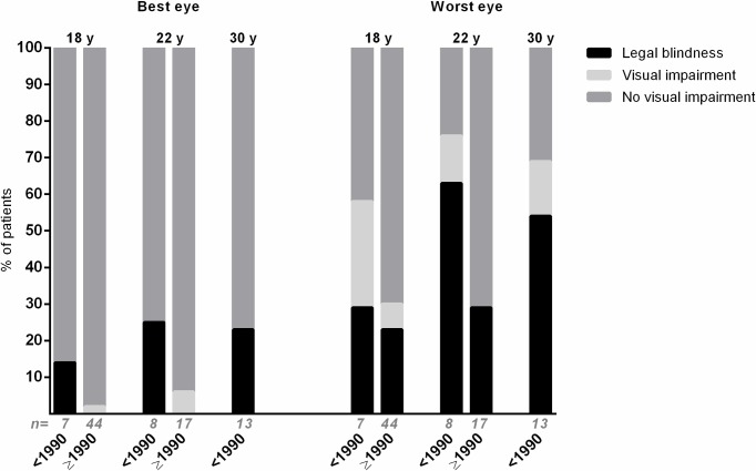
Data of the visual ability at the different ages are shown in Fig 1. At the age of 18 years, 2/51 patients (4%) were visually impaired or legally blind. One was diagnosed before 1990 and one after 1990. Seventeen out of 51 patients (33%) had at least one eye which had a visual acuity of 20/50 or worse and/or a visual field of ≤10° of which 13/17 patients were diagnosed after 1990 (which represents 30% of all 44 patients diagnosed after 1990) and 4/17 patients were diagnosed before 1990 (which represents 58% of all seven patients diagnosed before 1990).
Fig 1. Visual impairment of patients with juvenile idiopathic arthritis associated uveitis.
Proportion of patients with juvenile idiopathic arthritis associated uveitis with no visual impairment, visual impairment or legal blindness of the best (left) and worst (right) eye at the age of 18, 22 and 30 years, with uveitis onset before (<) 1990/after (≥) 1990. Visual acuity better than 20/50 was defined as no visual impairment, visual acuity equal to or less than 20/50 was defined as visual impairment and visual acuity equal to or less than 20/200 was defined as legal blindness.[29] If patients had a visual field of 10° or less they were also classified as ‘legal blindness’, according to the criteria for visual impairment of the World Health Organization.[30]. Statistical analyses comparing the situation <1990 and ≥1990 were derived from the Fisher exact test, there were no statistically significant differences. ‘n = ‘ = total number of patients included in the bar.
Of all patients with an impaired visual ability of at least one eye, 10 out of 17 (59%) were diagnosed with uveitis before arthritis versus five out of 33 (15%) of the patients with a good visual ability (P = .001). Also at the age of 22 years, the two patients (100%) with visual impairment of the best eye were diagnosed with uveitis before arthritis, compared to three out of 22 patients (14%) with normal visual acuity (P = .036).
No differences were found for gender, uveitis onset before 1990 or after 1990, or total number of ocular surgeries since uveitis onset.
The course of visual acuity could be measured for 23 patients with follow-up data at both 18 and 22 years. The median BCVA between 18 and 22 years (n = 23 patients) appeared to be stable. Sixteen of these patients (70%) were diagnosed after 1990. Six patients had follow-up data at all age years (18, 22 and 30 years) and had a median BCVA of one eye of 20/63 (IQR 20/20-0.25/200) at the age of 18 years and 2/200 (IQR 20/22-0.25/200) at the age of 30 years (P = .465) and for the other eye a median BCVA of 20/25 at 18 years (IQR 20/16-20/100) and 20/40 at 30 years (IQR 20/25-0.02/200; P = .116). All these six patients were diagnosed before the year 1990.
Uveitis activity and treatment
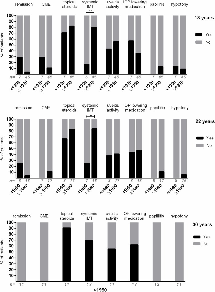
Of the 52 patients with available data at the age of 18 years, four patients (8%) were in remission during the entire year (Fig 2). Almost half of the patients (n = 28/52, 54%) had an episode of active uveitis during their 18th life year, 37/51 patients (73%) were on systemic IMT and 42/52 (81%) used topical steroids. Four of the 37 (11%) 18 year-old patients on systemic IMT had no uveitis activity and did not use topical steroids (all diagnosed ≥1990). All others in the group ‘without remission’ were at least on topical corticosteroids or had uveitis activity.
Fig 2. Uveitis activity, treatment and complications of patients with uveitis associated with juvenile idiopathic arthritis in adulthood.
Outcomes of patients with uveitis onset before (<) and after (≥) the year 1990. At the top: outcomes at age 18; middle: age 22 years; bottom: age 30 years. Note that all data at age 30 years were from patients with uveitis onset before the year 1990. Remission was defined as inactive disease for ≥ three months after discontinuing all treatments for eye disease.[29] Systemic immunomodulatory treatment (IMT) was defined ‘Yes’ when used for more than six months. Uveitis activity was defined ‘Yes’ when there were at least 1+ cells in the anterior chamber, as determined by the grading system of the Standardization of Uveitis Nomenclature (SUN) working group.[29]. CME = cystoid macular edema; IOP = intraocular pressure. *: P < .05; **: P < .005. ‘n = ‘ = total number of patients included in the bar.
The majority of 18 year-old patients diagnosed after 1990 were on systemic IMT (n = 36/45, 80%), in contrast to the patients diagnosed before 1990, where only 1/6 patients (17%) was on systemic IMT during the 18th life year (P = .004). A comparable result was found at the age of 22 years (P = .017, Fig 2). No other statistically significant differences were found for uveitis activity or treatment between the patients diagnosed before and after 1990.
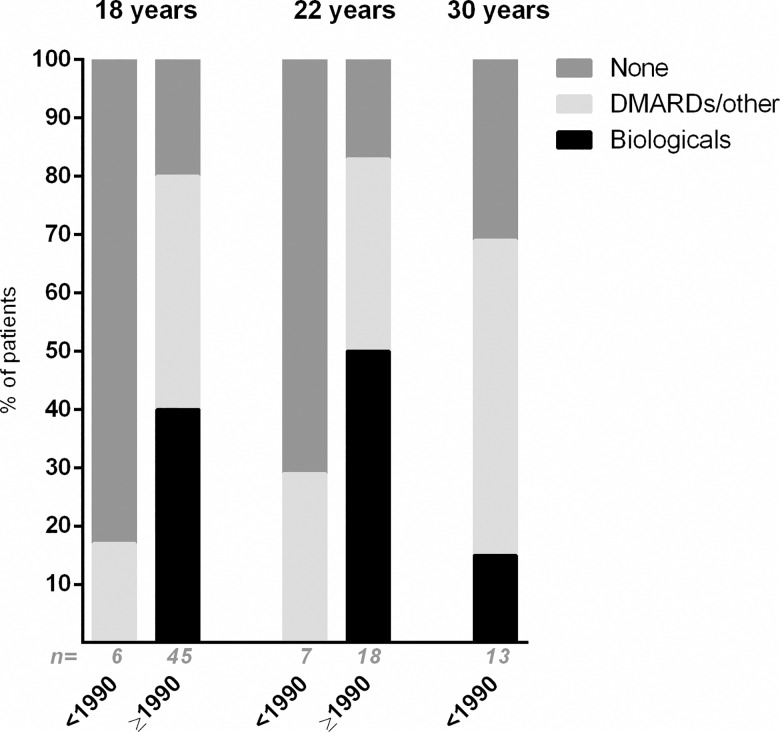
The different treatment strategies are presented in Fig 3. The figure shows that patients diagnosed after 1990 were treated with biologicals more often than patients diagnosed with uveitis before 1990.
Fig 3. The use of systemic immunomodulatory treatment in patients with juvenile idiopathic arthritis associated uveitis.
Treatment at the age of 18, 22 and 30 years with uveitis onset before (<) and after (≥) the year 1990. DMARD = disease modifying anti-rheumatic drugs. A biological was always given combined with a DMARD (in the figure noted as ‘Biologicals’), a DMARD/other drugs were usually given as combination therapy. ‘n = ‘ = total number of patients included in the bar. The exact treatment, with number of patients using this treatment in brackets, was: Treatment 18 years, <1990: Corticosteroids (n = 1). Treatment 18 years, ≥ 1990: Corticosteroids (n = 1), Methotrexate (n = 31), Mycophenolate motefil (n = 2), Azathioprine (n = 2), Cyclosporine (n = 1), Adalimumab (n = 16), Etanercept (n = 1), Infliximab (n = 2). Treatment 22 years, < 1990: Corticosteroids (n = 1), Methotrexate (n = 1).Treatment 22 years, ≥ 1990: Corticosteroids (n = 2), Methotrexate (n = 9), Mycophenolate motefil (n = 3), Azathioprine (n = 1), Hydroxychloroquine (n = 2), Adalimumab (n = 4), Etanercept (n = 2), Tocilizumab (n = 2), Infliximab (n = 1).Treatment 30 years, <1990: Corticosteroids (n = 4), Methotrexate (n = 5), Mycophenolate motefil (n = 1), Hydroxychloroquine (n = 1), Cyclosporine (n = 1), Sulfasalazine (n = 1), Tocilizumab (n = 1), Etanercept (n = 1).
Course of complications and surgery
Ten of the 24 patients (42%) with follow-up data available at the age of 18 as well as at the age of 22 years underwent surgery between their 18th and 22nd year of age. Three/24 patients (14%) underwent cataract surgery and 9/24 (38%) underwent glaucoma surgery between their 18th and 22nd birthday.
Seventeen/24 patients (71%) had already had surgery by the time they became 18 years old (6/24 patients had had surgery for cataract and glaucoma, 10/24 for cataract only, 1/24 for glaucoma only). Four/24 patients (17%) had their first surgery after the age of 18. Three/24 patients (13%) never underwent surgery before their 22nd life year. Note that all of these 24 patients had bilateral uveitis and the majority of patients (n = 17/24, 71%) was diagnosed with uveitis after 1990.
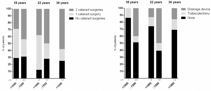
Fig 4 presents the number of cataract surgeries and type of glaucoma surgeries performed in the complete study population. Though not statistically significant, these results suggest that patients underwent glaucoma surgery more often when having an uveitis onset after 1990.
Fig 4. Cataract and glaucoma surgery of patients with uveitis associated with juvenile idiopathic arthritis in adulthood.
Proportion of patients who had undergone cataract (left) and glaucoma (right) surgery by the time they were 18, 22 and 30 years old, with uveitis onset before (<) and after (≥) the year 1990. Many patients had multiple glaucoma surgeries; in this figure patients with at least one glaucoma surgery versus no glaucoma surgeries are presented. Some of the patients who received a drainage device, also had a trabeculectomy in their medical history and are here included in the ‘Drainage device’ group. There were no statistically significant differences between the groups with uveitis onset before and after 1990. ‘n = ‘ = total number of patients included in the bar.
None of the patients developed posterior synechiae between the age of 18 and 22 years, though one patient (4%) developed band keratopathy.
Active inflammation in the anterior chamber was less frequent at the 22nd life year (8/23, 35%) compared to the 18th life year (15/23, 65%; P = .039). No differences were found for complete remission of uveitis, presence of CME, use of topical steroids, use of systemic IMT, use of IOP lowering medication, papillitis or hypotony.
Discussion
Our results document that JIA is not solely a disease of childhood, but that its activity, accompanied use of systemic medications, complications and additional visual loss continues in early adulthood. Only 4% of the JIA-uveitis patients were in remission at 18 years of age.
Previous literature, as presented in Table 2, described patients with JIA-uveitis in adulthood, but broad ranges of follow-up make it difficult to interpret their results. In our study we studied different outcome measurements at fixed time-points. Further, to get a good impression of the visual ability of the patients, visual acuity cannot be seen apart from visual field outcomes. Only one previous study of 12 adult JIA-uveitis patients also studied visual field outcomes.[14]
Table 2. Juvenile idiopathic arthritis associated uveitis in adulthood, a literature overview.
| Author | Nr patients | Mean/median age years (range) | Follow-up | Visual acuity | Definition active uveitis | Uveitis activity | Complications, treatment |
|---|---|---|---|---|---|---|---|
| Packham et al 2002[13] | 54 | 35 (19–78) (= age of total JIA group with 246 patients) | Situation at the end of follow-up. | NA | Not defined | NA | 66% of the patients had glaucoma during follow-up, 55% had cataract, 69% had had eye-surgery (not specifically during adulthood). |
| Zak et al 2003[14] | 12 | 32 (22–49) | Situation at the end of follow-up. | Studied visual field outcomes.a 1 patient with bilateral severe visual field loss. 1 patient with unilateral VA <0.02. | Not defined | NA | Almost all patients had anterior segment findings as sequelae. |
| Ozdal et al 2005[15] | 18 (30 eyes) | 30 (18–48) | Situation at the end of follow-up. Minimum follow-up = 2 years. | 9 patients (50%) had a BCVA <20/150 of at least one eye. | Presence of cells or keratic precipitates, with or without flare. | 19 (63%) of the eyes had active uveitis. | 3 eyes with phtisis. 73% (22 eyes) had cataract extraction, 4 eyes (13%) had glaucoma surgery by drainage device or trabeculectomy (not specifically during adulthood). 11 patients (61.1%) required the use of a systemic immunosuppresive agent. 16/18 patients were on topical steroids. |
| Kotaniemi et al 2005[16] | 19 | 24 (22–26) | One consult for evaluation. | 100% binocular normal BCVA, 3 patients had unilateral BCVA <0.1. | Use of topical corticosteroids and/or at least 3 cells in the anterior chamber. | 8 patients had active uveitis. | 4 patients had glaucoma, 5 had cataract. 53% of the patients were on systemic IMT. 10/19 patients used treatment (systemic and/or topical). |
| Camuglia et al 2009[17] | 17 | 30 (21–43) | Situation at the end of follow-up. | 20% of the eyes had visual loss up to 6/12, 13.3% of the eyes had visual loss up to 6/60. | Not defined | NA | 53% of the patients (9 patients, 13 eyes) had new complications of cataract or glaucoma after their 16th birthday. Two eyes had glaucoma surgery and 10 eyes had cataract surgery after the age of 16. 30% of the patients had synechiae during their uveitis course. 10/17 patients used systemic treatment, all patients had topical treatment. |
| Skarin et al 2009[35] | 55 | NA | Follow-up at 24 years (range 18–46) after uveitis onset. | NA | Not defined | 49% of the 55 patients had signs of active uveitis or were receiving topical corticosteroids. | 12 patients (33%) had glaucoma, 28 patients (78%) had cataract. |
| Oray et al 2016[18] | 77 (135 eyes) | 29.7 (±11) | Situation at the end of follow-up. | 37 eyes (28%) had a visual acuity of ≤20/50. 20 eyes (15%) had a visual acuity of ≤20/200. | ≥0.5+ cells in the anterior chamber. | 78 eyes (58%). | Ocular surgery in 68 eyes. 13 patients (17%) were treated with conventional IMT (e.g. MTX), 52 patients (68%) were treated with biologicals. At least one complication in 95 eyes (72%). |
This table describes all previous literature on the course of juvenile idiopathic arthritis associated uveitis in adulthood.
aThis was the only study which described visual field outcomes.
NA = not applicable; IMT = immunomodulatory treatment; MTX = Methotrexate.
Our study showed a trend towards a slightly better visual outcome for patients diagnosed after 1990 than for patients diagnosed before 1990. Also, a higher percentage of patients diagnosed after 1990 used systemic IMT, which suggests an improvement of visual outcome due to novel and more intensive immunomodulating treatment strategies since 1990.[19–22] It is likely that the use of ophthalmologic screening guidelines for patients with JIA has also contributed to improved visual outcome.[23–25] Furthermore, the awareness of glaucoma increased during the last decades with earlier and improved interventions and more prevention of severe glaucomatous visual field loss, as also displayed by Fig 4.[31,32] Also, cataract extractions are nowadays performed at an earlier stage, preventing development of amblyopia. Previous literature described a delay of cataract formation in patients treated with MTX, which means that, since the introduction of MTX as a treatment for JIA-uveitis, children have less chance to develop cataract at an age vulnerable for developing amblyopia.[33]
Better visual outcome in relation to systemic treatment was also shown by the study of Gregory et al.[4] Unfortunately, we could not correlate use of systemic IMT to visual outcome, since our study had data at three time-points rather than continuous data over the years. Additionally, the prescription of IMT might have been influenced by uveitis as well as arthritis activity. Anyhow, only four patients were solely on IMT without uveitis activity or topical steroids for their uveitis, suggesting that most patients in this study received systemic IMT at least partially for their uveitis.
Though our results suggest that most patients with JIA-uveitis have a fairly good binocular visual prognosis, with a rather stable visual outcome during the adolescent years, about one third of the patients with uveitis onset after the year 1990 had at least one visually impaired eye (Fig 1). In line with previous literature, risk factors for an impaired visual outcome were onset of uveitis before arthritis and lower age at onset of uveitis.[34] Because of the relatively small sample size in our patient groups, we cannot exclude that other factors which were described previously, such as male gender, are also possible risk factors for visual impairment.[6]
Despite the slight improvement of visual acuity and the increased use of systemic IMT after 1990, uveitis in early adulthood was active in approximately half of the patients. This is an important finding to be aware of, since a previous study described uveitis activity to be associated with an increased risk of visual loss.[4] It is extremely important for ophthalmologists to keep a close eye on these patients and treat them accurately, to prevent them from more bi- or unilateral visual impairment due to uveitis activity in their future lives. A longer follow-up and a larger cohort are necessary to be able to document their visual functioning over the years.
Our study cohort presents a representative JIA-uveitis population, as the male-to-female ratio, median age of arthritis onset and presence of ANA are similar to previous literature.[12,14–17,35,36]
Because of its retrospective character our study has limitations. There might have been a tertiary referral bias, since patients were selected from a cohort of tertiary centers. Though, this was minimized as much as possible by retrieving medical charts from patients being followed also by their own ophthalmologists outside the tertiary centers. Therefore, the percentage of patients with active uveitis and/or complications during adulthood might be lower than presented in this study. The sample size of patients evaluated at the age of 30 years is also limited, and moreover, the data of these patients at their 18th and 22nd year of life were not available, because it was not possible to retrieve their old medical charts.
Overall results on visual outcomes in this study are quite promising, with an improvement in visual acuity, probably also due to systemic IMT. Several studies have shown that that the introduction of MTX and anti-TNFα therapies have been important in the prevention of complications or even the occurrence of uveitis in JIA patients.[4,37] But these treatment strategies might have significant impact on the quality of life of patients. For example, use of systemic IMT will result in more visits to the ophthalmologists or rheumatologists, also, these young adult women might wish to become pregnant which conflicts with the use of most immunosuppressive medications. For future research it would be interesting to investigate the effect of the use of systemic IMT on these aspects, including quality of life of these adult patients.
In conclusion, the results of this study imply that binocular visual outcome in adulthood is fairly good in most patients with JIA-uveitis and that binocular visual ability seems to be stable in the first years after puberty. Despite the apparent improvement in visual prognosis in the last decades, still up to 30% of all adult JIA-uveitis patients have developed severe visual impairment or even blindness of at least one eye. Also, uveitis activity is continuing in early adulthood, and the majority of patients need ongoing treatment. Additionally, uveitis associated complications still arise in early adulthood and a relevant proportion of the patients need cataract or glaucoma surgery during adulthood. Awareness of these findings is important for ophthalmologists and rheumatologists treating patients with JIA-uveitis, as well as for the patients themselves, in order to prevent these patients from becoming visually disabled even many years after their typical childhood onset of this disease.
Acknowledgments
Funding/Support: This work was supported by the combined grants from the Dr. F.P. Fischer Stichting, Amersfoort; The ODAS Stichting, the Landelijke Stichting Voor Blinden en Slechtzienden, Utrecht; the Stichting Nederlands Oogheelkundig Onderzoek (SNOO), Rotterdam, the Netherlands. Data access, responsibility and analysis: Drs. Haasnoot and Dr. de Boer had full access to all the data in the study and take responsibility for the integrity of the data and the accuracy of the data analysis.
Data Availability
All relevant data are within the paper.
Funding Statement
This study was funded by the combined grants from the Dr. F.P. Fischer Stichting, Amersfoort; The ODAS Stichting, the Landelijke Stichting Voor Blinden en Slechtzienden, Utrecht; the Stichting Nederlands Oogheelkundig Onderzoek (SNOO), Rotterdam, the Netherlands. The funders had no role in study design, data collection and analysis, decision to publish, or preparation of the manuscript.
References
- 1.Sen ES, Dick AD, Ramanan AV. Uveitis associated with juvenile idiopathic arthritis. Nat Rev Rheumatol 2015. June;11(6):338–348. 10.1038/nrrheum.2015.20 [DOI] [PubMed] [Google Scholar]
- 2.Kanski JJ. Uveitis in juvenile chronic arthritis: incidence, clinical features and prognosis. Eye (Lond) 1988;2 (Pt 6)(Pt 6):641–645. 10.1038/eye.1988.118 [DOI] [PubMed] [Google Scholar]
- 3.Kesen MR, Setlur V, Goldstein DA. Juvenile idiopathic arthritis-related uveitis. Int Ophthalmol Clin 2008. Summer;48(3):21–38. 10.1097/IIO.0b013e31817d998f [DOI] [PubMed] [Google Scholar]
- 4.Gregory AC 2nd, Kempen JH, Daniel E, Kacmaz RO, Foster CS, Jabs DA, et al. Risk factors for loss of visual acuity among patients with uveitis associated with juvenile idiopathic arthritis: the Systemic Immunosuppressive Therapy for Eye Diseases Study. Ophthalmology 2013. January;120(1):186–192. 10.1016/j.ophtha.2012.07.052 [DOI] [PMC free article] [PubMed] [Google Scholar]
- 5.Ozdal PC, Vianna RN, Deschenes J. Visual outcome of juvenile rheumatoid arthritis-associated uveitis in adults. Ocul Immunol Inflamm 2005. February;13(1):33–38. 10.1080/09273940590909220 [DOI] [PubMed] [Google Scholar]
- 6.Kalinina Ayuso V, Ten Cate HA, van der Does P, Rothova A, de Boer JH. Male gender and poor visual outcome in uveitis associated with juvenile idiopathic arthritis. Am J Ophthalmol 2010. June;149(6):987–993. 10.1016/j.ajo.2010.01.014 [DOI] [PubMed] [Google Scholar]
- 7.Dana MR, Merayo-Lloves J, Schaumberg DA, Foster CS. Visual outcomes prognosticators in juvenile rheumatoid arthritis-associated uveitis. Ophthalmology 1997. February;104(2):236–244. 10.1016/s0161-6420(97)30329-7 [DOI] [PubMed] [Google Scholar]
- 8.Edelsten C, Lee V, Bentley CR, Kanski JJ, Graham EM. An evaluation of baseline risk factors predicting severity in juvenile idiopathic arthritis associated uveitis and other chronic anterior uveitis in early childhood. Br J Ophthalmol 2002. January;86(1):51–56. 10.1136/bjo.86.1.51 [DOI] [PMC free article] [PubMed] [Google Scholar]
- 9.Kotaniemi K, Kautiainen H, Karma A, Aho K. Occurrence of uveitis in recently diagnosed juvenile chronic arthritis: a prospective study. Ophthalmology 2001. November;108(11):2071–2075. 10.1016/s0161-6420(01)00773-4 [DOI] [PubMed] [Google Scholar]
- 10.Gare BA, Fasth A. Epidemiology of juvenile chronic arthritis in southwestern Sweden: a 5-year prospective population study. Pediatrics 1992. December;90(6):950–958. [PubMed] [Google Scholar]
- 11.Kotaniemi K, Kaipiainen-Seppanen O, Savolainen A, Karma A. A population-based study on uveitis in juvenile rheumatoid arthritis. Clin Exp Rheumatol 1999. Jan-Feb;17(1):119–122. [PubMed] [Google Scholar]
- 12.Hoeve M, Kalinina Ayuso V, Schalij-Delfos NE, Los LI, Rothova A, de Boer JH. The clinical course of juvenile idiopathic arthritis-associated uveitis in childhood and puberty. Br J Ophthalmol 2012. June;96(6):852–856. 10.1136/bjophthalmol-2011-301023 [DOI] [PubMed] [Google Scholar]
- 13.Packham JC, Hall MA. Long-term follow-up of 246 adults with juvenile idiopathic arthritis: functional outcome. Rheumatology (Oxford) 2002. December;41(12):1428–1435. 10.1093/rheumatology/41.12.1428 [DOI] [PubMed] [Google Scholar]
- 14.Zak M, Fledelius H, Pedersen FK. Ocular complications and visual outcome in juvenile chronic arthritis: a 25-year follow-up study. Acta Ophthalmol Scand 2003. June;81(3):211–215. 10.1034/j.1600-0420.2003.00066.x [DOI] [PubMed] [Google Scholar]
- 15.Ozdal PC, Vianna RN, Deschenes J. Visual outcome of juvenile rheumatoid arthritis-associated uveitis in adults. Ocul Immunol Inflamm 2005. February;13(1):33–38. 10.1080/09273940590909220 [DOI] [PubMed] [Google Scholar]
- 16.Kotaniemi K, Arkela-Kautiainen M, Haapasaari J, Leirisalo-Repo M. Uveitis in young adults with juvenile idiopathic arthritis: a clinical evaluation of 123 patients. Ann Rheum Dis 2005. June;64(6):871–874. 10.1136/ard.2004.026955 [DOI] [PMC free article] [PubMed] [Google Scholar]
- 17.Camuglia JE, Whitford CL, Hall AJ. Juvenile idiopathic arthritis associated uveitis in adults: a case series. Ocul Immunol Inflamm 2009. Sep-Oct;17(5):330–334. 10.3109/09273940903118626 [DOI] [PubMed] [Google Scholar]
- 18.Oray M, Khachatryan N, Ebrahimiadib N, Abu Samra K, Lee S, Foster CS. Ocular morbidities of juvenile idiopathic arthritis-associated uveitis in adulthood: results from a tertiary center study. Graefes Arch Clin Exp Ophthalmol 2016. September;254(9):1841–9. 10.1007/s00417-016-3340-z [DOI] [PubMed] [Google Scholar]
- 19.Truckenbrodt H, Hafner R. Methotrexate therapy in juvenile rheumatoid arthritis: a retrospective study. Arthritis Rheum 1986. June;29(6):801–807. 10.1002/art.1780290616 [DOI] [PubMed] [Google Scholar]
- 20.Lovell DJ, Ruperto N, Goodman S, Reiff A, Jung L, Jarosova K, et al. Adalimumab with or without methotrexate in juvenile rheumatoid arthritis. N Engl J Med 2008. August 21;359(8):810–820. 10.1056/NEJMoa0706290 [DOI] [PubMed] [Google Scholar]
- 21.Lovell DJ, Giannini EH, Reiff A, Cawkwell GD, Silverman ED, Nocton JJ, et al. Etanercept in children with polyarticular juvenile rheumatoid arthritis. Pediatric Rheumatology Collaborative Study Group. N Engl J Med 2000. March 16;342(11):763–769. 10.1056/NEJM200003163421103 [DOI] [PubMed] [Google Scholar]
- 22.Giannini EH, Brewer EJ, Kuzmina N, Shaikov A, Maximov A, Vorontsov I, et al. Methotrexate in resistant juvenile rheumatoid arthritis. Results of the U.S.A.-U.S.S.R. double-blind, placebo-controlled trial. The Pediatric Rheumatology Collaborative Study Group and The Cooperative Children's Study Group. N Engl J Med 1992. April 16;326(16):1043–1049. 10.1056/NEJM199204163261602 [DOI] [PubMed] [Google Scholar]
- 23.Kanski JJ. Screening for uveitis in juvenile chronic arthritis. Br J Ophthalmol 1989. March;73(3):225–228. 10.5353/th_b4171059 [DOI] [PMC free article] [PubMed] [Google Scholar]
- 24.American Academy of Pediatrics Section on Rheumatology and Section on Ophthalmology: Guidelines for ophthalmologic examinations in children with juvenile rheumatoid arthritis. Pediatrics 1993. August;92(2):295–296. [PubMed] [Google Scholar]
- 25.Cassidy J, Kivlin J, Lindsley C, Nocton J, Section on Rheumatology, Section on Ophthalmology. Ophthalmologic examinations in children with juvenile rheumatoid arthritis. Pediatrics 2006. May;117(5):1843–1845. 10.1542/peds.2006-0421 [DOI] [PubMed] [Google Scholar]
- 26.Petty RE, Southwood TR, Manners P, Baum J, Glass DN, Goldenberg J, et al. International League of Associations for Rheumatology classification of juvenile idiopathic arthritis: second revision, Edmonton, 2001. J Rheumatol 2004. February;31(2):390–392. [PubMed] [Google Scholar]
- 27.Berntson L, Fasth A, Andersson-Gare B, Kristinsson J, Lahdenne P, Marhaug G, et al. Construct validity of ILAR and EULAR criteria in juvenile idiopathic arthritis: a population based incidence study from the Nordic countries. International League of Associations for Rheumatology. European League Against Rheumatism. J Rheumatol 2001. December;28(12):2737–2743. [PubMed] [Google Scholar]
- 28.Bloch-Michel E, Nussenblatt RB. International Uveitis Study Group recommendations for the evaluation of intraocular inflammatory disease. Am J Ophthalmol 1987. February 15;103(2):234–235. 10.1016/s0002-9394(14)74235-7 [DOI] [PubMed] [Google Scholar]
- 29.Jabs DA, Nussenblatt RB, Rosenbaum JT, Standardization of Uveitis Nomenclature (SUN) Working Group. Standardization of uveitis nomenclature for reporting clinical data. Results of the First International Workshop. Am J Ophthalmol 2005. September;140(3):509–516. 10.1016/j.ajo.2005.03.057 [DOI] [PMC free article] [PubMed] [Google Scholar]
- 30.International Statistical Classification of Diseases and Related Health Problems-10.; 2014. p. chapter VII: diseases of the eye and adnexa; H54: visual impairment including blindness (binocular or monocular).
- 31.Foster CS, Havrlikova K, Baltatzis S, Christen WG, Merayo-Lloves J. Secondary glaucoma in patients with juvenile rheumatoid arthritis-associated iridocyclitis. Acta Ophthalmol Scand 2000. October;78(5):576–579. 10.1034/j.1600-0420.2000.078005576.x [DOI] [PubMed] [Google Scholar]
- 32.Kotaniemi K, Sihto-Kauppi K. Occurrence and management of ocular hypertension and secondary glaucoma in juvenile idiopathic arthritis-associated uveitis: An observational series of 104 patients. Clin Ophthalmol 2007. December;1(4):455–459. [PMC free article] [PubMed] [Google Scholar]
- 33.Sijssens KM, Rothova A, Van De Vijver DA, Stilma JS, De Boer JH. Risk factors for the development of cataract requiring surgery in uveitis associated with juvenile idiopathic arthritis. Am J Ophthalmol 2007. October;144(4):574–579. 10.1016/j.ajo.2007.06.030 [DOI] [PubMed] [Google Scholar]
- 34.Angeles-Han ST, Yeh S, Vogler LB. Updates on the risk markers and outcomes of severe juvenile idiopathic arthritis-associated uveitis. Int J Clin Rheumtol 2013. February 1;8(1): 10.2217/ijr.12.83 [DOI] [PMC free article] [PubMed] [Google Scholar]
- 35.Skarin A, Elborgh R, Edlund E, Bengtsson-Stigmar E. Long-term follow-up of patients with uveitis associated with juvenile idiopathic arthritis: a cohort study. Ocul Immunol Inflamm 2009. Mar-Apr;17(2):104–108. 10.1080/09273940802650398 [DOI] [PubMed] [Google Scholar]
- 36.Haasnoot AJ, van Tent-Hoeve M, Wulffraat NM, Schalij-Delfos NE, Los LI, Armbrust W, et al. Erythrocyte sedimentation rate as baseline predictor for the development of uveitis in children with juvenile idiopathic arthritis. Am J Ophthalmol 2015. February;159(2):372–7. 10.1016/j.ajo.2014.11.007 [DOI] [PubMed] [Google Scholar]
- 37.Tappeiner C, Schenck S, Niewerth M, Heiligenhaus A, Minden K, Klotsche J. Impact of Antiinflammatory Treatment on the Onset of Uveitis in Juvenile Idiopathic Arthritis: Longitudinal Analysis From a Nationwide Pediatric Rheumatology Database. Arthritis Care Res (Hoboken) 2016. January;68(1):46–54. 10.1002/acr.22649 [DOI] [PMC free article] [PubMed] [Google Scholar]
Associated Data
This section collects any data citations, data availability statements, or supplementary materials included in this article.
Data Availability Statement
All relevant data are within the paper.


