Huang and Rasband discuss Albrecht et al.’s new model for the axon initial segment’s diffusion barrier.
Abstract
What prevents the movement of membrane molecules between axonal and somatodendritic domains is unclear. In this issue, Albrecht et. al. (2016. J. Cell Biol. http://dx.doi.org/10.1083/jcb.201603108) demonstrate via high-speed single-particle tracking and superresolution microscopy that lipid-anchored molecules in the axon initial segment are confined to membrane domains separated by periodically spaced actin rings.
A protein’s location reflects its function. This is especially apparent in neurons because of their complex morphologies and highly specialized membrane domains. For example, the most basic division in neuronal morphology is the distinction between dendrites and axons. A neuron may have many dendrites that receive synaptic input, but only a single long axon that is responsible for propagation of action potentials and communication among neurons in a circuit. In axons, cytoskeletal proteins, such as microtubules, neurofilaments, and actin microfilaments, are particularly important and must be well organized to maintain axon integrity over distances that can be many thousands of times longer than the diameter of the neuronal cell body. The actin-based cytoskeleton is connected to the plasma membrane through spectrins and ankyrins. A submembranous network of actin, spectrin, and ankyrin was first described in erythrocytes and proposed to resist the mechanical stresses faced by erythrocytes as they move through vessels and capillaries (Bennett and Baines, 2001). However, the detailed structure of the axonal actin/spectrin/ankyrin submembranous cytoskeleton was largely unknown until it was recently revealed by superresolution microscopy. In axons, actin filaments are arranged in periodic rings separated by spectrin tetramers; the spectrin tetramers are ∼190 nm long, which corresponds to the distance between actin rings (Xu et al., 2013; Zhong et al., 2014; Leterrier et al., 2015). Consistent with a structural role for spectrins, axons break easily in β-spectrin mutant nematodes. Remarkably, breakage is prevented by paralyzing the mutant worms (Hammarlund et al., 2007). Together, these observations suggest that a major role for the actin/spectrin/ankyrin cytoskeleton in axons may be to maintain membrane integrity and to withstand the mechanical strain experienced by long axons.
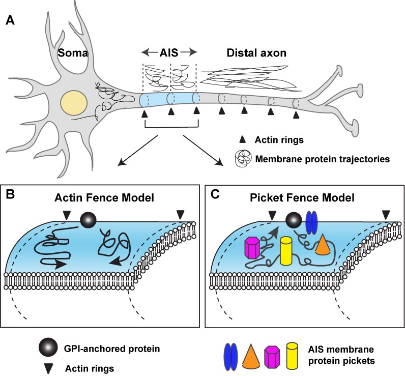
Different kinds of spectrins and ankyrins are further restricted to specialized axonal domains. For example, the axon initial segment (AIS) is located in the proximal axon and is enriched with voltage-gated Na+ and K+ channels responsible for action potential initiation. AIS ion channels are clustered by the scaffolding protein ankyrin G (ankG) which is linked to the actin cytoskeleton by βIV spectrin. In contrast, the distal axonal cytoskeleton, composed of αII-spectrin, βII spectrin, and ankyrin B (ankB), functions as an intra-axonal boundary to restrict ankG to the proximal axon (Galiano et al., 2012). Despite different spectrins and ankyrins, both AIS and distal axon cytoskeletons have a common periodic organization (Xu et al., 2013). Besides firing action potentials, the AIS also maintains neuronal polarity. Loss of the scaffolding protein ankG in the AIS dismantles the AIS and causes axons to acquire dendritic properties. Without ankG, somatodendritic molecules (both membrane and cytosolic) redistribute into the former axon, indicating the AIS functions as both a cytoplasmic and membrane diffusion barrier to prevent mixing of somatodendritic and axonal proteins (Hedstrom et al., 2008; Fig. 1 A). Previous studies showed the mobility of membrane proteins is significantly reduced at the AIS compared with the distal axon (Winckler et al., 1999; Nakada et al., 2003). The AIS barrier develops when actin/βIV spectrin/ankG and their associated proteins become enriched at the AIS, but is disrupted after actin depolymerization. These observations suggested a “picket fence” model where the mobility of membrane proteins is impeded because of crowding and steric hindrance resulting from the high density of transmembrane proteins tethered to ankG. Both the remarkable ability of the AIS to limit membrane protein diffusion and the striking periodic organization of the actin cytoskeleton prompted Albrecht et al. (2016) to examine the relationship between membrane protein properties and the periodic axonal cytoskeleton. In this issue, Albrecht et al. propose a new function for the AIS actin rings: to assemble a fence, or barrier, that restricts the diffusion of membrane proteins in the AIS to regions between the actin rings (Fig. 1 B). This new model for the AIS diffusion barrier is conceptually different than the picket fence model, which instead relies on high densities of membrane proteins.
Figure 1.
Models for the restricted diffusion of membrane proteins in the AIS. (A) Somatodendritic proteins are excluded from the axon by the AIS. Membrane proteins in the AIS are highly stable and are confined between actin rings located at regularly spaced intervals along the axon. Membrane proteins in the distal axon are freely mobile and diffuse across actin rings. (B) The actin fence model proposes that the actin, or its associated proteins, functions as fence to constrain GPI-GFP proteins to regions between adjacent actin rings. (C) The picket fence model proposes that a high density of membrane proteins at the AIS (including Na+ channels and NF186 anchored to ankG between the actin rings) impedes the diffusion of GPI-GFP because of molecular crowding.
To arrive at the “actin fence” model, in an experimental tour-de-force, Albrecht et al. (2016) measured the trajectories of single glycosylphosphatidylinositol-anchored GFP (GPI-GFP) molecules within the AIS using high-density single-particle tracking (SPT) on primary rat hippocampal neurons at different developmental time points. They observed a dramatic reduction in the mobility of GPI-GFP between day in vitro 3 (DIV 3) and DIV 5. The reduced mobility was observed at all later time points. Surprisingly, the reduction in mobility was only observed in the AIS, whereas transmembrane proteins in the distal axon remained highly motile at all times. Further analysis of the trajectories of GPI-GFP at the AIS revealed that GPI-GFP proteins were restricted to specific regions and formed a striped pattern perpendicular to the long axes of axons and spaced with a periodicity of ∼190 nm. This observation suggested the involvement of the periodic actin cytoskeleton. To further interrogate the relationship between the reduced mobility of GPI-GFP and the submembranous actin-spectrin cytoskeleton, the SPT trajectories of GPI-GFP were reconstructed and overlaid on superresolution images of the axonal cytoskeleton. The GPI-GFP trajectories overlapped with the spectrin periodic pattern, but alternated with the actin rings, suggesting that the actin rings may function as fences to limit the diffusion of the GPI-GFP molecules.
The available evidence suggests two alternative (but not mutually exclusive) models that could explain both the limited and periodic diffusion of GPI-GFP. The first model, as suggested here by Albrecht et al. (2016), implicates actin as a key component of a fence that restricts the mobility of membrane proteins between adjacent actin rings (Fig. 1 B). Support for this model includes the correlation between the SPT trajectories and the location of the periodic actin cytoskeleton as revealed by superresolution microscopy. Furthermore, modeling of the diffusion of proteins in the membrane by Albrecht et al. (2016) also supported the feasibility of this mechanism. However, there are several caveats and limitations that must be considered. First, no diffusion barrier was observed in the distal axon despite the existence of a periodic actin/spectrin/ankyrin cytoskeleton like the one that exists in the AIS. Thus, the actin rings by themselves are not sufficient to limit diffusion and instead the results suggest that actin is but one component of a large protein complex that constitutes the fence. Second, no direct evidence was provided demonstrating the dependence of the restricted trajectories on actin. Third, it is not clear how a cytoplasmic actin–dependent fence could translate into a diffusion barrier for GPI-GFP, which is anchored only in the outer leaflet of the plasma membrane. Fourth, the pattern of spaced GPI-GFP trajectories was seen as early as DIV 4. This is important because the authors’ interpretation depends on the idea that there is not a high density of AIS membrane proteins at the AIS by DIV4, thereby excluding molecular crowding as a mechanism responsible for the reduced diffusion of GPI-GFP. However, although there is experimental support for this developmental sequence (Boiko et al., 2007; Jones et al., 2014), other studies report high densities of AIS membrane proteins by DIV4 (Nakada et al., 2003; Yang et al., 2007). These discrepancies may be explained by different culture conditions or different sensitivities of the antibodies used to detect the clustering of AIS membrane proteins.
The second model posits that membrane proteins found at the AIS would limit the diffusion of GPI-GFP because of their very high density and the molecular crowding that occurs. This model is supported by the observation that membrane proteins (e.g., Na+ channels and NF186), both anchored to ankG and enriched at the AIS, are found in a periodic pattern that alternates with actin rings (Leterrier et al., 2015). Thus, high densities of these membrane proteins would impede the diffusion of GPI-GFP and display an apparent confinement between actin rings. The fact that GPI-GFP trajectories are not confined between actin rings in the distal axon supports the idea that a high density of membrane proteins at the AIS contributes to the reduced diffusion in the membrane between the actin rings.
Additional experiments will be required to distinguish between these models of membrane protein diffusion at the AIS. For example, disruption of the actin cytoskeleton by treatment with latrunculin has been shown to perturb the periodic organization of both actin and βII spectrin in the distal axon, but not βIV spectrin in the AIS (Leterrier et al., 2015). Because AIS membrane proteins are anchored through ankG–βIV spectrin protein complexes, treatment with latrunculin should not disrupt their periodic organization. Thus, freely diffusable AIS GPI-GFP in latrunculin-treated neurons would strongly support the actin fence model. Alternatively, depletion of AIS membrane proteins through shRNA-mediated silencing (Hedstrom et al., 2007) would test whether protein crowding at the AIS is responsible for reduced diffusion. No change in the confinement of GPI-GFP to membrane stripes after loss of AIS membrane proteins would support the actin fence model. Finally, a previous study has shown that overexpression of βII spectrin is sufficient to induce the formation of a periodic cytoskeleton in dendrites (Zhong et al., 2014). It will be interesting to determine if a periodic cytoskeleton in dendrites is sufficient to confine GPI-GFP between actin rings or if it remains unconstrained as in axons.
In conclusion, it is important to note that the two contrasting models limiting membrane protein diffusion in the AIS are not mutually exclusive. Indeed, the necessity to maintain neuronal polarity for proper nervous system function may be supported by multiple molecular mechanisms. The results reported by Albrecht et al. (2016) provide additional evidence for actin/spectrin/ankyrin-dependent mechanisms that control the precise distribution and location of proteins in axons.
Acknowledgments
This work was supported by National Institutes of Health grants NS044916 and NS069688 to M.N. Rasband.
The authors declare no competing financial interests.
References
- Albrecht D., Winterflood C.M., Sadeghi M., Tschager T., Noe F., and Ewers H.. 2016. Nanoscopic compartmentalization of membrane protein motion at the axon initial segment. J. Cell Biol. 10.1083/jcb.201603108 [DOI] [PMC free article] [PubMed] [Google Scholar]
- Bennett V., and Baines A.J.. 2001. Spectrin and ankyrin-based pathways: metazoan inventions for integrating cells into tissues. Physiol. Rev. 81:1353–1392. [DOI] [PubMed] [Google Scholar]
- Boiko T., Vakulenko M., Ewers H., Yap C.C., Norden C., and Winckler B.. 2007. Ankyrin-dependent and -independent mechanisms orchestrate axonal compartmentalization of L1 family members neurofascin and L1/neuron-glia cell adhesion molecule. J. Neurosci. 27:590–603. 10.1523/JNEUROSCI.4302-06.2007 [DOI] [PMC free article] [PubMed] [Google Scholar]
- Galiano M.R., Jha S., Ho T.S., Zhang C., Ogawa Y., Chang K.J., Stankewich M.C., Mohler P.J., and Rasband M.N.. 2012. A distal axonal cytoskeleton forms an intra-axonal boundary that controls axon initial segment assembly. Cell. 149:1125–1139. 10.1016/j.cell.2012.03.039 [DOI] [PMC free article] [PubMed] [Google Scholar]
- Hammarlund M., Jorgensen E.M., and Bastiani M.J.. 2007. Axons break in animals lacking β-spectrin. J. Cell Biol. 176:269–275. 10.1083/jcb.200611117 [DOI] [PMC free article] [PubMed] [Google Scholar]
- Hedstrom K.L., Xu X., Ogawa Y., Frischknecht R., Seidenbecher C.I., Shrager P., and Rasband M.N.. 2007. Neurofascin assembles a specialized extracellular matrix at the axon initial segment. J. Cell Biol. 178:875–886. 10.1083/jcb.200705119 [DOI] [PMC free article] [PubMed] [Google Scholar]
- Hedstrom K.L., Ogawa Y., and Rasband M.N.. 2008. AnkyrinG is required for maintenance of the axon initial segment and neuronal polarity. J. Cell Biol. 183:635–640. 10.1083/jcb.200806112 [DOI] [PMC free article] [PubMed] [Google Scholar]
- Jones S.L., Korobova F., and Svitkina T.. 2014. Axon initial segment cytoskeleton comprises a multiprotein submembranous coat containing sparse actin filaments. J. Cell Biol. 205:67–81. 10.1083/jcb.201401045 [DOI] [PMC free article] [PubMed] [Google Scholar]
- Leterrier C., Potier J., Caillol G., Debarnot C., Rueda Boroni F., and Dargent B.. 2015. Nanoscale architecture of the axon initial segment reveals an organized and robust scaffold. Cell Reports. 13:2781–2793. 10.1016/j.celrep.2015.11.051 [DOI] [PubMed] [Google Scholar]
- Nakada C., Ritchie K., Oba Y., Nakamura M., Hotta Y., Iino R., Kasai R.S., Yamaguchi K., Fujiwara T., and Kusumi A.. 2003. Accumulation of anchored proteins forms membrane diffusion barriers during neuronal polarization. Nat. Cell Biol. 5:626–632. 10.1038/ncb1009 [DOI] [PubMed] [Google Scholar]
- Winckler B., Forscher P., and Mellman I.. 1999. A diffusion barrier maintains distribution of membrane proteins in polarized neurons. Nature. 397:698–701. 10.1038/17806 [DOI] [PubMed] [Google Scholar]
- Xu K., Zhong G., and Zhuang X.. 2013. Actin, spectrin, and associated proteins form a periodic cytoskeletal structure in axons. Science. 339:452–456. 10.1126/science.1232251 [DOI] [PMC free article] [PubMed] [Google Scholar]
- Yang Y., Ogawa Y., Hedstrom K.L., and Rasband M.N.. 2007. βIV spectrin is recruited to axon initial segments and nodes of Ranvier by ankyrinG. J. Cell Biol. 176:509–519. 10.1083/jcb.200610128 [DOI] [PMC free article] [PubMed] [Google Scholar]
- Zhong G., He J., Zhou R., Lorenzo D., Babcock H.P., Bennett V., and Zhuang X.. 2014. Developmental mechanism of the periodic membrane skeleton in axons. eLife. 3:04581 10.7554/eLife.04581 [DOI] [PMC free article] [PubMed] [Google Scholar]


