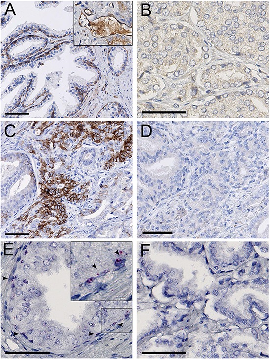
Figure 1.

A. MET protein staining in basal epithelial and endothelial cells (inset), in the normal prostate. B. MET protein absence in localized prostate cancer. C. N-cadherin positive area in localized prostate cancer. D. MET protein absence in the N-cadherin positive area. E. MET RNA expression in normal prostate basal epithelial cells (arrowheads). F. Absence of MET RNA in localized prostate cancer. Scale bars represent 50 μm at 20x (A, C, D) or 40x (B, E, F) magnification.
