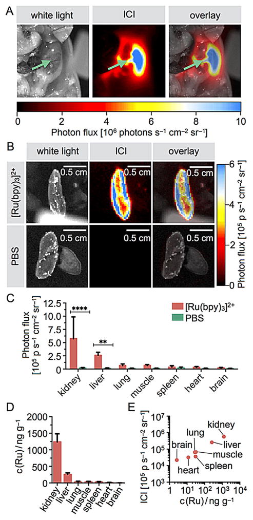
Figure 4.
In vivo abdominal surface chemiluminescence of [Ru(bpy)3]2+. The ICI agent was injected intravenously into healthy mice (26 nmol in 100 μL PBS), and the agent was allowed to clear from the bloodstream within 10 min. A) White light (left), ICI (center), and overlay (right) images of a mouse body cavity injected with 33 nmol of [Ru(bpy)3]2+ in 100 μL PBS. The green arrows point toward the right kidney. B) Images of excised kidneys are clearly visible in mice injected with the ICI agent, but not when injected with PBS. C) Quantification of the imaging results shown in panel A, with imaging quantification performed on excised organs. D) Quantification of ruthenium metal concentrations in various tissues using ICP-MS. E) Correlation of ICI photon flux and ICP-MS determined ruthenium concentrations.

