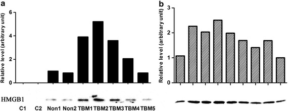
Fig. 2.

Western blotting and grayscale analysis of CSF HMGB1. a Samples from 2 controls, 2 no-TBM patients and 5 TBM patients; b Samples from 9 definite TBM patients. 30 μl CSF was loaded and separated by SDS-PAGE, and HMGB1 was detected using a monoclonal anti-HMGB1 antibody. The grayscale value of protein bands were analyzed by ImageJ software
