

Abstract
The nut weevil (Curculio nucum) is one of the most important and widespread pests in hazelnut orchards. In order to screen entomopathogenic fungal strains with high virulence against C. nucum, the growth rate, sporulation, and cumulative mortality of different Metarhizium anisopliae and Beauveria bassiana strains were investigated, and the process by which M. anisopliae CoM 02 infects C. nucum larvae was observed using scanning electron microscopy. The results indicated that the growth rate and sporulation of different fungal strains significantly differed. Thirteen days after inoculation with M. anisopliae CoM 02, the cumulative mortality of C. nucum larvae reached 100 %, which was considerably higher than that of the other five strains. As the most virulent of the six test strains, the cadaver rate, LT50, and LT90 of M. anisopliae CoM 02 were 93.4 %, 7.05 and 11.90 days, respectively. Analysis of the infection process by scanning electron microscopy showed that the spore attachment, hyphal germination, hyphal rapid growth, and sporulation of M. anisopliae CoM 02 occurred on the 3rd, 5th, 7th, and 11th day after inoculation, respectively, indicating that the infection cycle takes approximately 11 days. This finding suggests that the highly virulent M. anisopliae plays an important role in the biocontrol of C. nucum in China.
Electronic supplementary material
The online version of this article (doi:10.1007/s12088-016-0614-4) contains supplementary material, which is available to authorized users.
Keywords: Metarhizium anisopliae, Beauveria bassiana, Hazelnut weevil
Introduction
The nut weevil (Curculio nucum) is a medium-sized beetle, with characteristics of the Curculionini tribe of the weevil family (Curculionidae). It is one of the most important and widespread pests of hazelnut orchards; its larvae develop in hazelnuts, causing 15 % to more than 30 % nut yield losses [1–3]. C. nucum larvae, which feed on hazelnuts (Fig. 1), are found throughout most of Europe and Asia [3, 4]. In recent years, C. nucum has developed medium to high levels of resistance to the main chemical pesticides used in northeast China, resulting in a need for increased dosage and repeated applications of these pesticides. Frequent applications of pesticides increase the production cost as well as the risk of chemical pollution [5]. Thus, the environment-friendly methods of C. nucum control should be developed in China, as this would result in both food safety and economic benefits.
Fig. 1.
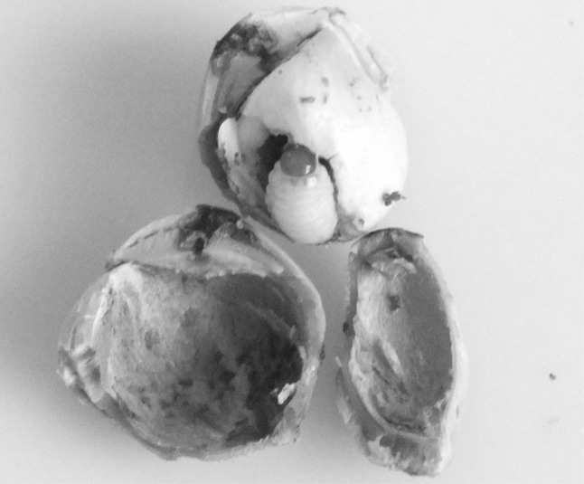
Curculio nucum larvae feeding on a hazelnut
Metarhizium anisopliae and Beauveria bassiana are fungi that grow naturally in soils throughout the world and, as parasitoids, are capable of causing disease in various insects. M. anisopliae and B. bassiana and their related species are used as biological pesticides to control a number of pests such as termites [6], thrips [7], and locusts [8]. M. anisopliae and B. bassiana do not appear to infect humans or other animals and are, therefore, considered safe for use as pesticides.
After egg hatching and feeding in the nut, the mature C. nucum larvae leave the nuts though round holes and burrow into the ground, where they undergo metamorphosis over the next year [2, 3]. Thus, pesticide treatment on mature larvae without hard and thick nutshell protection may be an effective strategy to control their populations. M. anisopliae and B. bassiana are very suitable for culture in vitro, and are expected to be used in the development of a potential formulation for C. nucum biocontrol. However, effective pathogenic strains against C. nucum are lacking in China. Our study aimed to screen highly virulent strains and subsequently test their infectiousness. The findings of this study will be beneficial for developing applicable biocontrol fungal agents for C. nucum.
Materials and Methods
Tested Insects
In 2014, approximately 300 kg of fresh hazelnuts infected by C. nucum was harvested from the area surrounding Siping City, Jilin Province, China. After they were bagged and transported to the laboratory, the infected nuts were piled up and covered by plastic sheeting, to maintain indoor relatively 65 humidity at 25 °C. C. nucum larvae were collected every morning over a 2-week period and were placed in a 1000-mL beaker containing humus with 75 % humidity. The weak C. nucum larvae, which were incapable of burrowing, were excluded from the study. The remaining C. nucum larvae in the beakers were then stored at 4 °C for further experiments.
Tested Fungal Strains
Test strains included four strains of M. anisopliae and two of B. bassiana; their origins are listed in Table S1. Before usage, the strains were transferred to SDY liquid medium (Sabouraud’s dextrose agar, supplemented with 1 % [wt/vol] yeast extract), at a culture temperature and oscillation speed of 25 °C and 100 rpm respectively. The activated strains were then transferred to purple sweet potato dextrose agar (PPDA) and cultured for 10 days. After sporulation, the spores were collected and stored for further usage. The spore germination ratio of the six tested strains was more than 95 % according to the spore germination experiment.
Measurement of Mycelia Growth Speed and Sporulation
Each fungal strain was inoculated on three separate PPDA plates, which were incubated at 25 °C. Their colony diameters were measured every day, and their initial day of sporulation was recorded. The strains were cultured for 15 days. The colony growth rates of the different strains were compared. At the end of the experiment, three holes of 1-cm diameter were created in PPDA using a sterile hole punch. The spores present on each of the PPDA discs produced as a result were oscillated and dispersed in 20 mL of 0.3 % Tween 80 solution. Subsequently, the spore concentration was determined using a blood cell counter, and the spore amount per unit area was calculated.
Measurement of the Cumulative Mortality of C. nucum Larvae After Inoculation
A certain amount of fungal spores was scraped from each of the colonies and suspended into a solution of sterilized 0.3 % Tween 80 at a concentration of 1 × 10⁷ spores/mL. C. nucum larvae were isolated from the humus using a sieve and tweezers. Each larva was immersed in 100 mL of the prepared spore suspension for 8 s. The surplus spore suspension liquid was absorbed using sterilized filter paper. The inoculated larvae were then placed in sterilized insect-rearing boxes containing 1-cm-deep sterilized humus, which were incubated at 25 °C and 75 % relative humidity in total darkness. The same treatment was repeated with each fungal strain. The sterilized 0.3 % Tween 80 solution was used as a control. In total, seven treatments were included in the study, and five independent biological replicates were used for each of the treatment and ten larvae were used in each of the replicates. The number of dead larvae was counted and recorded every day, and any larvae that died within 24 h after inoculation were not counted. The final number of larvae used in each treatment varied from 40 to 48. Mycelium growth and sporulation on the cuticles of larvae were observed with a dissecting microscope to determine whether B. bassiana or M. anisopliae caused the death. The number of dead larvae with visible mycelium and spores was used to calculate the larval mortality rate [9].
Sample Preparation for Scanning Electron Microscopy (SEM)
For primary fixation, the C. nucum larvae were individually immersed in 2.5 % glutaraldehyde solution on the 3rd, 5th, 7th, and 11th days after inoculation at 4 °C for 24 h. The specimens were then washed thrice with 0.1 mol/L phosphate-buffered saline for 15 min, and fixed with 1 % osmium tetroxide (OsO4) solution for 1.5 h. The specimens were again washed thrice with 0.1 mol/L phosphate-buffered saline for 15 min. Subsequently, the larvae were dehydrated in a graded series of ethanol (50, 70, 80, 90, and 95 %), each for 15 min, and then in 100 % ethanol for 20 min, three times. The dehydrated specimens were treated with a mixed solution of ethanol and isoamyl acetate (1:1) for 30 min, and then treated with pure isoamyl acetate for 2 h [10]. Critical point drying was performed with a K850 Critical Point Dryer (Quorum Technologies, Ashford, UK). After platinum ion plating with IS12/IS13 Sputter Coater, the specimens were observed with a KYKY-1000B Scanning Electron Microscope (KYKY technology development Ltd., Beijing, China).
Statistical Analysis
The data were analyzed using the analysis of variance (ANOVA) and (correlation coefficient) CORR features of the software SAS version 8.01 (SAS Institute, Inc., Cary, NC, USA). Means were compared via t-tests with a designated significance level of 5 %. The values expressed as percentages were arcsine-transformed before analysis. The following formula was used to calculate the corrected mortality: Corrected mortality (%) = (mortality of treatment − control mortality) × 100/(100 − control mortality). The calculation of lethality rates LT50 and LT90 was performed as described by Cui et al. [11].
Results
Growth Rate and Sporulation of Different Entomopathogenic Fungal Strains
The growth rates and spore yields of the tested fungal strains are shown in Table 1. We observed significant differences in the growth rates and sporulation of the different strains. After 15 days of culturing on PPDA medium, the colony diameter of M. anisopliae var. acridum CoM 02 strain was found to be the largest at 6.2 cm. This was followed by the diameters recorded for M. anisopliae 3.4607 and M. anisopliae var. acridum 3.7986. The colony diameter of B. bassiana CoB 04 was the smallest of all the strains. These findings also indicated that the fastest growing strain always produced the spores earlier (Table 1).
Table 1.
Growth rate and sporulation of different entomopathogenic fungal strains
| Strain code | Colony diameter (cm) | Initial sporulation time (days) | Sporulation (×108/cm2) | ||
|---|---|---|---|---|---|
| 5 days | 10 days | 15 days | |||
| M. anisopliae var. acridum 3.7986 | 2.0 b | 3.6 ab | 5.7 bc | 3 | 1.5 b |
| M. anisopliae 3.4607 | 2.2 a | 3.7 ab | 6.0 ab | 3 | 1.4 bc |
| M. anisopliae CoM 01 | 1.9 b | 3.4 b | 5.4 c | 3 | 1.2 d |
| M. anisopliae var. acridum CoM 02 | 2.3 a | 3.9 a | 6.2 a | 3 | 1.7 a |
| B. bassiana CoB 03 | 2.0 b | 3.6 ab | 5.6 bc | 3 | 1.3 cd |
| B. bassiana CoB 04 | 1.5 c | 2.8 c | 4.6 d | 5 | 1.3 cd |
Data followed by different letters within the same column are significantly different at P = 0.05 level
Pathogenicity of M. anisopliae and B. bassiana on C. nucum larvae
The cumulative mortality of C. nucum larvae increased gradually with time; however, a significant difference in the cumulative mortality was found after the inoculation of larvae with different fungal strains (Fig. 2; Table S2). Among them, the cumulative mortality of larvae inoculated with M. anisopliae CoM 01 and M. anisopliae var. acridum CoM 02 was significantly higher than that of the other four strains. The cumulative mortality of larvae inoculated with M. anisopliae var. acridum 3.7986, M. anisopliae 3.4607, B. bassiana CoB 03, and B. bassiana CoB 04 strains was relatively lower, and their corrected mortality was less than 60 % on the 15th day after inoculation (Table 2 and Table S2). The cumulative mortality rate of the larvae inoculated with M. anisopliae var. acridum CoM 02 reached 100 % on the 13th day after inoculation, and their death ratio was found to be significantly higher than that of the other five strains. Thus, of the six tested fungal strains, the efficiency of causing host-death was found to be the highest in M. anisopliae var. acridum CoM 02.
Fig. 2.

Cumulative mortality of C. nucum larvae after inoculation with differing strains of Metarhizium anisopliae and Beauveria bassiana. Note: CK, treatment with sterilized 0.3 % Tween 80 solution was used as a control. Key: 3.7986, M. anisopliae var. acridum 3.7986; 3.4607, M. anisopliae 3.4607; CoM 01, M. anisopliae CoM 01; CoM 02, M. anisopliae var. acridum CoM 02; CoB 03, B. bassiana CoB 03; and CoB 04, B. bassiana CoB 04
Table 2.
LT50 and LT90 values of different fungal strains infecting C. nucum larvae
| Strain code | LT50 | LT90 |
|---|---|---|
| M. anisopliae var. acridum 3.7986 | 12.58 b | 23.30 b |
| M. anisopliae 3.4607 | 16.14 a | 28.76 a |
| M. anisopliae CoM 01 | 8.73 c | 15.60 c |
| M. anisopliae var. acridum CoM 02 | 7.05 d | 11.90 d |
| B. bassiana CoB 03 | 17.06 a | 30.49 a |
| B. bassiana CoB 04 | 16.47 a | 28.98 a |
Data followed by different letters within the same column are significantly different at P = 0.05 level
LT50 and LT90 values of the six test strains are shown in Table 2. The results showed that the LT50 and LT90 of M. anisopliae CoM 01 and CoM 02 were significantly lower than those of the other 4 strains tested here, implying that they were the most efficient in causing host-death. The LT50 and LT90 values of M. anisopliae var. acridum CoM 02 were 7.05 and 11.90 days respectively, which were significantly lower than those of the other five strains studied. This finding is consistent with the larvae cumulative mortality curve shown in Fig. 2.
Attachment, Germination, and Sporulation of M. anisopliae CoM 02 on the Cuticles of C. nucum Larvae
The SEM results revealed detail information about the attachment, germination, and sporulation of M. anisopliae var. acridum CoM 02 on the cuticles of C. nucum larvae. Three days after treatment with spores, abundant conidia were present on the depression of the intersegmental cuticle, as shown in Fig. 3a. During this period, little germination of the conidia was observed. Five days after conidial inoculation, many conidia on the cuticle of C. nucum larvae began to germinate, forming long germ tubes (Fig. 3b). Seven days after inoculation, the germ tubes elongated and formed a complicated interlaced net, and the next generation of conidia began to form at the top of the germ tubes (Fig. 3c). Eleven days after inoculation, large amounts of conidia formed on the cuticle of C. nucum larvae (Fig. 3d). Thus, M. anisopliae var. acridum CoM 02 was observed to take approximately 11 days to complete one infection cycle on the cuticle of C. nucum larvae.
Fig. 3.

SEM images showing the process of M. anisopliae CoM 02 infection in C. nucum larvae. a Fungal spores were adhering to the C. nucum larvae cuticle, 3 days after inoculation. b Hyphae germinated from spores were penetrating the C. nucum larvae cuticle, 5 days after inoculation. c Large amount of hyphae formed on the cuticular surface of the C. nucum larvae, 7 days after inoculation. d Large amount of spores had formed on the cuticular surface of C. nucum larvae, 11 days after inoculation. Scale bars a, b = 3.0 μm; c = 6.0 μm; d = 8.0 μm
Discussion
In the recent 5 years, the area of artificial hazelnut culture has increased rapidly in China [12–14], and the yield loss resulting from C. nucum infestation has become more serious. Prevalence of C. nucum has resulted in yield losses in major hazelnut producing countries such as Turkey [15]. Some ideal characteristics of fungal pesticides include cuticle infection, environmental friendliness, and the potential to be produced in large batches [16]. Strains with a high degree of pathogenicity and good growth characteristics are the basis of the C. nucum biocontrol. In general, mortality, rate of host-death, and the median lethal time (LT50) can reflect the pathogenicity of strains on target pests. The greater mortality rate and lower LT50 imply a higher degree of pathogenicity [17]. The mortality rate of M. anisopliae var. acridum CoM 02 reached 93.4 %; its LT50 and LT90 values of 7.05 and 11.90 days respectively were significantly lower than that of the other five tested strains. Furthermore, its sporulation ability was the highest of the six strains tested here. The comparison of its growth speed and mortality rate implied that M. anisopliae var. acridum CoM 02 was the most effective biocontrol strain for C. nucum.
The attachment of fungal conidia to the host cuticle is the prerequisite for establishing a parasitic relationship between fungi and C. nucum larvae. Therefore, the unsuitable host cuticle nutrition and surface bacteria might hinder the infection process of the fungi. Thus, fungal conidia are capable of taking advantage of host nutrition and germination, as they are the key factors for successful fungal infection [18–20]. In this study, the inoculation of the other five strains, besides M. anisopliae var. acridum CoM 02, also caused varying degrees of larval death. In all cases, the cumulative mortality remained stable from the 15th day after inoculation, which implied that the parasitic relationship of these strains with C. nucum larvae was unstable. In some previous studies, M. anisopliae has been found to attach to and germinate on the host cuticle within a few hours after inoculation [21, 22]. However, our SEM results indicated that M. anisopliae var. acridum CoM 02 took much longer, approximately 11 days, to complete its life cycle. This might be due to the difference in the experimental conditions, such as culture moisture and the amount of fungus inoculated. Nevertheless, M. anisopliae var. acridum CoM 02 caused the death of all larvae on the 13th day after inoculation. Further studies are required to determine whether M. anisopliae var. acridum CoM 02 could be successfully used to biocontrol C. nucum in the field.
Electronic supplementary material
Below is the link to the electronic supplementary material.
Acknowledgments
This work was funded by the following grant: International Advanced Agricultural Science and Technology Plan (2015-4-23), State Forestry Bureau of China.
Author’s Contribution
Cheng Y. Q. and Liu J. F. conceived the study and designed the experiments. Chen L. T. provided intellectual advice on the project and experimental design. Liu T., Zhao Y. X. and Geng W. T. performed SEM and pathogencity experiments. Cheng Y. Q. and Liu J. F. wrote the manuscript.
Compliance with Ethical Standards
Conflict of interest
The authors declare no conflicts of interests.
References
- 1.Akça I, Tuncer C. Biological control and morphological studies on nut weevil (curculio nucum l. col., curculionidae) Acta Hortic. 2005;686:413–420. doi: 10.17660/ActaHortic.2005.686.57. [DOI] [Google Scholar]
- 2.Saruhan I, Tuncer C, Mehlenbacher SA. Population densities and seasonal fluctuations of hazelnut pests in Samsun, Turkey. Acta Hortic. 2001;556:495–502. doi: 10.17660/ActaHortic.2001.556.72. [DOI] [Google Scholar]
- 3.Wojciechowicz E. Infestation of hazel nuts by hazelnut weevil (Curculio nucum L., coleoptera, curculionidae) in Poland. J Plant Prot Res. 2005;45:59–61. [Google Scholar]
- 4.Batalla-Carrera L, Morton A, García-Del-Pino F. Field efficacy against the hazelnut weevil, Curculio nucum and short-term persistence of entomopathogenic nematodes. Span J Agric Res. 2013;11:1112–1119. doi: 10.5424/sjar/2013114-4210. [DOI] [Google Scholar]
- 5.Greenfield M, Gómez-Jiménez MI, Ortiz V, Vega FE, Kramer M, Parsa S. Beauveria bassiana, and Metarhizium anisopliae, endophytically colonize cassava roots following soil drench inoculation. Biol Control. 2016;95:40–48. doi: 10.1016/j.biocontrol.2016.01.002. [DOI] [PMC free article] [PubMed] [Google Scholar]
- 6.Maniania NK, Ekesi S, Songa JM. Managing termites in maize with the entomopathogenic fungus Metarhizium anisopliae. Int J Trop Insect Sci. 2002;22:41–46. doi: 10.1017/S1742758400015046. [DOI] [Google Scholar]
- 7.Ekesi S, Maniania NK, Lwande W. Susceptibility of the legume flower thrips to Metarhizium anisopliae on different varieties of cowpea. Biocontrol. 2012;45:79–95. doi: 10.1023/A:1009927302916. [DOI] [Google Scholar]
- 8.Ouedraogo RM, Cusson M, Goettel MS, Brodeur J. Inhibition of fungal growth in thermoregulating locusts, Locusta migratoria, infected by the fungus Metarhizium anisopliae var acridum. J Invertebr Pathol. 2003;82:103–109. doi: 10.1016/S0022-2011(02)00185-4. [DOI] [PubMed] [Google Scholar]
- 9.He XY, Cai SP, Tong YH, Xiong Y, Huang Y, Xie JD, Chen SL. Pathogenicity evaluation of the entomopathogenic fungi Beauveria bassiana and Metarhizium anisopliae against adults of Basilepta melanopus (Coleoptera: Eumolpidae) Acta Entomol Sin. 2011;54:1281–1287. [Google Scholar]
- 10.Zhou X, Wang DW, Guo K, Mao SF. Germination and sporulation of Pandora delphacis (Entomophthoromycota: Entomophthorales) on the rice pest Nilaparvata lugens: SEM observation. Mycosystema. 2014;33:819–826. [Google Scholar]
- 11.Cui Q, Zhang Y, Zang Y, Nong X, Wang G, Zhang Z. Screening of high toxic Metarhizium strain against Plutella xylostella and its marking with green fluorescent protein. World J Microb Biot. 2014;30:2767–2773. doi: 10.1007/s11274-014-1700-6. [DOI] [PubMed] [Google Scholar]
- 12.Liu JF, Zhang HD, Cheng YQ, Wang J, Zhao YX, Geng WT. Comparison of ultrastructure, pollen tube growth pattern and starch content in developing and abortive ovaries during the progamic phase in hazel. Front Plant Sci. 2014;5:528. doi: 10.3389/fpls.2014.00528. [DOI] [PMC free article] [PubMed] [Google Scholar]
- 13.Cheng YQ, Wang J, Liu JF, Zhao YX, Geng WT, Zhang HD. Analysis of ovary DNA methylation during delayed fertilization in hazel using the methylation-sensitive amplification polymorphism technique. Acta Physiol Plant. 2015;37:231. doi: 10.1007/s11738-015-1984-7. [DOI] [Google Scholar]
- 14.Cheng YQ, Liu JF, Zhang HD, Wang J, Zhao YX, Geng WT. Transcriptome analysis and gene expression profiling of abortive and developing ovules during fruit development in hazelnut. PLoS ONE. 2015;10:e0122072. doi: 10.1371/journal.pone.0122072. [DOI] [PMC free article] [PubMed] [Google Scholar]
- 15.Akça I, Tuncer C, Saruhan I. Sex discrimination of adult Curculio nucum L. (coleoptera: curculionidae), a pest of hazelnuts in Turkey. Entomol News. 2007;118:87–89. doi: 10.3157/0013-872X(2007)118[87:SDOACN]2.0.CO;2. [DOI] [Google Scholar]
- 16.Zimmermann G. Review on safety of the entomopathogenic fungus Metarhizium anisopliae. Biocontrol Sci Tech. 2017;17:879–920. doi: 10.1080/09583150701593963. [DOI] [Google Scholar]
- 17.Wekesa VW, Maniania NK, Knapp M, Boga HI. Pathogenicity of Beauveria bassiana and Metarhizium anisopliae to the tobacco spider mite Tetranychus evansi. Exp Appl Acaro. 2005;36:41–50. doi: 10.1007/s10493-005-0508-3. [DOI] [PubMed] [Google Scholar]
- 18.Mosbach A, Leroch M, Mendgen KW, Hahn M. Lack of evidence for a role of hydrophobins in conferring surface hydrophobicity to conidia and hyphae of Botrytis cinerea. BMC Microbiol. 2011;11:10. doi: 10.1186/1471-2180-11-10. [DOI] [PMC free article] [PubMed] [Google Scholar]
- 19.Braun EJ, Howard RJ. Adhesion of fungal spores and germlings to host plant surfaces. Protoplasma. 1994;181:202–212. doi: 10.1007/BF01666396. [DOI] [Google Scholar]
- 20.Hajek AE, Eastburn CC. Attachment and germination of Entomophaga maimaiga conidia on host and non-host larval cuticle. J Invertebr Pathol. 2003;82:12–22. doi: 10.1016/S0022-2011(02)00198-2. [DOI] [PubMed] [Google Scholar]
- 21.Pires LM, Marques EJ, Valéria WT, Teixeira AAC, Alves LC, Alves ESB. Ultrastructure of Tuta absoluta parasitized eggs and the reproductive potential of females after parasitism by Metarhizium anisopliae. Micron. 2009;40:255–261. doi: 10.1016/j.micron.2008.07.008. [DOI] [PubMed] [Google Scholar]
- 22.Vestergaard S, Gillespie AT, Butt TM, Schreiter G, Eilenberg J. Pathogenicity of the hyphomycete fungi Verticillium lecanii and Metarhizium anisopliae to the western flower thrips, Frankliniella occidentalis. Biocontrol Sci Techn. 1995;5:185–192. doi: 10.1080/09583159550039909. [DOI] [Google Scholar]
Associated Data
This section collects any data citations, data availability statements, or supplementary materials included in this article.