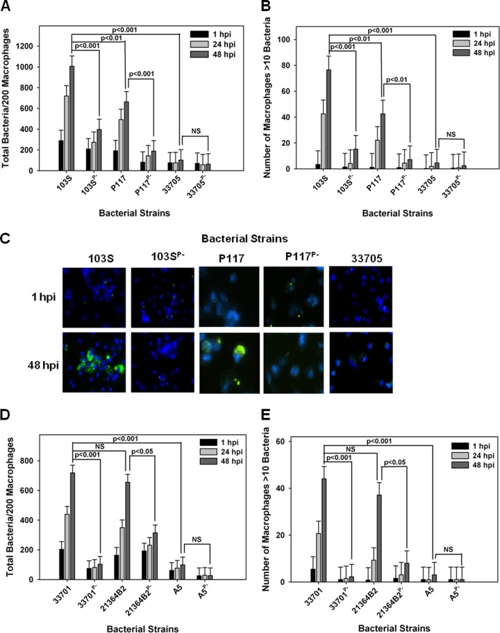
FIG 2 .
pVAPB-type plasmid-containing R. equi isolates display variable replication ability in equine macrophages. The intracellular growth of R. equi in equine alveolar macrophages infected with strains 103S, 103SP−, P117, P117P−, 33705, 33705P−, 33701, 33701P−, 21364B2, 21364B2P−, A5, and A5P− at an MOI of 5:1 was assessed. Following a 1-h incubation allowing for phagocytosis, monolayers were washed and medium supplemented with amikacin was added to the monolayers to kill any extracellular bacteria. Triplicate monolayers were fixed at 1 h, 24 h, and 48 h postinfection (hpi), stained as described in Materials and Methods, and examined under fluorescence microscopy. The number of bacteria per 200 macrophages (A and D) and the number of macrophages with greater than 10 bacteria per 200 macrophages (B and E) were counted. Representative images of the infected macrophage monolayers are shown (×100 magnification) (C). In these representative microscopy images, R. equi displays green fluorescence and the macrophage nucleus is DAPI stained. Error bars represent standard deviations from the means. Data and statistical analysis are a compilation of 4 individual experiments. NS, not significant.

