Abstract
Aim:
The aim of this study is to compare the retention force of three different types of overdenture attachment systems used in implant-retained mandibular complete overdentures.
Materials and Methods:
Twenty-one similar acrylic resin blocks were prepared and divided into three study groups: Group A (snap attachment) - 10 specimens, Group B (locator attachment) - 1 specimen, and Group C (syncone attachment) - 10 specimens. A single rectangular heat cure acrylic resin block with two implant analogs 22 mm apart was used with all specimens. Each specimen was subjected to 5500 cycles of insertion and removal in the presence of artificial saliva, representing 5 years of usage. Retention was measured three times for each specimen using universal testing machine. Data were analyzed using one-way and two-way analysis of variance at 95% level of confidence.
Results:
Locator attachment group (Group B) showed the greatest retention level throughout the study, followed by snap attachment (Group A), and syncone attachment (Group C) showed the lowest retention level.
Conclusion:
Regardless of the initial retention level of overdenture attachment, gradual loss of retention values is inevitable. However, the rate of retention loss in overdenture attachments is higher in types which comprised plastic parts within their components, rather than those totally made up of noble metals.
Keywords: Fatigue strength, locator, overdenture attachment, retention force, snap, syncone
INTRODUCTION
Many edentulous patients are satisfied with their conventional complete dentures although some often encounter problems with this design. These problems included poor stability and retention of the prosthesis, pain during mastication, and decreased chewing efficiency. Several clinical studies have shown that these problems can be successfully addressed using a dental prosthesis employed in conjunction with dental implants in implant-supported or implant-retained overdentures.[1] The concept of overdentures originally involved fixing mechanical attachments to teeth roots to enhance retention and stability of conventional dentures. In recent years, various attachments systems have been successfully used with removable implant overdentures.[2,3,4] Snap attachment is one of the ball and socket types of attachment. The abutment of snap attachment (the male part) is screwed over the implant fixture. on the other hand, the female part is fixed in the fitting surface of the overdentue. The locator attachment was introduced in 2001 and does not use the splinting of implants. This attachment is self-aligning, has dual retention through both external and internal mating surfaces,[5,6] resilient, durable, and has some built-in angulation compensation.[7] Telescopic type of removable prostheses with immediate loading has been used in the edentulous or partially edentulous mandible.[8,9] Previous investigations of attachments are in agreement that loss of retentive force over time is inevitable. This loss of retention has been attributed to wear of attachment components.[2] Comparison of the retentive capacity of the previously mentioned overdenture attachments over time will be discussed in details in our study. In our null hypothesis, there is no difference in the retention among the three types of attachment.
MATERIALS AND METHODS
Preparation of analog base block
In our study, a single rectangular heat cure acrylic resin (Acrostone Manufacturing and Import Co, Cairo, Egypt), base block with the dimensions of 4 cm in length, 2 cm in width, and 3 cm in height, had been constructed. The analog holes were prepared 22 mm apart from each other by the aid of an acrylic stent to ensure parallelism and avoid discrepancies arise from misalignment of attachment components which accelerates wear mechanisms.[3,10,11] It was made of clear heat cure acrylic resin (Acrostone Manufacturing and Import Co., Cairo, Egypt) with the same length (4 cm), width (2 cm), but of a lower thickness (1 cm) with two vertical metal sleeves installed parallel to each other by aid of the surveyor (Ney Surveyor, Dentsply, NY, USA), 22 mm apart from each other. Holes were made so that the top of the two implant analogs (Ankylos, Dentsply Friadent, Mannheim, Germany) 4 mm in diameter, 12 mm in length was 1 mm below the top surface of the base block. A cold cure acrylic resin (Acrostone Manufacturing and Import Co., Cairo, Egypt) was used to seal the space between the analog and side walls of the drilled hole.
Heat cure acrylic resin cylinder 5 cm length, 1 cm diameter was prepared. A wide hole was prepared in the undersurface of the base block to accommodate dimensions of the acrylic resin cylinder with a sufficient room for cold cure acrylic resin for fixation of the acrylic cylinder in the base block [Figure 1].
Figure 1.
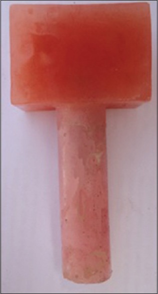
The acrylic cylinder fixed to the bottom of the acrylic base block
Preparation and grouping of overdenture simulating blocks
Twenty-one heat cure acrylic resin blocks 4 cm in length, 2 cm in width, and 2 cm in height were prepared and divided into as follows.
Group A: Including ten blocks that were used for snap attachment (Ankylos, Dentsply Friadent, Mannheim, Germany)
Group B: Including a single block used for locator attachment (Zest Anchors Inc., for Ankylos, Dentsply Friadent, Manhiem Germany) group, due to the interchanging capacity of the nylon male, this single block had been used for ten samples by changing the nylon male after each full cycle of measurement
Group C: Including ten blocks that were used in syncone attachment (Ankylos, Dentsply Friadent, Mannheim, Germany), Telescopic crown, group (contained secondary crowns of syncone attachment).
Following manufacturer instructions of different attachment types, direct pick-up technique was used for installing of attachment components (abutments and denture part) in the three study groups.
Retention force measurement
We adopted the methodology utilized by many other studies as Gamborena et al.,[12] Fromentin et al.,[13] Botega et al.,[14] da Fontoura Frasca et al.,[15] Branchi et al.,[16] and Evtimovska et al.[6] for evaluation of retention force of the selected attachments. The Instron universal testing machine was used to measure the retention forces. The analog base block was attached to the lower compartment of the universal testing machine while overdenture simulating blocks were attached to the upper compartment of the machine. To maintain the presence of saliva throughout the testing process, the base block remained in the bottom of a plastic deep pool while the vertical cylinder penetrates the bottom of the pool to be attached to the lower compartment of the universal testing machine. Adhesive silicone was used to seal minor spaces between the cylinder base block joint and the bottom of the pool. The pool was filled by artificial saliva (Xerotin Artificial Saliva, UK, 1.5 mm Ca, 3.0 mm P, 20.0 mm NaHCO3, pH 7.0) to a level above the top of the base block by about 2 mm [Figure 2a and b].
Figure 2.
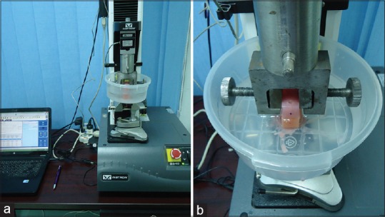
(a) Overview of a specimen mounted in the Instron. (b) A closer view
Dislodgement of the blocks containing denture parts of the three attachments was performed using the universal testing machine parallel to the long axis of the implant analogs. The crosshead speed was adjusted at 50 mm/min as this has been reported to approximate the speed of the movement of the denture away from the ridge in vivo.[17,18]
The Instron universal testing machine controlled by a computer to interface the (Bluehill, ITW Inc., England) software was used to apply maximum seating and dislodging forces for each specimen at a cross head speed of 50 mm/s. It was programmed to apply 5500 cycles of displacement and insertion of 4 mm magnitude of movement at frequency of 12 cycles/min. Retention forces were calculated three times (initially, after 3000 and 5500 cycles).
A specimen was considered failed if showed separation between attachment components and over-denture simulating blocks, or separation of attachment components itself.
Digital light microscopy examination
Using digital light microscope (Celestron, LLC., Torrance, CA, USA), a randomly selected denture parts of all attachments had been examined using the same object-lens distance (13 mm) and the same magnification (×40) had been used for all specimens.
Statistical analysis
Data are presented as means and standard deviations (SDs); the level of significance was set at 0.05 for all tests. Data analysis was performed using the statistical software SPSS 16.0 (SPSS Inc., Chicago, IL, USA) for Windows. Statistical analysis was performed using one-way analysis of variance at 95% level of confidence whenever a statistical significant difference was recorded among different tested groups, Tukey–Kramer post hoc test was performed to make pairwise comparisons between each two significant difference groups or sections.
RESULTS
Table 1 shows mean ± SD of retention levels recorded at different number of cycling in each study group and percent change of retention values from retention value recorded initially within the same group [Figure 3]. Neither of the specimens had reported failure at any stage of the study.
Table 1.
Mean±standard deviation of retention levels recorded throughout the study and percent change in retention throughout the study
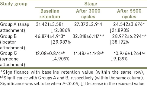
Figure 3.
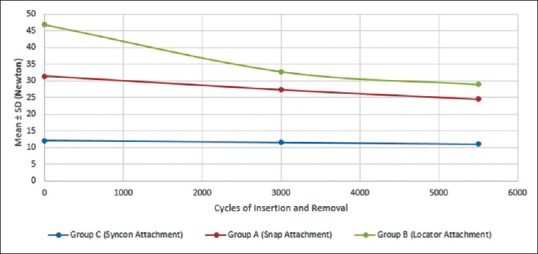
Changes in retention level during different stages of the study
In comparison of retention values of different systems at the same number of cycling, all systems were significantly different from each other, where locator attachment group showed the greatest retention values followed by snap attachment, and syncone attachment group showed the lowest retention values throughout the study.
In comparison of retention level changes in each group after different number of cycling, the retention of snap attachment group significantly decreased by 21.893% from retention levels recorded initially after 5500 cycles. Retention of locator attachment group significantly decreased by 29.987% and 38.192% from retention levels recorded initially after 3000 and 5500 cycles, respectively. However, there was no statistically significant difference in retention levels recorded at different stages of measurement. Digital light microscopy examination revealed distortion of nylon insert of locator attachment [Figure 4] loss of surface texture of snap attachment [Figure 5] and minimal changes in surface characteristics of syncone attachment system [Figure 6].
Figure 4.

Nylon male part (a) before cyclic removal and insertion, (b) after 3000 cycles, (c) after 5500 cycles
Figure 5.
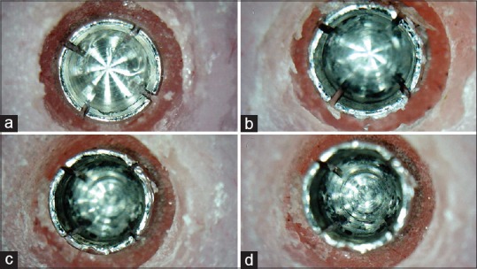
Changes in snap female at different levels of cycling: (a) Snap female at the beginning, (b) after 3000 cycle, (c) after 5500 cycles, (d) after 5500 cycles a closer view from inside
Figure 6.
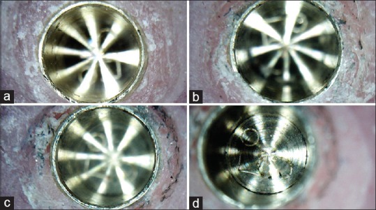
Changes in syncone secondary crown at different levels of cycling: (a) Syncone secondary crown at the beginning, (b) after 3000 cycle, (c) after 5500 cycles, (d) after 5500 cycles a closer view from inside
DISCUSSION
Using complete dentures is a challenge because of decrease of force and muscle coordination and difficulty in achieving an acceptable level of prosthesis retention and stability due to bone resorption.[19,20]
In this study, we simulate the use of only two implant analogs installed in acrylic resin block for stabilization of the lower denture to provide reliable and predictable treatment outcomes. It is regarded as the minimum standard of care for completely edentulous patients.[21,22]
These analogs were 22 mm apart as it had been reported that natural canines are separated by the same distance.[23] On the other hand, Doukas et al. stated that there are no standardized rules for the ideal distance between attachments to achieve optimum retention.[24]
Traditional Instron (IS) testing machines are the most common instrument used to replicate the vertical separation of the denture from the mouth.[25] It had been accepted as reliable and valid instrument to test peak load forces in vitro.[17,26,27,28]
There is no consensus in the literature considering number of cycles should different specimens subjected to, where different number of cycles had been used in different studies; for example, 10,000[29] 5500[12,14,30] 3000,[15,31] or 14,600 cycles.[32]
In this study, the number of cycles to which specimens were subjected was 5500 cycles corresponds to 5 years usage of dentures considering average three removals per day, which is considered long enough for prosthesis replacement.[28]
Fatigue or failure of overdenture attachments adversely affects their function, maintenance, and patient satisfaction.[33] After an appropriate number of insertion removal cycles, attachments obtain more stable retentive properties, which represent the postinsertion period.[34] Accordingly, this study compared retentive properties of the attachments in this postinsertion period and not to limit it to the initial assessment only.[25]
The presence of artificial saliva at room temperature was maintained throughout the study to simulate a wet oral environment to sufficiently form a protective and lubricant layer between attachment components.[35] On the other hand, the principle of the use of a lubricant is an accepted standard for wear simulation systems.[29,30,36]
On the other hand, the composition and temperature of saliva could also influence the results, mainly if one considers the susceptibility of polymeric components to absorb water.[14]
The three attachment systems under evaluation met the minimum value of 5 N, required for the stability of overdenture throughout the study.[14,15] On the other hand, the locator attachment showed a significantly higher retention values than the other systems.
The locator attachment greater retention had been reported in many previous studies.[15,37,38] This could be attributed to that locator attachment has dual retention feature that means the male part will retain on the inside and outside of the abutment.[39] Moreover, the greater cross-section of the locator attachment increases surface area available for frictional contact between components of these attachments.[39]
However, regardless of the high retention values recorded at the beginning of the study, locator attachment showed the greatest decrease of retention overtime which is in agreement with many previous reports.[18,32,40] This could be attributed to wear and plastic deformation of the nylon inserts.[32,41] Such findings had been proved by digital microscopy examination of nylon male parts at different stages of the study [Figure 4].
Snap attachment showed a lower retention level than locator attachment. This could be attributed to greater resiliency of ball attachment, resulting in a lower initial retention forces.[15] An additional mechanism described to interpret the greater retention forces of locator attachment is that ball attachments are inserted with minimum pressure while in case of locator, a higher effort for proper position of overdenture is needed and patients biting force is not sufficient; hence, a bimanual press on is necessary. Consequently, a higher forces to draw off the overdenture is required.[38]
However, despite the lower retention of snap attachment, it did not decrease significantly except after 5 years of usage which had been confirmed by light microscopy [Figure 5]. This could be attributed to that the female part is made of noble metal alloy with high percentage of gold that has great malleability and ductility and promotes the adaptation and maintenance of the retention forces, being in accordance with Bayer et al., who also found greater retention forces for metal components that maintained for a longer period.[36]
The lowest retention values of syncone overdenture attachment as shown in Table 1 could be attributed to that it depends only on friction of facing smooth surfaces[42] with no additional means of retention like the other two types of attachment in our study. Our results are supported by studies of many authors who stated that the tapered telescopes exhibit friction only when completely seated; as soon as the outer crown is dislodged even slightly from the terminal position, retention of the system is lost avoiding undue forces on the abutment.[42,43]
In contrast to locator attachment and snap attachment, syncone overdenture attachment system showed a stable retentive properties in concurrence with Zhang et al. in 2008, who found that the retention forces of ankylose syncone conical crown system where almost constant during the entire testing which involved 5000 insertion removal cycle.[37] Another in vivo study confirmed that after 30.3 months of usage, conical crown concept offers a stable complete denture retention.[44]
The statistically similar values obtained from the beginning to end of the tests in this case resulted from the combination of both abutment and a retention component made of noble metal alloy. Further confirmation of such finding had been revealed form light microscopy examination of secondary crowns of syncone attachment system revealed a very little change in surface characters and borders [Figure 6].
CONCLUSION
Within the limitations of the present in vitro study, the results suggested that
All systems evaluated showed retention values higher than 5 N, the minimum required for stabilizing the prosthesis, being considered suitable for clinical use
The locator attachment system showed higher retention values than snap and syncone attachment
Retention loss is inevitable in all types of attachment due to different mechanisms; however, the rate of loss varies according to attachment components as well as the mode of attachment retention
The rate of retention loss in overdenture attachments was higher in attachment types which comprised plastic parts within their components, rather than those totally made up of noble metals.
Financial support and sponsorship
Nil.
Conflicts of interest
There are no conflicts of interest.
REFERENCES
- 1.Chen YF, Yang YH, Lee JH, Chen JH, Lee HE, Chou TM. Tongue support of complete dentures in the elderly. Kaohsiung J Med Sci. 2012;28:273–8. doi: 10.1016/j.kjms.2011.11.005. [DOI] [PubMed] [Google Scholar]
- 2.Alsabeeha NH, Payne AG, Swain MV. Attachment systems for mandibular two-implant overdentures: A review of in vitro investigations on retention and wear features. Int J Prosthodont. 2009;22:429–40. [PubMed] [Google Scholar]
- 3.Dileep Nag V, Ravindra P, Thirupathi Reddy B. A simplified chair-side technique with pre-fabricated directional rings in a case of divergent root retained overdenture. J Indian Prosthodont Soc. 2011;11:130–2. doi: 10.1007/s13191-011-0070-0. [DOI] [PMC free article] [PubMed] [Google Scholar]
- 4.John J, Rangarajan V, Savadi RC, Satheesh Kumar KS, Satheesh Kumar P. A finite element analysis of stress distribution in the bone, around the implant supporting a mandibular overdenture with ball/o ring and magnetic attachment. J Indian Prosthodont Soc. 2012;12:37–44. doi: 10.1007/s13191-012-0114-0. [DOI] [PMC free article] [PubMed] [Google Scholar]
- 5.Trakas T, Michalakis K, Kang K, Hirayama H. Attachment systems for implant retained overdentures: A literature review. Implant Dent. 2006;15:24–34. doi: 10.1097/01.id.0000202419.21665.36. [DOI] [PubMed] [Google Scholar]
- 6.Evtimovska E, Masri R, Driscoll CF, Romberg E. The change in retentive values of locator attachments and hader clips over time. J Prosthodont. 2009;18:479–83. doi: 10.1111/j.1532-849X.2009.00474.x. [DOI] [PubMed] [Google Scholar]
- 7.Büttel AE, Bühler NM, Marinello CP. Locator or ball attachment: A guide for clinical decision making. Schweiz Monatsschr Zahnmed. 2009;119:901–18. [PubMed] [Google Scholar]
- 8.May D, Romanos ME. Immediate implant-supported mandibular overdentures retained by conical crowns: A new treatment concept. Quintessence Int. 2001;52:283–90. [Google Scholar]
- 9.Romanos GE, May S, May D. Immediate loading of tooth-implant-supported telescopic mandibular prostheses. Int J Oral Maxillofac Implants. 2012;27:1534–40. [PubMed] [Google Scholar]
- 10.Misch CE. Dental Implant Prosthetics. 3rd ed. Vol. 1. USA: Elsevier Mosby; 2005. [Google Scholar]
- 11.Atashrazm P, Dashti MH, Alavijeh LZ, Azarmaeh S, Mahdizadeh S. The influences of implant angulations in one and two directions on the retentive properties of overdenture attachments: An in vitro study. J Indian Prosthodont Soc. 2014;14:72–7. doi: 10.1007/s13191-013-0272-8. [DOI] [PMC free article] [PubMed] [Google Scholar]
- 12.Gamborena JI, Hazelton LR, NaBadalung D, Brudvik J. Retention of ERA direct overdenture attachments before and after fatigue loading. Int J Prosthodont. 1997;10:123–30. [PubMed] [Google Scholar]
- 13.Fromentin O, Picard B, Tavernier B. In vitro study of the retention and mechanical fatigue behavior of four implant overdenture stud-type attachments. Pract Periodontics Aesthet Dent. 1999;11:391–7. [PubMed] [Google Scholar]
- 14.Botega DM, Mesquita MF, Henriques GE, Vaz LG. Retention force and fatigue strength of overdenture attachment systems. J Oral Rehabil. 2004;31:884–9. doi: 10.1111/j.1365-2842.2004.01308.x. [DOI] [PubMed] [Google Scholar]
- 15.da Fontoura Frasca LC, Castro Mattia PR, Botega DM, Rivaldo EG. Evaluation of retention forces and resistance to fatigue of attachment systems for overdentures: Plastic and metal components. Implant Dent. 2014;23:451–5. doi: 10.1097/ID.0000000000000093. [DOI] [PubMed] [Google Scholar]
- 16.Branchi R, Vangi D, Virga A, Guertin G, Fazi G. Resistance to wear of four matrices with ball attachments for implant overdentures: A fatigue study. J Prosthodont. 2010;19:614–9. doi: 10.1111/j.1532-849X.2010.00613.x. [DOI] [PubMed] [Google Scholar]
- 17.Williams BH, Ochiai KT, Hojo S, Nishimura R, Caputo AA. Retention of maxillary implant overdenture bars of different designs. J Prosthet Dent. 2001;86:603–7. doi: 10.1067/mpr.2001.120838. [DOI] [PubMed] [Google Scholar]
- 18.Rutkunas V, Mizutani H. Retentive and stabilizing properties of stud and magnetic attachments retaining mandibular overdenture. An in vitro study. Stomatol Baltic Dent Maxillofac J. 2004;6:85–90. [Google Scholar]
- 19.Lahori M, Kaul AS, Chandra S, Nagrath R, Gupta H. Comparative evaluation of bone in mandibular implant retained overdentures using delayed and immediate loading protocol: An in-vivo study. J Indian Prosthodont Soc. 2013;13:113–21. doi: 10.1007/s13191-012-0240-8. [DOI] [PMC free article] [PubMed] [Google Scholar]
- 20.Cristache CM, Muntianu LA, Burlibasa M, Didilescu AC. Five-year clinical trial using three attachment systems for implant overdentures. Clin Oral Implants Res. 2014;25:e171–8. doi: 10.1111/clr.12086. [DOI] [PubMed] [Google Scholar]
- 21.Thomason JM, Feine J, Exley C, Moynihan P, Müller F, Naert I, et al. Mandibular two implant-supported overdentures as the first choice standard of care for edentulous patients – The York consensus statement. Br Dent J. 2009;207:185–6. doi: 10.1038/sj.bdj.2009.728. [DOI] [PubMed] [Google Scholar]
- 22.Koole S, De Bruyn H. Contemporary undergraduate implant dentistry education: A systematic review. Eur J Dent Educ. 2014;18(Suppl 1):11–23. doi: 10.1111/eje.12076. [DOI] [PubMed] [Google Scholar]
- 23.Takeshita S, Kanazawa M, Minakuchi S. Stress analysis of mandibular two-implant overdenture with different attachment systems. Dent Mater J. 2011;30:928–34. doi: 10.4012/dmj.2011-134. [DOI] [PubMed] [Google Scholar]
- 24.Doukas D, Michelinakis G, Smith PW, Barclay CW. The influence of interimplant distance and attachment type on the retention characteristics of mandibular overdentures on 2 implants: 6-month fatigue retention values. Int J Prosthodont. 2008;21:152–4. [PubMed] [Google Scholar]
- 25.Chung KH, Chung CY, Cagna DR, Cronin RJ., Jr Retention characteristics of attachment systems for implant overdentures. J Prosthodont. 2004;13:221–6. doi: 10.1111/j.1532-849X.2004.04042.x. [DOI] [PubMed] [Google Scholar]
- 26.Breeding LC, Dixon DL, Schmitt S. The effect of simulated function on the retention of bar-clip retained removable prostheses. J Prosthet Dent. 1996;75:570–3. doi: 10.1016/s0022-3913(96)90465-5. [DOI] [PubMed] [Google Scholar]
- 27.Petropoulos VC, Smith W, Kousvelari E. Comparison of retention and release periods for implant overdenture attachments. Int J Oral Maxillofac Implants. 1997;12:176–85. [PubMed] [Google Scholar]
- 28.Fromentin O, Lassauzay C, Abi Nader S, Feine J, de Albuquerque Junior RF. Testing the retention of attachments for implant overdentures – Validation of an original force measurement system. J Oral Rehabil. 2010;37:54–62. doi: 10.1111/j.1365-2842.2009.02020.x. [DOI] [PubMed] [Google Scholar]
- 29.Wichmann MG, Kuntze W. Wear behavior of precision attachments. Int J Prosthodont. 1999;12:409–14. [PubMed] [Google Scholar]
- 30.Pigozzo MN, Mesquita MF, Henriques GE, Vaz LG. The service life of implant-retained overdenture attachment systems. J Prosthet Dent. 2009;102:74–80. doi: 10.1016/S0022-3913(09)60112-8. [DOI] [PubMed] [Google Scholar]
- 31.Nagaoka E, Nagayasu Y, Yamashita H, Matsushiro H, Okuno Y. Study of retention in attachments for overdenture. (II) O-ring attachment. J Osaka Univ Dent Sch. 1980;20:215–26. [PubMed] [Google Scholar]
- 32.Kobayashi M, Srinivasan M, Ammann P, Perriard J, Ohkubo C, Müller F, et al. Effects of in vitro cyclic dislodging on retentive force and removal torque of three overdenture attachment systems. Clin Oral Implants Res. 2014;25:426–34. doi: 10.1111/clr.12156. [DOI] [PubMed] [Google Scholar]
- 33.Payne AG, Solomons YF. Mandibular implant-supported overdentures: A prospective evaluation of the burden of prosthodontic maintenance with 3 different attachment systems. Int J Prosthodont. 2000;13:246–53. [PubMed] [Google Scholar]
- 34.Besimo CE, Guarneri A. In vitro retention force changes of prefabricated attachments for overdentures. J Oral Rehabil. 2003;30:671–8. doi: 10.1046/j.1365-2842.2003.01140.x. [DOI] [PubMed] [Google Scholar]
- 35.Bayer S, Keilig L, Kraus D, Grüner M, Stark H, Mues S, et al. Influence of the lubricant and the alloy on the wear behaviour of attachments. Gerodontology. 2011;28:221–6. doi: 10.1111/j.1741-2358.2009.00352.x. [DOI] [PubMed] [Google Scholar]
- 36.Bayer S, Steinheuser D, Grüner M, Keilig L, Enkling N, Stark H, et al. Comparative study of four retentive anchor systems for implant supported overdentures – retention force changes. Gerodontology. 2009;26:268–72. doi: 10.1111/j.1741-2358.2009.00286.x. [DOI] [PubMed] [Google Scholar]
- 37.Zhang RG, Hannak WB, Roggensack M, Freesmeyer WB. Retentive characteristics of Ankylos SynCone conical crown system over long-term use in vitro . Eur J Prosthodont Restor Dent. 2008;16:61–6. [PubMed] [Google Scholar]
- 38.Kleis WK, Kämmerer PW, Hartmann S, Al-Nawas B, Wagner W. A comparison of three different attachment systems for mandibular two-implant overdentures: One-year report. Clin Implant Dent Relat Res. 2010;12:209–18. doi: 10.1111/j.1708-8208.2009.00154.x. [DOI] [PubMed] [Google Scholar]
- 39.Sadig W. A comparative in vitro study on the retention and stability of implant-supported overdentures. Quintessence Int. 2009;40:313–9. [PubMed] [Google Scholar]
- 40.Rutkunas V, Mizutani H, Takahashi H, Iwasaki N. Wear simulation effects on overdenture stud attachments. Dent Mater J. 2011;30:845–53. doi: 10.4012/dmj.2011-057. [DOI] [PubMed] [Google Scholar]
- 41.Abi Nader S, de Souza RF, Fortin D, De Koninck L, Fromentin O, Albuquerque Junior RF. Effect of simulated masticatory loading on the retention of stud attachments for implant overdentures. J Oral Rehabil. 2011;38:157–64. doi: 10.1111/j.1365-2842.2010.02145.x. [DOI] [PubMed] [Google Scholar]
- 42.Saito M, Miura Y, Notani K, Kawasaki T. Stress distribution of abutments and base displacement with precision attachment- and telescopic crown-retained removable partial dentures. J Oral Rehabil. 2003;30:482–7. doi: 10.1046/j.1365-2842.2003.01092.x. [DOI] [PubMed] [Google Scholar]
- 43.Wenz HJ, Lehmann KM. A telescopic crown concept for the restoration of the partially edentulous arch: The Marburg double crown system. Int J Prosthodont. 1998;11:541–50. [PubMed] [Google Scholar]
- 44.Eccellente T, Piombino M, Piattelli A, Perrotti V, Iezzi G. A new treatment concept for immediate loading of implants inserted in the edentulous mandible. Quintessence Int. 2010;41:489–95. [PubMed] [Google Scholar]


