Abstract
Ran, an important family of small GTP-binding proteins, has been shown to regulate a variety of important cellular processes in many eukaryotes. However, little is known about Ran function in pathogenic fungi. In this study, we report the identification and functional analysis of a Ran gene (designated PsRan) from Puccinia striiformis f. sp. tritici (Pst), an important fungal pathogen affecting wheat production worldwide. The PsRan protein contains all conserved domains of Ran GTPases and shares more than 70% identity with Ran proteins from other organisms, indicating that Ran proteins are conserved in different organisms. PsRan shows a low level of intra-species polymorphism and is localized to the nucleus. qRT-PCR analysis showed that transcript level of PsRan was induced in planta during Pst infection. Silencing of PsRan did not alter Pst virulence phenotype but impeded fungal growth of Pst. In addition, heterologous overexpression of PsRan in plant failed to induce cell death but suppressed cell death triggered by a mouse BAX gene or a Pst Ras gene. Our results suggest that PsRan is involved in the regulation of fungal growth and anti-cell death, which provides significant insight into Ran function in pathogenic fungi.
Small GTP-binding proteins in eukaryotes from yeast to human constitute a superfamily, which includes more than 100 members and is structurally classified into at least five families: Ras, Rho, Rab, Sar1/Arf, and Ran1. The Ran (Ras-related nuclear) protein was originally isolated as a homolog of Ras proteins and eukaryotes usually contain one to four Ran genes2. As the only known family of small GTP-binding proteins primarily localized inside the nucleus, Ran is originally thought to be devoted to the nuclear translocation of proteins3,4. However, Ran is now known to expand its important influence to nuclear assembly, mRNA processing, and cell cycle control5,6. Recent researches indicate that Ran also plays an important role in human cancer7 and apoptotic cell death8, animal immunity against virus infection9, animal development and reproduction10, and plant development and mediated responses to the environment11,12. Increased evidences suggest that Ran is involved in the regulation of a variety of important cellular processes in different eukaryotes.
As with other living organisms, pathogenic fungi that are the causes of deadly diseases in human, animals, and plants use numerous signal-transduction systems to sense and respond to their environments13. Small GTP-binding proteins are molecular switches in cellular signal transduction pathways14, and many members of the four families (Ras, Rho, Rab, and Sar1/Arf) in pathogenic fungi were proven to regulate a variety of important biological processes15,16,17,18. Noticeably, Ras proteins, the most well-known family of small GTP-binding proteins, act upstream of mitogen-activated protein kinase (MAPK) or cyclic adenosine monophosphate-protein kinase A (cAMP-PKA) pathways and appear to play important roles in fungal growth, asexual and sexual reproduction, virulence, and cell death of pathogenic fungi19,20,21,22. Some genes encoding putative fungal Ran proteins were identified in several pathogenic fungi (Fusarium graminearum, Colletotrichum acutatum, Puccinia striiformis f. sp. tritici, and Beauveria bassiana)23,24,25,26, but little is currently known about Ran function in pathogenic fungi. Thus, the identification and functional analysis of Ran genes from pathogenic fungi will lead to better understanding of their specific roles in pathogenic fungi.
As an important plant pathogenic fungus, Puccinia striiformis f. sp. tritici (Pst) can cause the wheat stripe rust disease that is one of the most important wheat diseases worldwide. Significant wheat yield losses caused by outbreaks of stripe rust have resulted in economic losses throughout human history27. Thus, the understanding of Pst pathogenesis and searching for novel pathogen control strategies are of great significance to durably control the wheat stripe rust disease. As an obligate biotroph pathogen, Pst grows only in planta and lacks an efficient and reliable system for stable transformation, which has long hindered the study of putative pathogenic genes. Recently, host-induced gene silencing (HIGS) has been developed and has proven to be a useful tool to study genes in obligate biotrophic pathogens28,29. Our recent study investigated the specific function of two Pst Ras genes using the barley stripe mosaic virus (BSMV)-mediated HIGS and heterologous expression assays, which showed that PsRas1 and PsRas2 are involved in rust pathogenicity and cell death, respectively30. The goals of the present study were to identify gene(s) encoding Ran protein(s) from Pst and to determine its or their specific functions. We found that Pst contains only one Ran gene and it plays an important role in the regulation of fungal growth and anti-cell death, which provides significant insight into Ran function in pathogenic fungi.
Results
Identification of one Ran gene from Pst
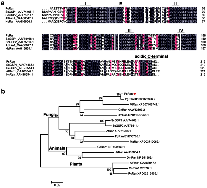
One cDNA clone (WRIC_343) encoding a putative fungal Ran protein was identified in the cDNA library of Pst-infected wheat leaves26. Mapping to Pst genome in the Broad Institute Puccinia database showed that the corresponding gene, PSTG_13752.1 (designated PsRan), has an open reading frame (ORF) of 651 bp that encodes a 216 aa protein with a predicted molecular mass of 24.07 kDa. Further sequence alignment revealed that PsRan shows high similarity (more than 70%) with Ran proteins from other organisms, including Saccharomyces cerevisiae, Arabidopsis thaliana, and Homo sapiens (Fig. 1a). The PsRan protein has four guanine nucleotide-binding domains, an effector domain, and an acidic C-terminal domain, which are the characteristics of Ran GTPases1,31 and are highly conserved during evolution (Fig. 1a). These results suggest that PsRan is a typical Ran gene.
Figure 1. Sequence alignment and phylogenetic analysis of PsRan and other Ran proteins in different organisms.
(a) Sequence alignment of PsRan and Ran proteins in three other organisms, including Saccharomyces cerevisiae (Sc), Arabidopsis thaliana (At) and Homo sapiens (Hs). Lines indicated six conserved domains for Ran GTPases, including four guanine nucleotide-binding domains (I–IV), an effector domain (E), and an acidic C-terminal domain. (b) Phylogenic analysis of PsRan and other Ran proteins in several fungi, animals, and plants. P. graminis f. sp. tritici (Pg), P. triticina (Pt), Melampsora larici-populina (Ml), Cryptococcus neoformans (Cn), Ustilago maydis (Um), S. cerevisiae (Sc), Aspergillus fumigatus (Af), F. graminearum (Fg), and Magnaporthe oryzae (Mo) are grouped as fungi; Caenorhabditis elegans (Ce), Homo sapiens (Hs), and Drosophila melanogaster (Dm) are grouped as animals; Arabidopsis thaliana (At), Oryza sativa (Os), and Ricinus communis (Rc) are grouped as plants. Phylogenetic analysis was carried out with the MEGA5 software by the neighbor-joining method and the red arrow indicates PsRan.
Because the model yeast S. cerevisiae contains two Ran proteins, including GSP1 and GSP232, a BLAST search using GSP1 and GSP2 as queries in the Broad Institute Puccinia database was done to make sure whether Pst contains other genes encoding Ran proteins besides PsRan. There is no other Ran GTPase-encoding gene besides PsRan (PSTG_13752.1) in Pst genome (Supplementary Figs S1 and S2), indicating Pst contains only one Ran gene, which is different from S. cerevisiae. Other two wheat rust fungi in the Broad Institute Puccinia database, including P. graminis f. sp. tritici (Pgt) and P. triticina (Pt), also contain only one Ran GTPase-encoding gene (Supplementary Figs S1 and S2).
In addition, a phylogenetic analysis of Ran proteins from various organisms resulted into two distinct phylogenetic clades (Fig. 1b). Ran proteins in plants are grouped into a clade and Ran proteins in fungi and animals are grouped into another clade (Fig. 1b), indicating a closer relationship of Ran in fungi and animals.
Low level of intra-species polymorphism in PsRan
To identify intra-species polymorphism in PsRan, we compared its ORF sequences in ten different Pst isolates, including five Chinese isolates (CYR23, CYR29, CYR31, CYR32, and Su11-4), three US isolates (PST-21, PST-43, and PST-130), and two UK isolates (PST-08-21 and PST-87-7). Only two single-nucleotide polymorphisms (SNPs) are observed for PsRan (Supplementary Fig. S3a). What’s more, all the two SNPs are synonymous and cannot cause the sequence change of amino acids (Supplementary Fig. S3b). Our results indicate that PsRan shows a low level of intra-species polymorphism and is highly conserved in different Pst isolates.
Nuclear localization of PsRan
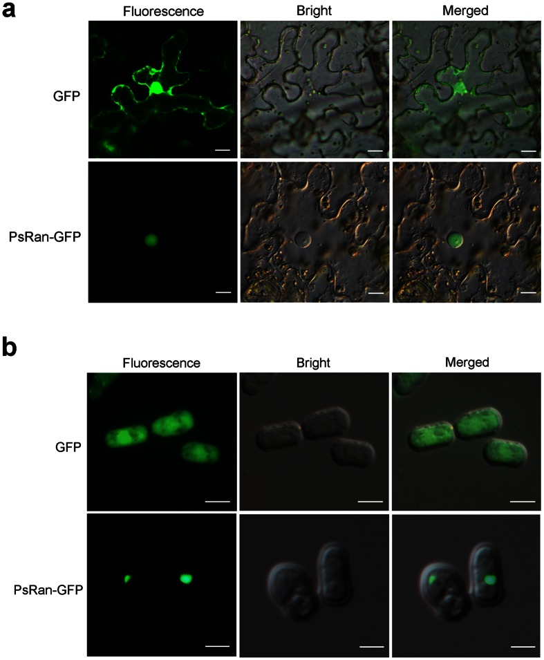
Previous studies have shown that Ran proteins primarily localize inside the nucleus33,34. Because there is no effective transformation system for Pst, we conducted localization experiments of PsRan in Nicotiana benthamiana and fission yeast Schizosaccharomyces pombe, which are easier to operate as a model plant and fungus, respectively. When a PsRan-GFP fusion protein was heterologously expressed in N. benthamiana leaves, its fluorescence was restricted to the nucleus, while the control expressing only GFP exhibited fluorescence throughout the cell (Fig. 2a). When we transiently expressed the PsRan-GFP fusion protein in S. pombe, we found that its fluorescence was again restricted to the nucleus (Fig. 2b). These results indicate the nuclear localization of PsRan when using a transient and distant system.
Figure 2. Subcellular localization of PsRan in N. benthamiana and S. pombe.
(a) Overexpression of PsRan-GFP fusion protein and only GFP (control) in N. benthamiana using A. tumefaciens. Bar = 20 μm. (b) Overexpression of PsRan-GFP fusion protein and only GFP (control) in S. pombe using the yeast expression vector pREP3X. Bar = 5 μm.
Transcript level of PsRan is induced in planta during Pst infection
To characterize transcript profiles of PsRan in different Pst infection stages, we assayed its transcript abundance by qRT-PCR. Compared with the transcript abundance in urediniospores, PsRan showed significantly increased transcript abundance in infected wheat leaves at 18, 24, and 48 hours post-inoculation (hpi), which belong to the “parasitic/biotrophic” phase of Pst (Fig. 3). At 18 hpi, the transcript of PsRan was peaked (up to 21-fold) (Fig. 3). Our results reveal that PsRan is an in planta induced gene and is preferentially expressed during the “parasitic/biotrophic” phase.
Figure 3. Transcription profiles of PsRan in different Pst infection stages by qRT-PCR.
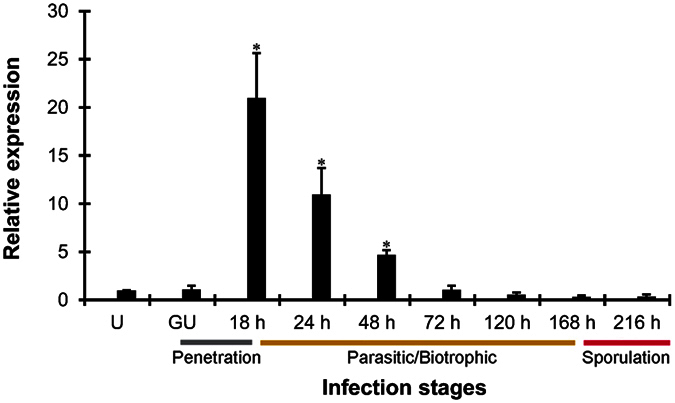
Pst development in wheat can be divided into three major stages, penetration stage (gray), parasitic/biotrophic stage (yellow), and sporulation stage (red). U, urediniospores; GU, in vitro germinated urediniospores; 18 h–216 h, wheat leaves infected with Pst at 18–216 hpi. Relative transcript levels were calculated by the comparative 2−ΔΔCT method and values are expressed relative to an endogenous Pst reference gene EF1. Results are composed of the means ± standard errors of three biological replications (each done in triplicates). Asterisks indicate a significant difference (P < 0.05) verus uredinospores using Student’s t test.
Silencing of PsRan does not alter Pst virulence phenotype
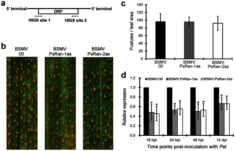
To investigate the role of PsRan in rust pathogenicity, we silenced it by using the BSMV-mediated HIGS system. Two different fragments (PsRan-1as and PsRan-2as) (Fig. 4a) were designed for specifically silencing of PsRan. Ten days after inoculation with BSMV, obvious photo bleaching was observed in BSMV:TaPDSas-inoculated plants that had the wheat phytoene desaturase (PDS) gene silenced (Supplementary Fig. S4), suggesting that the RNAi system is effective. The BSMV:00- (control), BSMV:PsRan-1as-, and BSMV:PsRan-2as-inoculated wheat plants were then inoculated with virulent Pst isolate CYR32, and their rust disease phenotypes were photographed at 14 days post-inoculation (dpi) with Pst. Both BSMV:PsRan-1as- and BSMV:PsRan-2as-inoculated wheat plants showed similar disease phenotypes as control wheat plants, with equivalent amounts of uredinia as control wheat plants (Fig. 4b,c). However, qRT-PCR analysis showed that transcript level of PsRan was significantly reduced in BSMV:PsRan-1as- and BSMV:PsRan-2as-inoculated wheat plants compared to that in control plants (Fig. 4d), indicating PsRan was partially knocked down by the RNAi. These results suggest that silencing of PsRan does not alter Pst virulence phenotype.
Figure 4. HIGS of PsRan did not alter Pst virulence phenotype.
(a) Two specific sequence regions for HIGS of PsRan. (b) Phenotypes of the fourth leaves of BSMV:00- (control), BSMV:PsRan-1as-, and BSMV:PsRan-2as-inoculated wheat plants at 14 dpi with Pst. (c) Quantification of uredinial density in BSMV:00-, BSMV:PsRan-1as-, and BSMV:PsRan-2as-inoculated wheat plants 14 dpi with Pst. Values represent mean ± standard errors of three biological replications (each done in triplicates). (d) Relative transcript levels of PsRan in BSMV:00-, BSMV:PsRan-1as-, and BSMV:PsRan-2as-inoculated wheat plants at 18 hpi, 24 hpi, 48 hpi, and 14 dpi with Pst. Values are expressed relative to endogenous Pst reference gene EF1, with the empty vector (BSMV:00) set at 1. Values represent mean ± standard errors of three biological replications (each done in triplicates). Differences were assessed using Student’s t-tests and asterisks indicate P < 0.05.
Silencing of PsRan impedes fungal growth of Pst
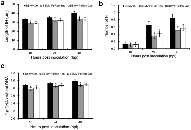
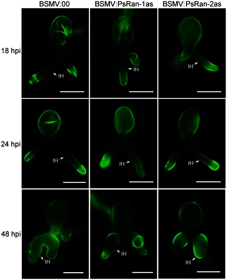
Despite of the unchanged virulence phenotype in PsRan-silenced wheat plants compared with control wheat plants (Fig. 4), we performed a cytological analysis of these wheat plants to investigate whether fungal growth of Pst is affected after silencing of PsRan. The number of haustoria and the length of infection hyphae were evaluated in PsRan-silenced and control wheat plants at 18, 24, and 48 hpi with Pst. Both the number of haustoria and the length of infection hyphae were significantly reduced in PsRan-silenced wheat plants compared with control wheat plants (Figs 5a,b and Fig. 6). In addition, qRT-PCR analysis showed that fungal biomass was also significantly decreased in PsRan-silenced wheat plants compared with control wheat plants (Fig. 5c), which is consistent with the cytological analysis. These results indicate that silencing of PsRan impedes fungal growth of Pst.
Figure 5. HIGS of PsRan impeded fungal growth of Pst.
(a) The average length of infection hyphae (IH) per infection unit in control (BSMV:00-inoculated) and PsRan-silenced (BSMV:PsRan-1as- and BSMV:PsRan-2as-inoculated) wheat plants. The length of IH was measured from the substomatal vesicle to the apex of the longest infection hyphae. (b) The average number of haustoria (H) per infection unit at control and PsRan-silenced wheat plants. (c) Fungal biomass measurements using qRT-PCR analysis of total DNA extracted from control (BSMV:00-inoculated) and PsRan-silenced (BSMV:PsRan-1as- and BSMV:PsRan-2as-inoculated) wheat plants. Ratio of total Pst DNA to total wheat DNA was assessed using the Pst gene PsEF1 and the wheat gene TaEF1. In (a–c), samples were taken at 18, 24, and 48 hpi with Pst, values represent mean ± standard errors of three biological replications (each done in triplicates), and differences were assessed using Student’s t-tests and asterisks indicate P < 0.05.
Figure 6. Micrographs of fungal growth in control (BSMV:00-inoculated) and PsRan-silenced (BSMV:PsRan-1as- and BSMV:PsRan-2as-inoculated) wheat plants.
Infected wheat leaves were sampled at 18, 24, and 48 hpi with Pst and then were examined under an Olympus BX-53 microscope after staining with wheat germ agglutinin conjugated to the fluorophore Alexa-488. IH: infection hypha. Bar = 20 μm.
Heterologous overexpression of PsRan in plant suppresses cell death triggered by a mouse BAX gene or a Pst Ras gene
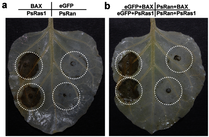
Because there are some similarities between fungal cell death and cell death in other higher eukaryotes35, we heterologously overexpressed PsRan in the model plant N. benthamiana to investigate the possible role of PsRan in cell death. Overexpression of PsRan in N. benthamiana did not trigger noticeable cell death as a mouse pro-apoptotic gene BAX or a Pst Ras gene PsRas1 that can induce strong cell death in N. benthamiana30,36 (Fig. 7a). However, overexpression of PsRan could suppress the cell death triggered by BAX or PsRas1 (Fig. 7b). These results indicate that PsRan is involved in anti-cell death.
Figure 7. Herologous overexpression of PsRan in plant failed to induce cell death but suppressed cell death triggered by BAX or PsRas1.
(a) Overexpression of PsRan and eGFP (negative control) could not trigger cell death in N. benthamiana as a mouse pro-apoptotic gene BAX and a Pst Ras gene PsRas1 that can induce strong cell death in N. benthamiana. (b) Overexpression of PsRan in N. benthamiana suppressed cell death triggered by BAX and PsRas1. N. benthamiana leaves were infiltrated with A. tumefaciens cells containing PVX carrying PsRan or a control gene (eGFP), followed after 24 h by A. tumefaciens cells carrying PVX:BAX/PsRas1.
Discussion
As molecular switches in cellular signal transduction pathways, Ran proteins have been shown to regulate a variety of important cellular processes in many eukaryotes. However, little is known about Ran function in pathogenic fungi. In the present study, we report the identification and functional analysis of a Ran gene from the important plant fungal pathogen Pst, which provides significant insight into Ran function in pathogenic fungi.
Only one Ran GTPase-encoding gene (PsRan) was identified in Pst. The same number of Ran-encoding gene is also observed in many other eukaryotes1. The PsRan protein contains all conserved domains of Ran GTPases and is localized to the nucleus as other known Ran proteins33,34, suggesting PsRan is a typical Ran gene. In addition, PsRan shares high (>70%) identity with other Ran proteins from fungi, plants, and animals. Previous studies showed that overexpression of various Ran proteins from plants, similarly to their mammalian/yeast homologues, suppressed the phenotype of the pim46-1 cell cycle mutant in yeast cells37,38. These observations indicate that Ran proteins in different organisms also have functional similarity besides conserved sequences.
qRT-PCR analysis showed that transcript level of PsRan was induced in planta during Pst infection, indicating PsRan may be important for plant infection of Pst. To investigate the specific role of PsRan in plant infection of Pst, we silenced PsRan using the BSMV-mediated HIGS system. The results showed that silencing of PsRan significantly decreased the number of Pst haustoria and the length of Pst infection hyphae, demonstrating the involvement of PsRan in fungal growth. Because little is currently known about Ran function in pathogenic fungi, this study is the first investigating of the role of Ran proteins from pathogenic fungi in fungal growth. In addition, other four families (Ras, Rho, Rab, and Sar1/Arf) in small GTP-binding proteins are also proven to regulate fungal growth15,16,17,18,39. These results highlight the great contribution of small GTP-binding proteins in fungal growth.
Successful Pst infection of wheat plants appears as a mass of uredinia arranged in long and narrow stripes on leaves40. However, PsRan-silenced wheat plants showed similar disease phenotypes as control wheat plants, with equivalent amounts of uredinia as control wheat plants, which indicate that silencing of PsRan did not alter Pst virulence phenotype. Silencing of many other rust genes from Pst, Pt, and Pgt also rarely changes their virulence phenotypes41,42. These results should not be taken as evidence that the majority of silenced genes are not involved in rust pathogenicity. The unchanged virulence phenotypes may be due to functional redundancy with other genes or that silencing was not sufficiently complete to knock down levels of encoded proteins to levels that would interfere with virulence phenotypes42.
Some small GTP-binding proteins are proven to be important regulators of cell death in different organisms43,44, and cell death is involved in several important biological processes in pathogenic fungi35. The possible roles of PsRan in cell death were investigated using heterologous systems because Pst lacks an efficient and reliable transformation system. Heterologous overexpression of PsRan in plant failed to induce cell death but suppressed cell death triggered by a mouse BAX gene or a Pst Ras gene, indicating that PsRan plays an important role in anti-cell death. Because there are some similarities between fungal cell death and cell death in other eukaryotes35, our results suggest that PsRan may truly function in the cell death of Pst. In addition, Ran proteins are also proven to be involved in human cell death, but as a death-promoting member8,45. These results indicate that Ran GTPases may function as positive or negative regulators of cell death in different organisms.
In conclusion, our study demonstrated that PsRan is involved in fungal growth and anti-cell death. Future studies should be directed toward the investigation of the specific mechanisms of PsRan in fungal growth and anti-cell death, which will contribute to the control of the wheat stripe rust disease.
Methods
Plant materials, strains and growth conditions
Wheat (Triticum aestivum) cv. Suwon 11, N. benthamiana, and five Chinese Pst isolates (CYR23, CYR29, CYR31, CYR32, and Su11-4) were used in this study. Wheat and N. benthamiana plants were grown at 20 °C and 25 °C, respectively. Escherichia coli JM109 was grown in a Luria-Bertani (LB) medium at 37 °C and used for plasmid construction. A. tumefaciens strain GV3101 was grown in LB medium at 28 °C and used for overexpression of PsRan in N. benthamiana. Antibiotics were used at final concentrations of 50 μg/ml ampicillin, 50 μg/ml kanamycin, 30 μg/ml rifampicin, and 25 μg/ml gentamycin.
Sequence analysis and polymorphism analysis
The conserved domains of PsRan and other Ran proteins in different organisms were deduced using PFAM (http://pfam.xfam.org/). Sequence alignment between PsRan and other Ran proteins in different organisms was created using DNAMAN 6.0. Phylogenetic analysis of PsRan and other Ran proteins in different organisms was carried out with the MEGA5 software by the neighbor-joining method.
To identify intra-species polymorphism in PsRan, PCR amplifications were performed using cDNAs of the five Chinese Pst isolates (CYR23, CYR29, CYR31, CYR32, and Su11-4). The amplicons were then amplified, cloned, and sequenced. The re-sequenced genomes of the three US isolates (PST-21, PST-43, and PST-130) and the two UK isolates (PST-08-21 and PST-87-7) were used directly46. Local blast searches using BioEdit were conducted to identify the corresponding sequences, and DNAMAN6.0 was then used to create multiple sequence alignments. At each nucleotide position in the alignment, if there were different bases (one or more), one SNP was counted. The sum of this count was then calculated over all of the positions in each gene.
Plasmid construction
The oligonucleotides used for plasmid construction in this study were documented in Supplementary Table S1. PsRan was cloned from the cDNA of Pst isolate CYR32 using FastPfu DNA Polymerase (TransGen Biotech, Beijing, China). To check the subcellular localization of PsRan in N. benthamiana and S. pombe, its ORF sequence was ligated into the plant binary expression vector pCAMBIA-1302 and the yeast expression vector pREP3X, respectively. To HIGS of PsRan, two specific partial cDNA regions were cloned into the BSMV gamma vector. For overexpression of PsRan in N. benthamiana, its ORF sequence was cloned into the PVX vector.
Total RNA extraction and qRT-PCR
The total RNAs of urediniospores, germinated urediniospores and infected wheat leaves at different time points (18, 24, 48, 72, 120, 168, and 216 hpi) were isolated using TRIzol reagent (Invitrogen, Carlsbad, CA, USA). After urediniospores were incubated for 10 hours in vitro, germinated urediniospores were then harvested. A 2.0-μg RNA aliquot of each sample was used for cDNA synthesis with an oligo(dT)18 primer using the Reverse Transcription PCR system (Promega, Madison, WI, USA). Subsequently, SYBR green qRT-PCR assays were performed using a 7500 Real-Time PCR System (Applied Biosystems, Foster City, CA). The Pst housekeeping gene EF1 was used as the endogenous reference to normalize the gene expression in Pst47. All reactions were performed in triplicate, and reactions without template were used as negative controls. The 2−ΔΔCT method was used to quantify the relative gene expression levels48.
Subcellular localization in N. benthamiana
The PsRan-GFP recombinant plasmid was introduced into the Agrobacterium tumefaciens strain GV3101 by electroporation. For infiltration of leaves, recombinant strains of A. tumefaciens were grown for 48 h, harvested, suspended in an infiltration media (10 mM MgCl2, 10 mM MES, pH 5.6, and 200 mM acetosyringone). Then A. tumefaciens suspensions were infiltrated at an OD600 of 0.8 into leaves of 4–6-week-old N. benthamiana plants using a syringe without a needle. Infiltrated plants were grown and maintained at 25 °C. Tissue samples were harvested at 2 or 3 d after infiltration and GFP signals were then observed at 2 dpi by an Olympus BX-53 microscope (Olympus Corporation, Tokyo, Japan) (excitation filter 485 nm, dichromic mirror 510 nm, and barrier filter 520 nm).
Subcellular localization in S. pombe
The GFP sequence and PsRan-GFP fusion sequence that was gotten using the overlapping PCR method were cloned into the yeast expression vector pREP3X as previously described49. The two recombinant plasmids were transformed into S. pombe by electroporation and transformed cells were cultured as previously described50. The GFP signals of yeast cells were also observed by an Olympus BX-53 microscope (Olympus Corporation, Tokyo, Japan) (excitation filter 485 nm, dichromic mirror 510 nm, and barrier filter 520 nm).
BSMV-mediated HIGS of PsRan
Capped in vitro transcripts were prepared from linearized plasmids containing the tripartite BSMV genome with the mMessage mMachine T7 in vitro transcription kit (Ambion, Austin, USA), following the manufacturer’s instructions. The second leaf of the wheat (Suwon 11) seedlings at the two-leaf stage was inoculated with BSMV transcripts by gently rubbing the surface with a gloved finger51. Three independent sets of plants were prepared for each of the four BSMV constructs (BSMV:00, BSMV:PsRan-1as, BSMV:PsRan-2as, and BSMV:TaPDS). The BSMV-infected plants were maintained in a growth chamber at 25 °C. Ten days after BSMV infection, the fourth leaves in BSMV:00-, BSMV:PsRan-1as- and BSMV:PsRan-2as-inoculated wheat plants were then inoculated with fresh virulent CYR32 urediniospores. The disease symptoms of the fourth leaves were recorded at 14 dpi with Pst. The inoculated fourth leaves were sampled at 18 hpi, 24 hpi, 48 hpi, and 14 dpi with Pst for RNA isolation to evaluate the silencing efficiencies of PsRan using qRT-PCR. These samples at 18, 24, and 48 hpi with Pst are also used for cytological observation of fungal growth and qRT-PCR analysis of fungal biomass.
Uredinia quantification
The Pst disease phenotype was quantified by counting the number of uredinia within a 1-cm2 area at 14 dpi with Pst. To avoid bias among the leaf samples, leaves from at least five treated plants were randomly selected. Interpretation of the results was determined by comparing the values of the silenced plants to those of the controls.
Cytological observation of Pst fungal growth in silenced wheat plants
Cytological analyses were performed to characterize Pst fungal growth in control and silenced wheat plants. Leaf segments (1.5 cm in length) cut from inoculated leaves were fixed and decolorized in ethanol/trichloromethane (3:1 v/v) containing 0.15% (w/v) trichloroacetic acid for 3–5 days. Decolorized leaf segments were stained with wheat germ agglutinin (WGA) conjugated to Alexa-488 (Invitrogen, Carlsbad, USA) as described previously52, and the stained samples were then examined under an Olympus BX-53 fluorescence microscope (Olympus Corporation, Tokyo, Japan) to observe Pst infection structures.
The number of haustoria and length of infection hyphae were measured as previously described53. For each wheat leaf sample in each biological replication, 30–50 infection sites from three leaves were examined to record number of haustoria and the length of infection hyphae per infection unit. The experiments were conducted in a completely randomized block design with three replications. Presence of a substomatal vesicle was defined as an established infection unit. The length of infection hyphae was measured from the substomatal vesicle to the apex of the longest infection hypha.
Fungal biomass by qRT-PCR
To measure fungal biomass in infected wheat leaves, relative quantification of the single-copy target genes PsEF1 (from Pst) and TaEF1 (from wheat) by qRT-PCR was carried out as previously described54,55. Total genomic DNA of the wheat cv. Suwon 11 or the Pst isolate CYR32 was used to prepare standard curves derived from seven serial dilutions and the correlation coefficients for the analysis of the dilution curves were above 0.99. The relative quantities of the PCR products of the Pst gene PsEF1 and the wheat gene TaEF1 in infected wheat leaves were then calculated using the gene-specific standard curves to quantify the Pst and wheat genomic DNA, respectively.
A. tumefaciens-mediated overexpression in N. benthamiana
The PVX:PsRan, PVX:eGFP, PVX:BAX, and PVX:PsRas1 recombinant plasmids were introduced into A. tumefaciens strain GV3101 by electroporation. Recombinant strains of A. tumefaciens were grown in LB liquid medium for 48 h, harvested, suspended in an infiltration medium (10 mM MgCl2) to an OD600 of 0.4.
To assay whether overexpression of PsRan in N. benthamiana can trigger cell death, A. tumefaciens suspensions carrying PVX:PsRan, PVX:eGFP (negative control), PVX:BAX (positive control), or PVX:PsRas1 (positive control) were infiltrated into the leaves of 4–6-week-old N. benthamiana plants using a syringe without a needle. The infiltrated N. benthamiana plants were grown and maintained in a cultivation room at 25 °C with a cycle of 16 h light/8 h darkness. Symptoms of plant cell death were monitored at 3 dpi with A. tumefaciens.
To assay suppression of BAX/PsRas1-triggered plant cell death, A. tumefaciens carrying PsRan were infiltrated initially, and then A. tumefaciens carrying BAX or PsRas1 were infiltrated into the same site 24 h later. Symptoms were monitored and photographed for 3 dpi with A. tumefaciens carrying BAX. A. tumefaciens cells carrying eGFP was infiltrated in parallel as controls.
Additional Information
How to cite this article: Cheng, Y. et al. Characterization of a Ran gene from Puccinia striiformis f. sp. tritici involved in fungal growth and anti-cell death. Sci. Rep. 6, 35248; doi: 10.1038/srep35248 (2016).
Supplementary Material
Acknowledgments
This study was financially supported by the National Basic Research Program of China (No. 2013CB127700), the National Natural Science Foundation of China (No. 31430069), the Key Grant Project of Chinese Ministry of Education (No. 313048), and the China Postdoctoral Science Foundation (No. 2015M580882).
Footnotes
The authors declare no competing financial interests.
Author Contributions Y.L.C. and Z.S.K. designed the research, Y.L.C., J.N.Y., Y.R.Z. and S.M.L. performed the experiments. Y.L.C. and J.N.Y. analyzed the data and wrote the manuscript.
References
- Takai Y., Sasaki T. & Matozaki T. Small GTP-binding proteins. Physiol Rev 81, 153–208 (2001). [DOI] [PubMed] [Google Scholar]
- Jiang S. Y. & Ramachandran S. Comparative and evolutionary analysis of genes encoding small GTPases and their activating proteins in eukaryotic genomes. Physiol Genomics 24, 235–251 (2006). [DOI] [PubMed] [Google Scholar]
- Ribbeck K., Lipowsky G., Kent H. M., Stewart M. & Gorlich D. NTF2 mediates nuclear import of Ran. EMBO J 17, 6587–6598 (1998). [DOI] [PMC free article] [PubMed] [Google Scholar]
- Smith A., Brownawell A. & Macara I. G. Nuclear import of Ran is mediated by the transport factor NTF2. Curr Biol 8, 1403–S1401 (1998). [DOI] [PubMed] [Google Scholar]
- Ciciarello M., Mangiacasale R. & Lavia P. Spatial control of mitosis by the GTPase Ran. Cell Mol Life Sci 64, 1891–1914 (2007). [DOI] [PMC free article] [PubMed] [Google Scholar]
- Fiore B. D., Ciciarello M. & Lavia P. Mitotic functions of the Ran GTPase network: the importance of being in the right place at the right time. Cell Cycle 3, 303–311 (2004). [PubMed] [Google Scholar]
- Sanderson H. S. & Clarke P. R. Cell biology: Ran, mitosis and the cancer connection. Curr Biol 16, R466–R468 (2006). [DOI] [PubMed] [Google Scholar]
- Moss D. K., Wilde A. & Lane J. D. Dynamic release of nuclear RanGTP triggers TPX2-dependent microtubule assembly during the apoptotic execution phase. J Cell Sci 122, 644–655 (2009). [DOI] [PMC free article] [PubMed] [Google Scholar]
- Han F. & Zhang X. Characterization of a ras-related nuclear protein (Ran protein) up-regulated in shrimp antiviral immunity. Fish Shellfish Immunol 23, 937–944 (2007). [DOI] [PubMed] [Google Scholar]
- Li K.-L., Wan P.-J., Wang W.-X., Lai F.-X. & Fu Q. Ran involved in the development and reproduction is a potential target for RNA-Interference-based pest management in Nilaparvata lugens. PLoS One 10, e0142142 (2015). [DOI] [PMC free article] [PubMed] [Google Scholar]
- Sinha V. B., Grover A., Singh S., Pande V. & Ahmed Z. Overexpression of Ran gene from Lepidium latifolium L. (LlaRan) renders transgenic tobacco plants hypersensitive to cold stress. Mol Biol Rep 41, 5989–5996 (2014). [DOI] [PubMed] [Google Scholar]
- Xu P. & Cai W. RAN1 is involved in plant cold resistance and development in rice (Oryza sativa). J Exp Bot. eru178 (2014). [DOI] [PMC free article] [PubMed] [Google Scholar]
- Bahn Y. S. et al. Sensing the environment: lessons from fungi. Nat Rev Microbiol 5, 57–69 (2007). [DOI] [PubMed] [Google Scholar]
- Weeks G. & Spiegelman G. B. Roles played by Ras subfamily proteins in the cell and developmental biology of microorganisms. Cell Signal 15, 901–909 (2003). [DOI] [PubMed] [Google Scholar]
- Lengeler K. B. et al. Signal transduction cascades regulating fungal development and virulence. Microbiol Mol Biol Rev 64, 746–785 (2000). [DOI] [PMC free article] [PubMed] [Google Scholar]
- Hogan D. A. & Sundstrom P. The Ras/cAMP/PKA signaling pathway and virulence in Candida albicans. Future Microbiol 4, 1263–1270 (2009). [DOI] [PubMed] [Google Scholar]
- Jiang L., Lee C. M. & Shen S. H. Functional characterization of the Candida albicans homologue of secretion-associated and Ras-related (Sar1) protein. Yeast 19, 423–428 (2002). [DOI] [PubMed] [Google Scholar]
- Mahlert M., Leveleki L., Hlubek A., Sandrock B. & Bolker M. Rac1 and Cdc42 regulate hyphal growth and cytokinesis in the dimorphic fungus Ustilago maydis. Mol Microbiol 59, 567–578 (2006). [DOI] [PubMed] [Google Scholar]
- Lee N. & Kronstad J. W. ras2 controls morphogenesis, pheromone response, and pathogenicity in the fungal pathogen Ustilago maydis. Eukaryot Cell 1, 954–966 (2002). [DOI] [PMC free article] [PubMed] [Google Scholar]
- Park G. et al. Multiple upstream signals converge on the adaptor protein Mst50 in Magnaporthe grisea. Plant Cell 18, 2822–2835 (2006). [DOI] [PMC free article] [PubMed] [Google Scholar]
- Bluhm B. H., Zhao X., Flaherty J. E., Xu J. R. & Dunkle L. D. RAS2 regulates growth and pathogenesis in Fusarium graminearum. Mol Plant Microbe Interact 20, 627–636 (2007). [DOI] [PubMed] [Google Scholar]
- Ramsdale M. Programmed cell death in pathogenic fungi. BBA-Mol Cell Res 1783, 1369–1380 (2008). [DOI] [PubMed] [Google Scholar]
- Hill-Ambroz K. et al. Expression analysis and physical mapping of a cDNA library of Fusarium head blight infected wheat spikes. Crop Sci 46, S-15-S-26 (2006). [Google Scholar]
- Mantilla J. G. et al. Transcriptome analysis of the entomopathogenic fungus Beauveria bassiana grown on cuticular extracts of the coffee berry borer (Hypothenemus hampei). Microbiology 158, 1826–1842 (2012). [DOI] [PubMed] [Google Scholar]
- Brown S. H. et al. Differential protein expression in Colletotrichum acutatum: changes associated with reactive oxygen species and nitrogen starvation implicated in pathogenicity on strawberry. Mol Plant Pathol 9, 171–190 (2008). [DOI] [PMC free article] [PubMed] [Google Scholar]
- Ma J. B. et al. Identification of expressed genes during compatible interaction between stripe rust (Puccinia striiformis) and wheat using a cDNA library. BMC Genomics 10, 586 (2009). [DOI] [PMC free article] [PubMed] [Google Scholar]
- Wellings C. R. Global status of stripe rust: a review of historical and current threats. Euphytica 179, 129–141 (2011). [Google Scholar]
- Yin C. & Hulbert S. Host induced gene silencing (HIGS), a promising strategy for developing disease resistant crops. Gene Technology 4, 130 (2015). [Google Scholar]
- Nowara D. et al. HIGS: host-induced gene silencing in the obligate biotrophic fungal pathogen Blumeria graminis. Plant Cell 22, 3130–3141 (2010). [DOI] [PMC free article] [PubMed] [Google Scholar]
- Cheng Y. et al. Two distinct Ras genes from Puccinia striiformis exhibit differential roles in rust pathogenicity and cell death. Environ Microbiol, doi: 10.1111/1462-2920.13379 (2016). [DOI] [PubMed] [Google Scholar]
- Scheffzek K., Klebe C., Fritzwolf K., Kabsch W. & Wittinghofer A. Crystal-structure of the nuclear Ras-related protein Ran in its Gdp-bound form. Nature 374, 378–381 (1995). [DOI] [PubMed] [Google Scholar]
- Belhumeur P. et al. Gsp1 and Gsp2, genetic suppressors of the Prp20-1 mutant in Saccharomyces cerevisiae: GTP-binding proteins involved in the maintenance of nuclear organization. Mol Cell Biol 13, 2152–2161 (1993). [DOI] [PMC free article] [PubMed] [Google Scholar]
- Melchior F., Paschal B., Evans J. & Gerace L. Inhibition of nuclear protein import by nonhydrolyzable analogues of GTP and identification of the small GTPase Ran/TC4 as an essential transport factor. J Cell Biol 123, 1649–1659 (1993). [DOI] [PMC free article] [PubMed] [Google Scholar]
- Bischoff F. R., Klebe C., Kretschmer J., Wittinghofer A. & Ponstingl H. RanGAP1 induces GTPase activity of nuclear Ras-related Ran. Proc Natl Acad Sci USA 91, 2587–2591 (1994). [DOI] [PMC free article] [PubMed] [Google Scholar]
- Ramsdale M. Programmed cell death in pathogenic fungi. BBA-Mol Cell Res 1783, 1369–1380 (2008). [DOI] [PubMed] [Google Scholar]
- Lacomme C. & Santa Cruz S. Bax-induced cell death in tobacco is similar to the hypersensitive response. Proc Natl Acad Sci USA 96, 7956–7961 (1999). [DOI] [PMC free article] [PubMed] [Google Scholar]
- Ach R. A. & Gruissem W. A small nuclear GTP-binding protein from tomato suppresses a Schizosaccharomyces pombe cell-cycle mutant. Proc Natl Acad Sci USA 91, 5863–5867 (1994). [DOI] [PMC free article] [PubMed] [Google Scholar]
- Merkle T. et al. Phenotype of the fission yeast cell cycle regulatory mutant pim1-46 is suppressed by a tobacco cDNA encoding a small, Ran-like GTP-binding protein. Plant J 6, 555–565 (1994). [DOI] [PubMed] [Google Scholar]
- Siriputthaiwan P., Jauneau A., Herbert C., Garcin D. & Dumas B. Functional analysis of CLPT1, a Rab/GTPase required for protein secretion and pathogenesis in the plant fungal pathogen Colletotrichum lindemuthianum. J Cell Sci 118, 323–329 (2005). [DOI] [PubMed] [Google Scholar]
- Chen W., Wellings C., Chen X., Kang Z. & Liu T. Wheat stripe (yellow) rust caused by Puccinia striiformis f. sp. tritici. Mol Plant Pathol 15, 433–446 (2014). [DOI] [PMC free article] [PubMed] [Google Scholar]
- Yin C., Jurgenson J. E. & Hulbert S. H. Development of a host-induced RNAi system in the wheat stripe rust fungus Puccinia striiformis f. sp. tritici. Mol Plant Microbe Interact 24, 554–561 (2011). [DOI] [PubMed] [Google Scholar]
- Yin C. et al. Identification of promising host-induced silencing targets among genes preferentially transcribed in haustoria of Puccinia. BMC Genomics 16, 1 (2015). [DOI] [PMC free article] [PubMed] [Google Scholar]
- Cox A. D. & Der C. J. The dark side of Ras: regulation of apoptosis. Oncogene 22, 8999–9006 (2003). [DOI] [PubMed] [Google Scholar]
- Kawasaki T. et al. The small GTP-binding protein Rac is a regulator of cell death in plants. Proc Natl Acad Sci USA 96, 10922–10926 (1999). [DOI] [PMC free article] [PubMed] [Google Scholar]
- Wilde A. & Zheng Y. Ran out of the nucleus for apoptosis. Nat Cell Biol 11, 11–12 (2009). [DOI] [PubMed] [Google Scholar]
- Cantu D. et al. Genome analyses of the wheat yellow (stripe) rust pathogen Puccinia striiformis f. sp. tritici reveal polymorphic and haustorial expressed secreted proteins as candidate effectors. BMC Genomics 14, 270 (2013). [DOI] [PMC free article] [PubMed] [Google Scholar]
- Yin C. et al. Generation and analysis of expression sequence tags from haustoria of the wheat stripe rust fungus Puccinia striiformis f. sp. tritici. BMC Genomics 10, 626 (2009). [DOI] [PMC free article] [PubMed] [Google Scholar]
- Livak K. J. & Schmittgen T. D. Analysis of relative gene expression data using real-time quantitative PCR and the 2− ΔΔCT method. Methods 25, 402–408 (2001). [DOI] [PubMed] [Google Scholar]
- Forsburg S. L. Comparison of Schizosaccharomyces pombe expression systems. Nucleic Acids Res 21, 2955–2956 (1993). [DOI] [PMC free article] [PubMed] [Google Scholar]
- Fu Y. P. et al. TaADF7, an actin-depolymerizing factor, contributes to wheat resistance against Puccinia striiformis f. sp. tritici. Plant J 78, 16–30 (2014). [DOI] [PubMed] [Google Scholar]
- Holzberg S., Brosio P., Gross C. & Pogue G. P. Barley stripe mosaic virus-induced gene silencing in a monocot plant. Plant J 30, 315–327 (2002). [DOI] [PubMed] [Google Scholar]
- Ayliffe M. et al. Nonhost resistance of rice to rust pathogens. Mol Plant Microbe Interact 24, 1143–1155 (2011). [DOI] [PubMed] [Google Scholar]
- Cheng Y. L. et al. Characterization of non-host resistance in broad bean to the wheat stripe rust pathogen. BMC Plant Biol 12, 96 (2012). [DOI] [PMC free article] [PubMed] [Google Scholar]
- Panwar V., McCallum B. & Bakkeren G. Endogenous silencing of Puccinia triticina pathogenicity genes through in planta-expressed sequences leads to the suppression of rust diseases on wheat. Plant J 73, 521–532 (2013). [DOI] [PubMed] [Google Scholar]
- Liu J. et al. An extracellular Zn-only superoxide dismutase from Puccinia striiformis confers enhanced resistance to host-derived oxidative stress. Environ Microbiol, doi: 10.1111/1462-2920.13451 (2016). [DOI] [PubMed] [Google Scholar]
Associated Data
This section collects any data citations, data availability statements, or supplementary materials included in this article.


