Abstract
Purpose
We have shown that cerium oxide nanoparticles (nanoceria), with unique characteristics and catalytic activities, are retained in the retina for more than 1 year after a single intravitreal injection and can be potentially used for the treatment of a variety of eye diseases. The objective of this study is to determine whether the retention of nanoceria in the eye causes inflammation or adverse side effects.
Methods
Wild-type (C57BL/6J) mice at P30 were intravitreally injected with several concentrations of nanoceria. The health of the photoreceptors was assessed by analyzing the expression of photoreceptor-specific genes, and the retinal structure and function. The effect of nanoceria was investigated by analyzing of the vascular system, the expression of inflammatory cytokines, and cellular infiltration into the eye.
Results
Our data showed that there were no changes in the retinal structure or function, or cytokine gene expression following a single intravitreal injection of nanoceria.
Conclusions
Nanoceria, at doses ranging from 17.2 ng to 1720 ng per eye, do not cause any damage to the retinal structure and function by 30 days post injection. No cellular infiltration and no increases in inflammatory responses were found in the eyes. Our data indicate that nanoceria are safe to use for treatment of a variety of eye diseases.
Introduction
An ideal therapeutic reagent for ocular disease treatment should be capable of passing the blood–retinal barrier, have prolonged retention in the ocular tissues, be selectively targeted to the expected sites, and have sustained effectiveness for long periods of time with a maximum benefit. This therapeutic reagent should also be safe with minimum damage to the tissue where the reagent is located and without adverse reactions and undesirable side effects. Selectivity, effectiveness, and safety are the three most important characteristics for an ideal pharmaceutical agent [1]. Unfortunately, thus far there are no such ideal drugs, and the currently available and potential drugs for the treatment of diseases need to have the dosage optimized to reduce their side effects.
We have been using cerium oxide nanoparticles (nanoceria) as therapeutics to treat a variety of ocular diseases in animal models. Nanoceria are catalytic antioxidants that mimic superoxide dismutase and catalase and as a new emerging nanomedicine have great advantages over other traditional antioxidants, such as unique physicochemical features of the surface structure for regenerative scavenging of free radicals that decreases repetitive doses. The tiny particle size (3–5 nm in diameter) on the atom-size scale enables nanoceria to easily cross cell and nuclear membranes. For years, we have used nanoceria as therapeutics to treat inherited and light-induced retinal degeneration [2-4] and to inhibit and regress neovascularization in a wet age-related macular degeneration (AMD) mouse model (vldlr−/−) [5,6]. We also showed storage of nanoceria at room temperature for 6 years does not reduce their effectiveness [7]. And nanoceria were shown to regulate the same antioxidative gene network as thioredoxin [8]. All these results indicated that nanoceria exert their function as a near “ideal” drug and can be used for a broad spectrum of diseases. However, data from our laboratory also demonstrated that nanoceria delivered to the rat eye by a single intravitreal injection are retained in the retina for more than 1 year [9]. Due to the slow elimination and clearance of nanoceria from the tissues [10], safety following long-term retention in the retinas will be a concern for the clinical application of nanoceria.
Although the published data from our laboratory show that nanoceria do not affect the retinal structure and function [9], there are no data to show whether the long-term presence of nanoceria in the eyes causes inflammation. To investigate the tolerance of ocular tissues and cells for nanoceria, we performed intravitreal injections of nanoceria, with a variety of doses, into P30 wild-type (WT) C57BL/6J mice. Assessment of the toxicity of nanoceria was conducted at post injection (PI) at 7 h and 3, 7, 15, and 30 day. We evaluated the retinal structure and function, photoreceptor-specific gene expression of mRNA and protein levels, and inflammatory responses, including the alteration of the vascular system and cytokine expression.
Methods
Animals
Wild-type (C57BL/6J) mice were purchased from the Jackson Laboratory (Bar Harbor, ME) and used as breeders for the colonies. Animal care and handling were performed according to the guidance of the ARVO Statement for the Use of Animals in Ophthalmic and Vision Research (ARVO), and the protocol for this study was approved by the Institutional Animal Care and Use Committee (IACUC) of the University of Oklahoma Health Sciences Center.
Intravitreal injection
Intravitreal injection was performed as previously reported [11,12]. Briefly, WT mice at P30 were anesthetized by intraperitoneal injection with ketamine (85 mg/kg) and xylazine (14 mg/kg; Henry Schein Animal Health, Dublin, OH). A puncture was made in the sclera just below the cornea with a 30 gauge needle; a 33 gauge needle attached to a Hamilton syringe was then inserted into the puncture, and 1 µl of saline or 1 µl of nanoceria in saline at the following concentrations, 0.1 mM (17.2 ng), 0.3 mM (51.6 ng), 1 mM (172 ng), 3 mM (516 ng), and 10 mM (1720 ng), were injected into the vitreous. After fully recovering from the anesthesia, the mice were returned to their original cages and maintained under the standard conditions. Age-matched mice were used as uninjected controls.
Fundoscopy and fluorescein angiography
Micron IV fundoscopy (Phoenix Research Labs, Pleasanton, CA) was performed, and evaluation of the fundus and neovascularization was the same as we previously reported [5]. The mice at the PI7 and PI30 day were anesthetized with ketamine and xylazine, the eyes were dilated, and the whiskers were trimmed. One drop of 2.5% Goniotaire (hypromellose, Altaire pharmaceuticals, Inc., Aquebogue, NY) was applied to the surface of the cornea. The mouse was placed on the bed of the Micron IV system, and the position of the eye and the objective of the funduscope were adjusted until the fundus was clearly seen. After a fundus image was taken, the mice were intraperitoneally injected with 40 µl of 5% AK-Fluor (Alcon, Fort Worth, TX). Then additional images were taken at 2 min and 4 min after injection using StreamPix software and fluorescein isothiocyanate (FITC) filters.
Electroretinography
Full-field electroretinography (ERG) was performed on the mice according to the procedure reported previously [2]. Briefly, mice at the PI30 day were dark adapted overnight, the eyes of the fully anesthetized mice were dilated, the whiskers were trimmed, and rod ERG was recorded by stimulating with a light flash of 600 cds/m2 intensity. Cone responses were recorded by stimulating with a light flash of an intensity of 1000 cds/m2 five times after 5 min of adaption to the light intensity of 100 cds/m2.
qRT-PCR and PCR array
Three to eight retinas from each group were collected and kept in TRIzol (Invitrogen, Carlsbad, CA) at −80 °C. Total RNA isolation, cDNA synthesis, and quantitative RT-PCR (qRT-PCR) were performed the same as previously reported [2]. Ten nanograms of cDNA was used in a 25 µl reaction volume for qRT–PCR. Primers for tumor necrosis factor α (TNF-α), interleukin 6 (IL-6), and macrophage migration inhibitory factor (MIF) were the same as previously reported [13]. The primers used for IL-1β were the forward primer: 5′-GGG CCT CAA AGG AAA GAA TC and the reverse primer: 5′-TAC CAG TTG GGG AAC TCT GCA. Calculation of the relative expression level of the target genes against the housekeeping gene (GAPDH) was the same as we previously reported [2]. Data shown are fold changes. PCR array assay using the “mouse common cytokines” array plates and retinas at the PI7 day was performed according to the instructions from SABiosciences. Data were analyzed with the array plate software (SABiosciences) and are shown as fold changes in the nanoceria-injected and saline-injected mice compared to the uninjected WT mice (CeO2/WT and saline/WT) with the p value indicated.
Immunohistochemistry
The immunohistochemistry procedure was the same as we previously published [2,5]. Briefly, paraffin sections were dewaxed and hydrated through a series of ethanol solutions, and then blocked with 5% bovine serum albumin (BSA). The slides were incubated with anti-rhodopsin antibody (1D4, 1:2,000, generous gift from Dr. Robert Molday, University of Columbia, Vancouver, Canada) and rabbit anti-M-opsin (1:500, Millipore) for 2 h at room temperature. After three washes, secondary antibody of Alexa-Flour 488 conjugated anti-mouse or anti-rabbit immunoglobulin G (IgG) was applied. The slides were coverslipped with mounting medium containing 4',6-diamidino-2-phenylindole (DAPI, Vector Laboratories, Inc., Burlingame, CA). Observation and image capture were performed using a Nikon Eclipse 800 fluorescence microscope (Tokyo, Japan) with proper filters.
Histology and quantitative histology
Eye enucleation, fixation, sectioning, and staining were the same as previously reported [2,5]. Representative retinal images from three to eight eyes stained with hematoxylin and eosin (H&E) were taken at 0.96 mm from the optic nerve head (ONH) at the superior hemisphere using Nikon Eclipse 800 microscopy under 20X and 40X objectives. For morphometric and quantitative histological analysis of the outer nuclear layer (ONL) thickness, blinded to each groups, five images were taken under 60X at a distance of every 0.32 mm from each side of the retina section starting from the ONH. The data are shown as mean ± standard error of the mean (SEM).
Western blot
Three to five individual eyecups, without the lens and cornea, from each group were homogenized, centrifuged, and 50 µg of the soluble protein were loaded on a 10% sodium dodecyl sulfate–polyacrylamide gel electrophoresis (SDS–PAGE) gel. The proteins were detected with the following primary antibodies: mouse anti-IL-1β (1:1,000, Millipore), rabbit anti-IL-6 (Proteintech, 1:1000), anti-TNF-α (1:1,000, Millipore) and anti-MIF (1:1000, Santa Cruz), mouse anti-rhodopsin (1D4, 1:4,000), rabbit anti-M-opsin (1:1,000, Millipore), goat anti-S-opsin (1:1,000, Santa Cruz), and rabbit anti-caspase 3 (1:1,000, Cell Signaling Technology). After stripping, the same membranes were probed with rabbit anti-actin-HRP (Horseradish peroxidase conjugate; 1:1,000, Cell Signaling Technology) or rabbit anti-GAPDH (1:2,500, Abcam). Development of bands, image capture, and the densitometric analysis of the bands were the same as we previously reported [5].
Statistical analysis
The unpaired Student t test for two group comparison or one-way ANOVA with the Bonferroni post hoc test for multiple comparisons was used to analyze the difference between groups. A p value of less than 0.05 was considered statistically significant and was indicated in each figure.
Results
In vivo observation of the overall appearance and movement of the eyeballs demonstrated that the injected eyes were normal sized without redness, with a clear cornea, normal iris response to light, and normal eyeball movement. These findings indicated that there were no ocular abnormalities in the injected eyes.
Retinal morphology and ONL thickness
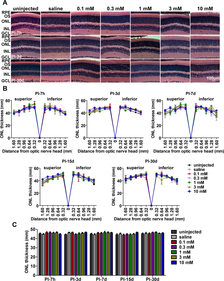
To evaluate the overall morphological changes in the retinas and any loss of photoreceptors, we performed histological and quantitative histological analysis on the H&E-stained retinal sections at the PI7 h and the PI3, PI7, PI15, and PI30 day. As shown in Figure 1, distinct and normal retinal layers were apparent. The eyes injected with various concentrations of nanoceria have 12–13 nuclear rows in the ONL of the retina which is the same number as in the uninjected controls, indicating that there is no alteration of the retinal structure in the injected eyes at any of the time points examined. Morphometric analysis of the ONL thickness across the entire retinas of each group demonstrated that no statistically significant difference in the ONL thickness was seen among the groups at all time points we tested. This finding demonstrated that nanoceria retention in the retina does not result in any damage to the normal tissue or to the photoreceptor cells. In addition, we did not see any retinal detachment at any of the time points.
Figure 1.
Nanoceria do not cause changes in retinal structure or morphology. A: Microscopic images were taken at 0.96 mm from the ONH from the superior side of the retinas using hematoxylin and eosin (H&E)-stained sections and are representative of three to eight eyes per group. B: Morphometric analysis of the ONL thickness across the entire retinas of each group. Each slide was measured at five points superiorly and inferiorly under 60X, and the average of the same point from three to eight eyes per group is shown. C: Quantitative histology of the average of 30–80 measurements per group demonstrated that no significant differences occurred among the groups. RPE, retinal pigment epithelium; OS, outer segment; ONL, outer nuclear layer; INL, inner nuclear layer; GCL, ganglion cell layer. Scale bar = 100 µm.
Expression of photoreceptor-specific genes and caspase 3
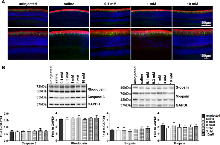
To assess retinal health, western blots were performed at the PI7 h (Figure 2) and the PI30 day (Appendix 1) to test photoreceptor-specific gene expression. The data showed that the protein levels of rhodopsin, M-opsin, and S-opsin were similar to those of the uninjected and the saline-injected controls. We also performed immunohistochemistry on the paraffin sections of the eyes to evaluate the localization and distribution of these proteins in the retinas. The data showed that rhodopsin, M-opsin, and S-opsin were properly localized in the photoreceptor cells in all groups. We also tested caspase 3 expression levels with western blot and demonstrated that the expression of caspase 3 within the group was similar (Figure 2B). These data indicated that nanoceria, even when present at high dosage, do not cause photoreceptor injury or mislocalization of these proteins.
Figure 2.
Nanoceria do not alter the amount or distribution of phototransduction proteins and caspase 3 level. A: Immunohistochemistry using paraffin sections at PI7 h showed that rhodopsin (red) and M-opsin (green) are properly localized in the outer segments of the retinas. n = 3–6 eyes per group. Scale bar = 100 µm. B: Western blots were performed at PI7 h to assess the protein levels of rhodopsin, M-opsin and S-opsin, and caspase 3. Densitometric analysis of the bands is shown as the mean ± standard error of the mean (SEM), and there are no statistically significant differences among the groups. n = 3–6 eyes per group.
Evaluation of retinal function
Full-field ERG was performed at PI30 day to assess retinal function by evaluating the responses of rods and cones to light stimulation. The ERG data showed that there were no statistically significant changes in the ERG amplitudes among the different groups at each time point tested (Figure 3), indicating that retention of nanoceria in the eye does not affect normal retinal function.
Figure 3.
Electroretinographic evaluation of retinal function. Rod and cone electroretinogram (ERG) performance (see detailed information in Methods) at the PI30 day demonstrated that the amplitudes from all nanoceria-injected groups are comparable to those of the uninjected mice. Data shown are mean ± standard error of the mean (SEM), and there are no statistically significant differences among the groups. n = 3–6 animals.
Analysis of proinflammatory cytokines
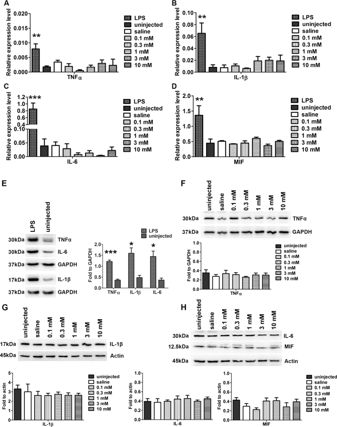
Infection induces innate and acute inflammation or chronic inflammatory responses, which involve activation and migration of various immune cells (macrophages, microglia, and neutrophils) to the site of inflammation and persistent release of proinflammatory cytokines [14,15]. To test the effects of nanoceria retention in the retina on the expression of proinflammatory cytokine genes, PCR array assays were performed using the “mouse common cytokines” array plates and the retinas from the uninjected, saline-injected, and 1 mM nanoceria-injected eyes at the PI7 day. A total of 89 genes were surveyed, and only 11 were upregulated (Bmp1, Bmp6, Fgf10, Il1a, Il1f5, Il1f9, Tnfrsf11b, Tnfsf9) or downregulated (Bmp2, Ctf1, Inhba; Table 1). However, these genes were also similarly upregulated or downregulated following saline injection, suggesting that the increases in the expression of these genes were most likely caused by the injury resulting from the injection procedure itself or from the saline in which the nanoceria were suspended. To determine whether any acute inflammatory responses were triggered by nanoceria, the expression of the mRNAs of the proinflammatory cytokines, TNF-α, IL-1β, IL-6, and MIF, was analyzed with qRT-PCR at an early time point (PI7 h). Compared to the higher levels of expression in the lipopolysaccharide (LPS)-induced inflammatory cytokines, none of the concentrations of nanoceria induced alterations in the expression of these cytokines compared to the uninjected controls (Figure 4A-D). Western blot assay at the PI7 h (Figure 4E-H) and the PI30 day (Appendix 2) demonstrated that the protein levels of TNF-α, IL-1β, IL-6, and MIF were similar among the groups injected with various concentrations of nanoceria versus the uninjected mice with no statistically significant differences seen.
Table 1. Mouse common cytokine genes with changes of expression at PI-7d (≥2.0 fold).
| Symbol | Gene name | Function | CeO2/wt fold | P value | Saline/wt fold | P value |
|---|---|---|---|---|---|---|
| Bmp1 |
Bone morphogenetic protein 1 |
TGFβ Superfamily |
3.22 |
0.0133* |
3.45 |
0.0146* |
| Bmp2 |
Bone morphogenetic protein 2 |
TGFβ Superfamily |
−2.39 |
0.2106 |
−2.68 |
0.2026 |
| Bmp6 |
Bone morphogenetic protein 6 |
TGFβ Superfamily |
2.19 |
0.0478* |
3.45 |
0.0108* |
| Ctf1 |
Cardiotrophin 1 |
Other Cytokines |
−2.91 |
0.0732 |
−4.62 |
0.0447* |
| Fgf10 |
Fibroblast growth factor 10 |
Growth Factors |
3.67 |
0.107 |
1.82 |
0.6488 |
| II1a |
Interleukin 1 alpha |
Interleukins |
2.41 |
0.4002 |
6.91 |
0.0313* |
| II1f5 |
Interleukin 1 family, member 5 (delta) |
Interleukins |
2.64 |
0.7914 |
2.93 |
0.718 |
| Il1f9 |
Interleukin 1 family, member 9 |
Interleukins |
4.19 |
0.4721 |
6.09 |
0.0156* |
| Inhba |
Inhibin alpha |
TGFβ Superfamily |
−2.23 |
0.252 |
−3.01 |
0.0828 |
| Tnfrsf11b |
Tumor necrosis factor receptor superfamily, member 11b (osteoprotegerin) |
TNF Superfamily |
1.93 |
0.641 |
2.76 |
0.2891 |
| Tnfsf9 | Tumor necrosis factor (ligand) superfamily, member 9 | TNF Superfamily | 2.54 | 0.8813 | 3.55 | 0.6702 |
(n=3–4, *p<0.05)
Figure 4.
Nanoceria do not change the mRNA or protein levels of inflammatory cytokines. A-D: Quantitative real-time PCR (qRT-PCR) analysis of the mRNA levels of acute and chronic inflammatory cytokines (tumor necrosis factor (TNF-α; A), interleukin 1β (IL-1β; B), IL-6 (C), and macrophage migration inhibitory factor (MIF; D) at the PI7 h. Injection of lipopolysaccharides (LPS) was used as a positive control. Uninjected and saline -injected wild-type (WT) mice served as the negative uninjected and the injected controls, respectively. No statistically significant differences in gene expression were seen among the groups of uninjected, saline-injected, and any of the nanoceria-injected groups. n = 3–8 eyes per group, **p<0.005, ***p<0.0001. E-H: Western blot assay to evaluate the protein levels of TNF-α (F), IL-1β (G), IL-6, and MIF (H) at PI7 h. Compared to the uninjected WT mice, the LPS-injected mice have two to four fold higher (n = 3–5 eyes per group, *p<0.05, ***p<0.0001) expression of these cytokines (E). However, none of the nanoceria- and saline-injected groups showed changes in the protein levels of these genes, and no statistically significant differences were seen among the groups. n = 3–5 eyes per group.
Assessment of the vascular system
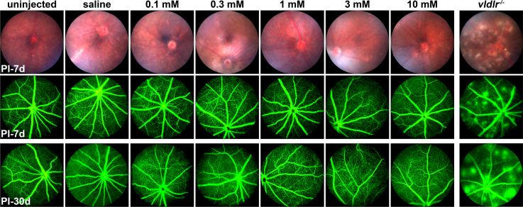
Acute and chronic inflammation causes increased vascular permeability and neovascularization [14]. To observe whether nanoceria in the eyes cause any abnormalities in the fundus appearance and organization of the retinal vascular system, we performed fundoscopy and fluorescein angiography using the eyes at the PI7 day and the PI30 day. Figure 5 shows that the patterns of the blood veins were similar among all the groups, and the fundus in all the eyes, either uninjected or injected, exhibited a normal appearance without noticeable flecks or spots. Fluorescein angiographic observation showed a well-organized vasculature without abnormal blood vessels and leakage in the uninjected mice, compared to the typical neovascularization in a wet AMD mouse model, the very low density lipoprotein receptor knockout (vldlr−/−) mouse, in which numerous hyper-fluorescence spots were seen and fluorescein leakage increased with time (the far right panel). In all mice injected with either saline or various doses of nanoceria, a clear and neat, uniform vascular structure and pattern were seen at these time points.
Figure 5.
Nanoceria do not cause vascular permeability or leakage. Fundoscopic and fluorescein angiographic images taken at the PI7 day and the PI30 day show that the eyes from the vldlr−/− mice exhibit an abnormal appearance of the fundus and have typical neovascularization and vascular leakage. None of the nanoceria- and saline-injected groups showed neovascularization or leakage and were similar to the uninjected mice. A representative image from each group is shown. n = 4–6 eyes per group.
Inflammatory cell infiltration
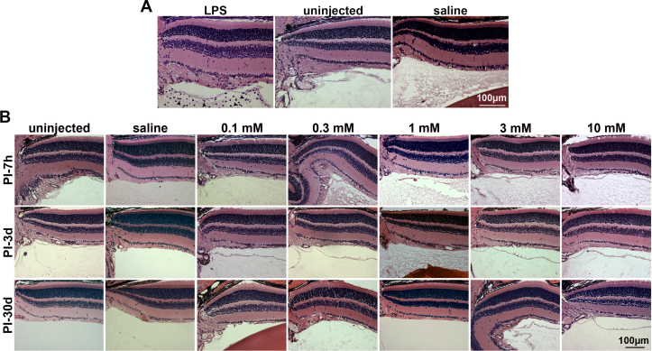
Inflammation also causes cellular infiltration into the vitreous. To investigate this, we performed histological analysis on the H&E-stained slides at five time points, with 0.5 µg of LPS in 1 µl of saline-injected eyes as a positive control [16]. Compared to the LPS-induced massive anterior segment and vitreous infiltration, we did not find any cells inside the vitreous of the eyes injected with nanoceria or saline (Figure 6).
Figure 6.
Nanoceria do not produce a cellular inflammatory response in the eye. Histological images were taken at the superior side of the eye under 20X from each group. A: Lipopolysaccharide (LPS)-injected eyes produced massive cellular infiltration (arrowhead) in the vitreous at 12 h after injection whereas no cellular infiltration was seen in the vitreous of the uninjected and saline-injected eyes. B: No cellular infiltration was seen in the eyes injected with saline, the eyes injected with various doses of nanoceria, or the uninjected eyes. Representatives from each group and only the PI7 h and the PI3 and PI30 day are shown. n = 3–6 eyes per group. Scale bar = 100 µm.
Discussion
Nanomaterials have attracted much attention over the past two decades. Because of their small sizes (usually less than 100 nm), surface structures, and unique physicochemical features, nanomaterials easily pass through membranes and are taken up into cells [17]. We and our colleagues have published a series of papers demonstrating the antioxidant properties of nanoceria in scavenging reactive oxygen species (ROS) and nitric oxide species (NOS), the enhancement of cellular survival, and the inhibition of apoptosis in numerous tissues and cells in vivo and in vitro [18,19]. In the eye, we showed that a single intravitreal injection of nanoceria produced sustained therapeutic effects in several mouse models of ocular diseases [2-6]. These results indicate the potential clinical benefit of nanoceria. It has been reported that intravitreal injection of many drugs (and biologic products) at a high dose usually produces ocular inflammation [20]. Although data from our laboratory showed that nanoceria at 1 mM did not alter the retinal structure and function in albino rats [9], obtaining acceptance of nanoceria as a therapeutic agent remains challenging [21], especially as the safety of the long-term retention of nanoceria at a maximum dose in the eye is largely unknown.
The toxicity of nanoceria has been assessed in a variety of cells and tissues, including the human neuroblastoma cell line (IMR32) [22], human lung cells [23,24], cultured human lung cancer cells [25], wild-type rats [26], human gastric cancer cells [27], and human hepatoma SMMC-7721 cells [28]. Interestingly, the data showed that nanosized CeO2 are more toxic than microparticles [22]. A similar report indicated that accumulation of nanoceria-caused toxicity is correlated with increased exposure time [29], and in a dose- and time-dependent manner that resulted from lipid peroxidation and cell membrane damage [25], DNA damage and apoptosis [23], inflammation [26], oxidative stress, and activation of the mitogen-activated protein kinase (MAPK) pathways [28]. In contrast to these reports, data documenting the protective functions of nanoceria have been reported by many research groups, and in a variety of cells and tissues with varied doses. Data showed that nanoceria decrease by 70% ischemia-induced 3-nitrotyrosine (indicator of protein damage) in a mouse model of cerebral ischemia [30], protect human tumor monocytes (U937 cells) against TNF-α and cycloheximide-induced alteration of calcium signals, ROS production, and apoptosis [31], and prevent apoptosis in primary cortical brain cultures [32]. Nanoceria attenuated the systemic inflammatory response associated with peritonitis and significantly improved survival rates [33], and selectively protected normal human cells, but not cancer cells, from ultraviolet (UV) radiation [34]. In another report, treatment with nanoceria (100 µg/ml) for 48 h did not cause growth or morphological changes in human lens epithelial cells, but exposure to a lower dosage (10 µg/ml) for a longer period of time (72 h) can harm cells [35]. Published data from our group and associated colleagues showed that pretreatment with varying concentrations of nanoceria protects normal cells from radiation-induced damage [36,37]. Similarly, nanoceria prevented tumor growth and invasion [38] and showed antiangiogenic properties [39], neuroprotection [19], cardioprotection [18], and anti-inflammatory activity [40,41] in multiple tissues or organs. Most importantly, our formulated nanoceria have been reported to be distributed in multiple organs and tissues of CD1 mice via various means of administration and do not cause overt toxicity or pathology, and no significant immune response was detected [10]. We think the differences observed in beneficial versus negative effects of nanoceria by various laboratories are primarily due to differences in the formulations, particle sizes, surface charges, concentrations, distribution and location of nanoceria inside the cells, the age of the animals at treatment, or the experimental conditions in general.
Currently, treatment of eye diseases caused by endogenous intraocular changes involves delivery of therapeutic agents (proteins, DNAs, and cells) into the back of the eye by subretinal or intravitreal injections that by themselves may cause infection and inflammation. In this paper, we experimentally demonstrated that nanoceria, at all doses used, are well tolerated by the ocular cells and do not induce detectable damage to the retinal health as determined with qRT-PCR, western blots, histology, and ERG. There were no changes in the levels of photoreceptor-specific proteins, distribution of visual pigments, retinal morphology, and response to the light. These results demonstrated that nanoceria exert their protective function without negative effects on retinal cells.
As nanoceria do not cause structural and functional alterations in the retina, we were especially interested in determining whether the retention of nanoceria in the eye was associated with increased expression of inflammatory cytokines. Foreign materials in the eye usually cause acute or chronic inflammatory responses [14], such as elevated cytokine gene expression within several hours or several days after the onset of inflammation. This is followed by cellular infiltration, immune cell activation, and migration to the sites of inflammation [14]. In this study, we performed a PCR array of the common mouse cytokines to assay 89 cytokines at the PI7 day. Of these cytokines, only 11 were either upregulated or downregulated by the nanoceria treatment, but they were similarly affected by saline injection alone (Table 1). Inflammation-associated cytokines, including TNF-α, IL-1β, IL-6, etc., are mainly produced by macrophages and monocytes at inflammatory sites [15] in response to acute and chronic inflammation. MIF, a multifunctional and ubiquitously expressed protein, is an upstream regulator of inflammatory-immune processes and is expressed at the site of inflammation where MIF primarily modulates macrophage and T cell function for host defense. qRT-PCR and western blots of IL-6, IL-1β, TNF-α, and MIF at PI7 h demonstrated that, compared to the high expression levels of these genes in the LPS-induced positive controls, there was no elevation of these proteins compared to the untreated group. Our previous study demonstrated that the retention of nanoceria in the retina for months after a single intravitreal injection did not alter the retinal structure and function [9]. Our current study suggests that there is no cytotoxicity of nanoceria to the retinal tissues and provides further, and direct, evidence that nanoceria could be a special therapeutic agent for the treatment of ocular diseases.
Acknowledgments
The authors thank the personnel at the animal, imaging and molecular modules of the Vision Research Core Facility at the Oklahoma University Health Sciences Center. This work was supported in part by NIH NEI P30 EY021725, R21EY018306, R01EY018724 and R01EY022111, National Science Foundation: CBET-0708172. Dr. Xue Cai (xue-cai@ouhsc.edu) and Dr. James F. McGinnis (james-mcginnis@ouhsc.edu) are co-corresponding authors for this paper. Conflict of Interest and Financial Disclosure Statements: University of Central Florida and University of Oklahoma Health Sciences Center hold patents with James F. McGinnis and Sudipta Seal listed as inventors.
Appendix 1.
Nanoceria do not change the levels of photoreceptor-specific proteins. Western blot assessment at PI30 day demonstrated that there are no significant differences on the amount of rhodopsin and S-opsin among groups. n=3–5 eyes per group. To access the data, click or select the words “Appendix 1.”
Appendix 2.
Nanoceria do not change the protein levels of inflammatory cytokines. Western blot assay at PI30 day demonstrated that no significant differences on the protein levels of TNF-α and MIF were seen. n=3–5 eyes per group. To access the data, click or select the words “Appendix 2.”
References
- 1.Petrak K. Essential properties of drug-targeting delivery systems. Drug Discov Today. 2005;10:1667–73. doi: 10.1016/S1359-6446(05)03698-6. [DOI] [PubMed] [Google Scholar]
- 2.Cai X, Sezate SA, Seal S, McGinnis JF. Sustained protection against photoreceptor degeneration in tubby mice by intravitreal injection of nanoceria. Biomaterials. 2012;33:8771–81. doi: 10.1016/j.biomaterials.2012.08.030. [DOI] [PMC free article] [PubMed] [Google Scholar]
- 3.Kong L, Cai X, Zhou X, Wong LL, Karakoti AS, Seal S, McGinnis JF. Nanoceria extend photoreceptor cell lifespan in tubby mice by modulation of apoptosis/survival signaling pathways. Neurobiol Dis. 2011;42:514–23. doi: 10.1016/j.nbd.2011.03.004. [DOI] [PMC free article] [PubMed] [Google Scholar]
- 4.Chen J, Patil S, Seal S, McGinnis JF. Rare earth nanoparticles prevent retinal degeneration induced by intracellular peroxides. Nat Nanotechnol. 2006;1:142–50. doi: 10.1038/nnano.2006.91. [DOI] [PubMed] [Google Scholar]
- 5.Cai X, Seal S, McGinnis JF. Sustained inhibition of neovascularization in vldlr−/− mice following intravitreal injection of cerium oxide nanoparticles and the role of the ASK1–P38/JNK-NF-kappaB pathway. Biomaterials. 2014;35:249–58. doi: 10.1016/j.biomaterials.2013.10.022. [DOI] [PMC free article] [PubMed] [Google Scholar]
- 6.Zhou X, Wong LL, Karakoti AS, Seal S, McGinnis JF. Nanoceria inhibit the development and promote the regression of pathologic retinal neovascularization in the Vldlr knockout mouse. PLoS ONE. 2011;6:e16733. doi: 10.1371/journal.pone.0016733. [DOI] [PMC free article] [PubMed] [Google Scholar]
- 7.Cai X, Sezate SA, McGinnis JF. Neovascularization: ocular diseases, animal models and therapies. Adv Exp Med Biol. 2012;723:245–52. doi: 10.1007/978-1-4614-0631-0_32. [DOI] [PubMed] [Google Scholar]
- 8.Cai X, Yodoi J, Seal S, McGinnis JF. Nanoceria and thioredoxin regulate a common antioxidative gene network in tubby mice. Adv Exp Med Biol. 2014;801:829–36. doi: 10.1007/978-1-4614-3209-8_104. [DOI] [PubMed] [Google Scholar]
- 9.Wong LL, Hirst SM, Pye QN, Reilly CM, Seal S, McGinnis JF. Catalytic nanoceria are preferentially retained in the rat retina and are not cytotoxic after intravitreal injection. PLoS ONE. 2013;8:e58431. doi: 10.1371/journal.pone.0058431. [DOI] [PMC free article] [PubMed] [Google Scholar]
- 10.Hirst SM, Karakoti A, Singh S, Self W, Tyler R, Seal S, Reilly CM. Bio-distribution and in vivo antioxidant effects of cerium oxide nanoparticles in mice. Environ Toxicol. 2013;28:107–18. doi: 10.1002/tox.20704. [DOI] [PubMed] [Google Scholar]
- 11.Cai X, Conley SM, Nash Z, Fliesler SJ, Cooper MJ, Naash MI. Gene delivery to mitotic and postmitotic photoreceptors via compacted DNA nanoparticles results in improved phenotype in a mouse model of retinitis pigmentosa. FASEB. 2010;24:1178–91. doi: 10.1096/fj.09-139147. [DOI] [PMC free article] [PubMed] [Google Scholar]
- 12.Cai X, Nash Z, Conley SM, Fliesler SJ, Cooper MJ, Naash MI. A partial structural and functional rescue of a retinitis pigmentosa model with compacted DNA nanoparticles. PLoS ONE. 2009;4:e5290. doi: 10.1371/journal.pone.0005290. [DOI] [PMC free article] [PubMed] [Google Scholar]
- 13.Zhu L, Shen J, Zhang C, Park CY, Kohanim S, Yew M, Parker JS, Chuck RS. Inflammatory cytokine expression on the ocular surface in the Botulium toxin B induced murine dry eye model. Mol Vis. 2009;15:250–8. [PMC free article] [PubMed] [Google Scholar]
- 14.Anderson JM. Biological responses to materials. Annu Rev Mater Res. 2001;31:81–110. [Google Scholar]
- 15.Gabay C, Kushner I. Acute-phase proteins and other systemic responses to inflammation. N Engl J Med. 1999;340:448–54. doi: 10.1056/NEJM199902113400607. [DOI] [PubMed] [Google Scholar]
- 16.Li X, Gu X, Boyce TM, Zheng M, Reagan AM, Qi H, Mandal N, Cohen AW, Callegan MC, Carr DJ, Elliott MH. Caveolin-1 increases proinflammatory chemoattractants and blood-retinal barrier breakdown but decreases leukocyte recruitment in inflammation. Invest Ophthalmol Vis Sci. 2014;55:6224–34. doi: 10.1167/iovs.14-14613. [DOI] [PMC free article] [PubMed] [Google Scholar]
- Cai X, Seal S, McGinnis J. Cerium oxide nanoparticle reduction of oxidative damage in retina. Stratton R, Hauswirth WW, Gardiner T, editors: Humana Press; 2012. 399–418. [Google Scholar]
- 18.Niu J, Azfer A, Rogers LM, Wang X, Kolattukudy PE. Cardioprotective effects of cerium oxide nanoparticles in a transgenic murine model of cardiomyopathy. Cardiovasc Res. 2007;73:549–59. doi: 10.1016/j.cardiores.2006.11.031. [DOI] [PMC free article] [PubMed] [Google Scholar]
- 19.Das M, Patil S, Bhargava N, Kang JF, Riedel LM, Seal S, Hickman JJ. Auto-catalytic ceria nanoparticles offer neuroprotection to adult rat spinal cord neurons. Biomaterials. 2007;28:1918–25. doi: 10.1016/j.biomaterials.2006.11.036. [DOI] [PMC free article] [PubMed] [Google Scholar]
- 20.Short BG. Safety evaluation of ocular drug delivery formulations: techniques and practical considerations. Toxicol Pathol. 2008;36:49–62. doi: 10.1177/0192623307310955. [DOI] [PubMed] [Google Scholar]
- 21.Walkey C, Das S, Seal S, Erlichman J, Heckman K, Ghibelli L, Traversa E, McGinnis JF, Self WT. Catalytic Properties and Biomedical Applications of Cerium Oxide Nanoparticles. Environ Sci Nano. 2015;2:33–53. doi: 10.1039/C4EN00138A. [DOI] [PMC free article] [PubMed] [Google Scholar]
- 22.Kumari M, Singh SP, Chinde S, Rahman MF, Mahboob M, Grover P. Toxicity study of cerium oxide nanoparticles in human neuroblastoma cells. Int J Toxicol. 2014;33:86–97. doi: 10.1177/1091581814522305. [DOI] [PubMed] [Google Scholar]
- 23.Mittal S, Pandey AK. Cerium oxide nanoparticles induced toxicity in human lung cells: role of ROS mediated DNA damage and apoptosis. Biomed Res Int. 2014;2014:891934. doi: 10.1155/2014/891934. [DOI] [PMC free article] [PubMed] [Google Scholar]
- 24.Ma JY, Mercer RR, Barger M, Schwegler-Berry D, Scabilloni J, Ma JK, Castranova V. Induction of pulmonary fibrosis by cerium oxide nanoparticles. Toxicol Appl Pharmacol. 2012;262:255–64. doi: 10.1016/j.taap.2012.05.005. [DOI] [PMC free article] [PubMed] [Google Scholar]
- 25.Lin W, Huang YW, Zhou XD, Ma Y. Toxicity of cerium oxide nanoparticles in human lung cancer cells. Int J Toxicol. 2006;25:451–7. doi: 10.1080/10915810600959543. [DOI] [PubMed] [Google Scholar]
- 26.Ma JY, Zhao H, Mercer RR, Barger M, Rao M, Meighan T, Schwegler-Berry D, Castranova V, Ma JK. Cerium oxide nanoparticle-induced pulmonary inflammation and alveolar macrophage functional change in rats. Nanotoxicology. 2011;5:312–25. doi: 10.3109/17435390.2010.519835. [DOI] [PubMed] [Google Scholar]
- 27.Li C, Zhao W, Liu B, Xu G, Liu L, Lv H, Shang D, Yang D, Damirin A, Zhang J. Cytotoxicity of ultrafine monodispersed nanoceria on human gastric cancer cells. J Biomed Nanotechnol. 2014;10:1231–41. doi: 10.1166/jbn.2014.1863. [DOI] [PubMed] [Google Scholar]
- 28.Cheng G, Guo W, Han L, Chen E, Kong L, Wang L, Ai W, Song N, Li H, Chen H. Cerium oxide nanoparticles induce cytotoxicity in human hepatoma SMMC-7721 cells via oxidative stress and the activation of MAPK signaling pathways. Toxicol In Vitro. 2013;27:1082–8. doi: 10.1016/j.tiv.2013.02.005. [DOI] [PubMed] [Google Scholar]
- 29.Yokel RA, Hussain S, Garantziotis S, Demokritou P, Castranova V, Cassee FR. The Yin: An adverse health perspective of nanoceria: uptake, distribution, accumulation, and mechanisms of its toxicity. Environ Sci Nano. 2014;1:406–28. doi: 10.1039/C4EN00039K. [DOI] [PMC free article] [PubMed] [Google Scholar]
- 30.Estevez AY, Pritchard S, Harper K, Aston JW, Lynch A, Lucky JJ, Ludington JS, Chatani P, Mosenthal WP, Leiter JC, Andreescu S, Erlichman JS. Neuroprotective mechanisms of cerium oxide nanoparticles in a mouse hippocampal brain slice model of ischemia. Free Radic Biol Med. 2011;51:1155–63. doi: 10.1016/j.freeradbiomed.2011.06.006. [DOI] [PubMed] [Google Scholar]
- 31.Gonzalez-Flores D, De Nicola M, Bruni E, Caputo F, Rodriguez AB, Pariente JA, Ghibelli L. Nanoceria protects from alterations in oxidative metabolism and calcium overloads induced by TNFalpha and cycloheximide in U937 cells: pharmacological potential of nanoparticles. Mol Cell Biochem. 2014;397:245–53. doi: 10.1007/s11010-014-2192-2. [DOI] [PubMed] [Google Scholar]
- 32.Arya A, Sethy NK, Das M, Singh SK, Das A, Ujjain SK, Sharma RK, Sharma M, Bhargava K. Cerium oxide nanoparticles prevent apoptosis in primary cortical culture by stabilizing mitochondrial membrane potential. Free Radic Res. 2014;48:784–93. doi: 10.3109/10715762.2014.906593. [DOI] [PubMed] [Google Scholar]
- 33.Manne ND, Arvapalli R, Nepal N, Thulluri S, Selvaraj V, Shokuhfar T, He K, Rice KM, Asano S, Maheshwari M, Blough ER. Therapeutic potential of cerium oxide nanoparticles for the treatment of peritonitis induced by polymicrobial insult in Sprague-Dawley rats. Crit Care Med. 2015;43:e477–89. doi: 10.1097/CCM.0000000000001258. [DOI] [PubMed] [Google Scholar]
- 34.Zhang L, Jiang H, Selke M, Wang X. Selective cytotoxicity effect of cerium oxide nanoparticles under UV irradiation. J Biomed Nanotechnol. 2014;10:278–86. doi: 10.1166/jbn.2014.1790. [DOI] [PubMed] [Google Scholar]
- 35.Pierscionek BK, Li Y, Schachar RA, Chen W. The effect of high concentration and exposure duration of nanoceria on human lens epithelial cells. Nanomedicine. 2012;8:383–90. doi: 10.1016/j.nano.2011.06.016. [DOI] [PubMed] [Google Scholar]
- 36.Colon J, Herrera L, Smith J, Patil S, Komanski C, Kupelian P, Seal S, Jenkins DW, Baker CH. Protection from radiation-induced pneumonitis using cerium oxide nanoparticles. Nanomedicine. 2009;5:225–31. doi: 10.1016/j.nano.2008.10.003. [DOI] [PubMed] [Google Scholar]
- 37.Colon J, Hsieh N, Ferguson A, Kupelian P, Seal S, Jenkins DW, Baker CH. Cerium oxide nanoparticles protect gastrointestinal epithelium from radiation-induced damage by reduction of reactive oxygen species and upregulation of superoxide dismutase 2. Nanomedicine. 2010;6:698–705. doi: 10.1016/j.nano.2010.01.010. [DOI] [PubMed] [Google Scholar]
- 38.Alili L, Sack M, von Montfort C, Giri S, Das S, Carroll KS, Zanger K, Seal S, Brenneisen P. Downregulation of tumor growth and invasion by redox-active nanoparticles. Antioxid Redox Signal. 2013;19:765–78. doi: 10.1089/ars.2012.4831. [DOI] [PMC free article] [PubMed] [Google Scholar]
- 39.Giri S, Karakoti A, Graham RP, Maguire JL, Reilly CM, Seal S, Rattan R, Shridhar V. Nanoceria: a rare-earth nanoparticle as a novel anti-angiogenic therapeutic agent in ovarian cancer. PLoS ONE. 2013;8:e54578. doi: 10.1371/journal.pone.0054578. [DOI] [PMC free article] [PubMed] [Google Scholar]
- 40.Hirst SM, Karakoti AS, Tyler RD, Sriranganathan N, Seal S, Reilly CM. Anti-inflammatory properties of cerium oxide nanoparticles. Small. 2009;5:2848–56. doi: 10.1002/smll.200901048. [DOI] [PubMed] [Google Scholar]
- 41.Kyosseva SV, Chen L, Seal S, McGinnis JF. Nanoceria inhibit expression of genes associated with inflammation and angiogenesis in the retina of Vldlr null mice. Exp Eye Res. 2013;116:63–74. doi: 10.1016/j.exer.2013.08.003. [DOI] [PMC free article] [PubMed] [Google Scholar]


