Abstract
Objective
Immune-related abnormalities are commonly reported in schizophrenia, including higher mRNA levels for the viral restriction factor interferon-induced transmembrane protein (IFITM) in the prefrontal cortex. The authors sought to clarify whether higher IFITM mRNA levels and other immune-related disturbances in the prefrontal cortex are the consequence of an ongoing molecular cascade contributing to immune activation or the reflection of a long-lasting maladaptive response to an in utero immune-related insult.
Method
Quantitative polymerase chain reaction was employed to measure mRNA levels for immune-related cytokines and transcriptional regulators, including those reported to regulate IFITM expression, in the prefrontal cortex from 62 schizophrenia and 62 healthy subjects and from adult mice exposed prenatally to maternal immune activation or in adulthood to the immune stimulant poly(I:C).
Results
Schizophrenia subjects had markedly higher mRNA levels for interleukin 6 (IL-6) (+379%) and interferon-β (+29%), which induce IFITM expression; lower mRNA levels for Schnurri-2 (−10%), a transcriptional inhibitor that lowers IFITM expression; and higher mRNA levels for nuclear factor-κB (+86%), a critical transcription factor that mediates cytokine regulation of immune-related gene expression. In adult mice that received daily poly(I:C) injections, but not in offspring with prenatal exposure to maternal immune activation, frontal cortex mRNA levels were also markedly elevated for IFITM (+304%), multiple cytokines including IL-6 (+493%), and nuclear factor-κB (+151%).
Conclusions
These data suggest that higher prefrontal cortex IFITM mRNA levels in schizophrenia may be attributable to adult, but not prenatal, activation of multiple immune markers and encourage further investigation into the potential role of these and other immune markers as therapeutic targets in schizophrenia.
Diverse lines of evidence from genetic, epidemiological, and biomarker studies suggest that immune- and inflammation-related abnormalities play an important role in the disease process of schizophrenia. For example, multiple genome-wide association studies have identified variants in genes involved in immune and inflammatory signaling pathways that associate with schizophrenia (1–6). In addition, exposure to infectious diseases in pregnancy (7, 8), including evidence of maternal response to infection such as higher serum levels of proinflammatory cytokines (9) and inflammatory biomarkers (10), have been linked to higher rates of schizophrenia in offspring (11). Furthermore, higher rates of autoimmune illnesses (12, 13) and higher levels of proinflammatory cytokines, such as interleukin 6 (IL-6) in the serum (14–16) and in the prefrontal cortex(17,18),have been reported in subjects with schizophrenia.
Consistent with these findings, we and others have reported evidence of immune activation in the prefrontal cortex in schizophrenia, including higher mRNA levels for the viral restriction factor interferon-induced transmembrane protein (IFITM), which inhibits the processes involved in viral entry and replication (19–21). Schizophrenia subjects with higher IFITM mRNA levels also had greater disturbances in markers of prefrontal cortex GABA neurons, suggesting that altered immune function may be involved in cortical circuitry alterations in the disorder. These findings suggest that investigating the pathogenesis of IFITM over-expression may provide a useful window into the role of altered prefrontal cortex immune markers in the pathophysiology of schizophrenia. However, the upstream factors that contribute to elevated mRNA levels for IFITM and other immune-related markers (17, 18) in the prefrontal cortex in schizophrenia are not known. For example, higher IFITM mRNA levels may be attributable to immune activation (18); however, it is not known whether higher levels of the cytokines and transcriptional regulators that regulate the expression of IFITM (Figure 1A) (22–24) and other immune-related markers are present in the prefrontal cortex of the same schizophrenia subjects who have higher IFITM mRNA levels (21). Furthermore, such correlative evidence of a relationship between IFITM and cytokine levels in the pre-frontal cortex, if present in schizophrenia, would require additional proof-of-principle testing of cause and effect using animal models of immune stimulation.
FIGURE 1. Schematic Illustration of the Relationships Among Immune Markers in the Healthy State and in Schizophreni a.
aPanel A illustrates factors regulating IFITM expression in the healthy state. Interleukin 6 (IL-6) and interferon-β induce IFITM expression. The transcriptional regulator nuclear factor-κB (NF-κB) plays a central role in regulating the expression of many immune-related genes, including IL-1B and IL-6, is itself regulated by immune-related cytokines (e.g., IL-1B), and increases expression of IFITM. The NF-κB site-binding protein Schnurri-2 inhibits NF-κB function. Panel Billustrates the molecular cascade of immune activation in schizophrenia. The markedly higher mRNA levels for IL-1B, IL-6, interferon-β, and NF-κB, and lower mRNA levels for Schnurri-2, in the prefrontal cortex of schizophrenia subjects all converge to increase levels of IFITM mRNAs.
It is also unknown whether higher IFITM mRNA levels in the prefrontal cortex in schizophrenia reflect a long-lasting maladaptive response to an insult that occurred much earlier, such as maternal immune activation in utero, as suggested by epidemiological findings (7–11). This hypothesis is supported by experimental findings that maternal immune activation using the immune stimulant poly(I:C) in mice leads to higher cytokine levels in fetal brain (25) that may persist to a varying extent postnatally (26). In addition, maternal immune activation has also been reported to produce epigenetic modifications to the promoter regions of genes that can alter gene expression postnatally (27).
To investigate these potential molecular and developmental mechanisms of higher IFITM mRNA levels in schizophrenia, we conducted postmortem brain tissue studies of mRNA levels of cytokines and transcriptional regulators that regulate levels of IFITM and other immune-related markers in the prefrontal cortex of a large cohort of schizophrenia subjects with elevated IFITM mRNA levels (21) and in the frontal cortex of mice exposed to immune stimulation prenatally or during adulthood using the immune stimulant poly(I:C). We also assessed for the impact of antipsychotic medications on the expression of immune markers using antipsychotic-exposed monkeys.
METHOD
Human Subjects
Brain specimens were obtained during routine autopsies conducted at the Allegheny County Office of the Medical Examiner (Pittsburgh) after consent was obtained from next of kin. An independent committee of experienced research clinicians made consensus DSM-IV diagnoses for each subject using structured interviews with family members and review of medical records. The same approach was used to confirm the absence of any psychiatric diagnoses in healthy comparison subjects (28). To control for experimental variance, subjects with schizophrenia or schizoaffective disorder (N=62) were matched individually to one nonpsychiatric comparison subject for sex and as closely as possible for age (see Table S1 in the data supplement that accompanies the online edition of this article) (29). Tissue samples from subjects in a pair were processed together throughout all stages of the study. The mean age, postmortem interval, RNA integrity number, and tissue freezer storage time did not differ between subject groups (Table 1). Mean brain pH differed between the schizophrenia and nonpsychiatric groups (mean=6.6 [SD=0.3] and mean=6.7 [SD=0.2], respectively; t=2.6, df=122, p=0.01), but the difference was quite small and of uncertain biological significance.
TABLE 1.
Demographic, Clinical, and Tissue Characteristics of Human Subjects
| Measure | Nonpsychiatric Comparison Group (N=62) | Schizophrenia Group (N=62) | ||
|---|---|---|---|---|
| N | % | N | % | |
|
| ||||
| Male | 47 | 75.8 | 47 | 75.8 |
| Race | ||||
| White | 52 | 83.9 | 46 | 74.2 |
| Black | 10 | 16.1 | 16 | 25.8 |
|
| ||||
| Mean | SD | Mean | SD | |
|
| ||||
| Age (years) | 48.7 | 13.8 | 47.7 | 12.7 |
| Postmortem interval (hours) | 18.8 | 5.5 | 19.2 | 8.5 |
| Freezer storage time (months) | 131.8 | 56.2 | 128.1 | 60.7 |
| Brain pHa | 6.7 | 0.2 | 6.6 | 0.3 |
| RNA integrity number | 8.2 | 0.6 | 8.1 | 0.6 |
|
| ||||
| N | % | N | % | |
|
| ||||
| Medications at time of death | ||||
| Antipsychotic | 54 | 87.1 | ||
| Antidepressant | 27 | 43.5 | ||
| Benzodiazepine or anticonvulsant | 24 | 38.7 | ||
| Nonsteroidal anti-inflammatory drug | 13 | 20.0 | 16 | 25.8 |
Significant difference between groups, p=0.01.
All procedures were approved by the University of Pittsburgh’s Committee for the Oversight of Research Involving the Dead and Institutional Review Board for Biomedical Research.
Quantitative Polymerase Chain Reaction
Frozen tissue blocks containing the middle portion of the right superior frontal gyrus were confirmed to contain pre-frontal cortex area 9 using Nissl-stained cryostat tissue sections for each subject (30). Gray matter was separately collected into a tube containing TRIzol reagent in a manner that ensured minimal white matter contamination and excellent RNA preservation (31, 32). Standardized dilutions of total RNA (10 ng/μL) for each subject were used to synthesize cDNA. All primer pairs (see Table S2 in the online data supplement) demonstrated high amplification efficiency (>90%) across a wide range of cDNA dilutions and specific single products in dissociation curve analysis. Quantitative polymerase chain reaction (PCR) was performed using the comparative cycle threshold (CT) method with Power SYBR Green dye and the ViiA-7 Real-Time PCR System (Applied Biosystems, Waltham, Mass.), as previously described (33). Three reference genes (beta actin, cyclophilin A, and glyceraldehyde-3-phosphate dehydrogenase [GAPDH]) that we previously reported to be stably expressed in the present cohort of schizophrenia and nonpsychiatric comparison subjects (29) were used to normalize target mRNA levels (34). The difference in CT (dCT) for each target transcript was calculated by subtracting the geometric mean CT for the three reference genes from the CT of the target transcript (mean of four replicate measures). Because dCT represents the log2-transformed expression ratio of each target transcript to the reference genes, the relative level of the target transcript for each subject is reported as 2−dCT (31, 35).
Poly(I:C)-Exposed Mice
Timed pregnant C57BL/6J mice (12 mice per condition; Jackson Laboratory, Bar Harbor, Maine) received intraperitoneal injections of polyriboinosinic-polyribocytidilic acid potassium salt (poly[I:C]; 20 mg/kg pure form; Sigma, St. Louis], a synthetic analogue of double-stranded RNA that acts as a viral mimic and induces an immune response (26, 36, 37), or an equivalent volume of normal saline daily for 3 days in middle (embryonic day 11–13) or late (embryonic day 15–17) gestation (for the study design, see Table S3 in the online data supplement). Pups were weaned at 25 days, separated by sex, then euthanized at 8 weeks of age by cervical dislocation with isoflurane as a general anesthetic. The brain was removed, frozen on dry ice, and stored at −80°C. Nonpregnant adult female mice received injections of poly(I:C) (20 mg/kg; N=8) or normal saline (N=8) daily for 3 days in parallel with the timed pregnant mice. The nonpregnant mice were euthanized 3 hours after the last injection (random estrous cycle); trunk blood was collected after decapitation, and IL-6 levels were quantified in the resulting serum using the Mouse IL-6 Quantikine ELISA (enzyme-linked immunosorbent assay) Immunoassay (R&D Systems, Minneapolis) in order to confirm the presence of an immune response (25, 38) (see Figure S1 in the data supplement). All animal studies followed the National Institutes of Health Guide for the Care and Use of Laboratory Animals and were approved by the Institutional Animal Care and Use Committee.
Fresh, frozen brains from one male and/or female offspring per poly(I:C)-injected mother (N=7–8 per sex per condition; see Table S3 in the data supplement) and the nonpregnant adult female mice that received daily poly(I:C) or normal saline injections for 3 days were included in the study. RNA was isolated from homogenates of frontal cortex tissue sections (12 μm) collected consecutively from the bregma +2.8 to +2.1 mm (excluding the olfactory tissue below the rhinal fissure [39]) into TRIzol. Quantitative PCR assessment of the three relevant variants of IFITM mRNA (IFITM1, IFITM2, and IFITM3; IFITM4 is a pseudogene, and IFITM5 is found only in osteoblasts [24]) and mRNA levels for immune system-related cytokines and transcriptional regulators was performed as described for the human studies (see Table S2 in the data supplement).
Antipsychotic-Exposed Monkeys
Young adult male long-tailed monkeys (Macaca fascicularis) received oral doses of haloperidol, olanzapine, or placebo (N=6 monkeys per group) twice daily for 17–27 months, as previously described (40). RNA was isolated from prefrontal cortex area 9, and quantitative PCR was conducted for the same three reference genes included in the human study and immune system-related cytokines and transcriptional regulators (see Table S2) with all monkeys from a triad processed together on the same plate.
Statistical Analysis
The analysis of covariance (ANCOVA) model we report for the schizophrenia study includes mRNA level as the dependent variable, diagnostic group as the main effect, and age, postmortem interval, brain pH, RNA integrity number, and freezer storage time as covariates. Because each schizophrenia subject was individually matched to a nonpsychiatric subject to account for the parallel processing of tissue samples from a pair and to balance diagnostic groups for sex and age, a second ANCOVA model with subject pair as a blocking factor and including postmortem interval, brain pH, RNA integrity number, and freezer storage time was also used. Because the two ANCOVA models produced similar results, we report only the first model. Subsequent analyses of differences in mRNA levels between schizophrenia subjects grouped by use of psychotropic medications, nonsteroidal anti-inflammatory drugs (NSAIDs) (either prescribed or over-the-counter use, indicated by presence of NSAIDs in toxicology screens of blood or urine), or smoking at time of death and immune/inflammation-related cause of death or history of autoimmune illness (12) were conducted using the first ANCOVA model. Spearman’s rank correlation coefficients (rs) were calculated to assess the relationships between the different immune marker mRNA levels. For the antipsychotic-exposed monkey study, an ANCOVA model with mRNA level as the dependent variable, treatment group as the main effect, and triad as a blocking factor was employed. For the mouse studies, measures of mRNA levels were compared between groups using t tests with an alpha of 0.05.
RESULTS
Immune Markers in the Prefrontal Cortex in Schizophrenia
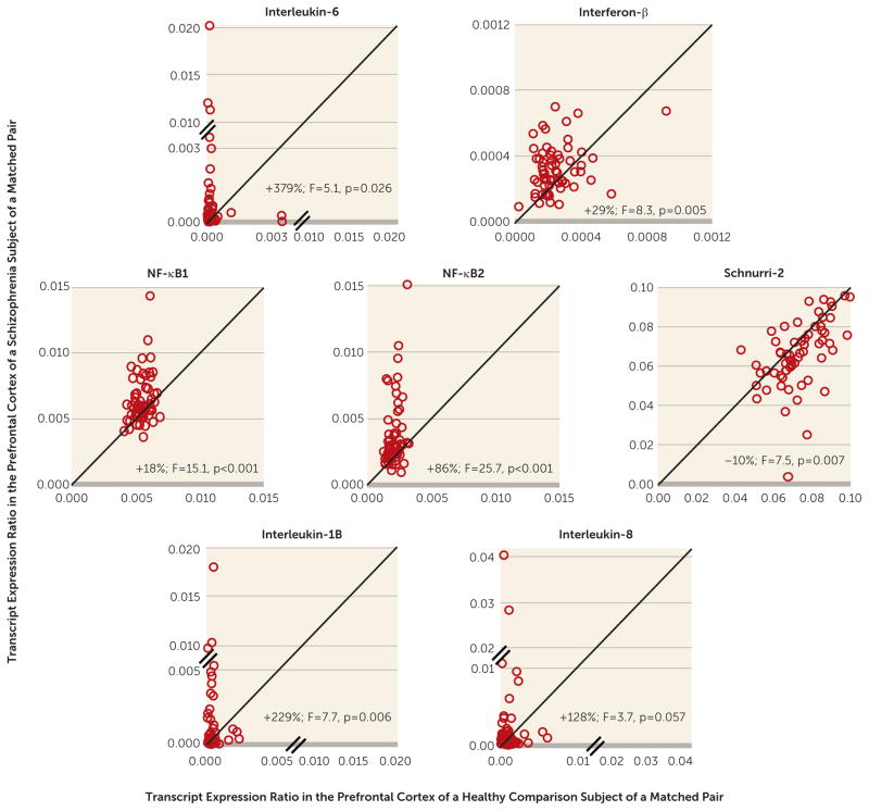
Multiple immune system-related cytokines (e.g., IL-6, interferon-β) have been reported to induce IFITM expression (Figure 1A) (22–24). Therefore, we first determined whether these immune-related markers were altered in the prefrontal cortex of the same cohort of schizophrenia subjects that we previously reported to have higher IFITM mRNA levels (i.e., IFITM1 and IFITM2/3) using the same quantitative PCR approach (21). Schizophrenia subjects had markedly higher mean mRNA levels for IL-6 (+379%; F=5.1, df=1, 117, p=0.026) and interferon-β (+29%; F=8.3, df=1, 117, p=0.005) (Figure 2). When subject pairs with a potential outlier (i.e., mRNA level more than three standard deviations from the mean of the respective diagnostic group) were excluded from the analysis, the differences in mRNA levels in the schizophrenia subjects for IL-6 (F=10.0, df=1, 107, p=0.002; N=5 pairs removed) and interferon-β(F=10.4, df=1, 115, p=0.002; N=1 pair removed) remained statistically significant. Across all subjects, transcript levels for IL-6 and interferon-β were positively correlated with mRNA levels for IFITM1 (rs=0.49, p<0.0001 and rs=0.38, p<0.0001, respectively) and IFITM2/3 (rs=0.52, p<0.0001 and rs=0.26, p=0.004, respectively), consistent with the reported roles of IL-6 and interferon-β in stimulating IFITM expression (22–24).
FIGURE 2. Quantitative PCR Analysis of mRNA Levels of Immune System-Related Cytokines and Transcriptional Regulators in the Prefrontal Cortex in Schizophrenia a.
aPCR=polymerase chain reaction. Transcript levels for each schizophrenia subject relative to the matched nonpsychiatric comparison subject are indicated by open circles. Data points to the left of the unity line indicate higher mRNA levels in the schizophrenia subject relative to the nonpsychiatric comparison subject. Percent difference in diagnostic group means and primary statistical analysis results are provided for each quantified mRNA (df=1, 117 in all analyses).
The transcription factor nuclear factor-κB (NF-κB) plays a central role in regulating the expression of many immune-related genes (41, 42). Furthermore, mice with deficits in the NF-κB site-binding protein Schnurri-2, which inhibits NF-κB function, have higher cortical levels of IFITM, suggesting an important role for NF-κB in regulating IFITM expression (Figure 1A) (43). Quantification of these transcriptional regulators revealed higher mean mRNA levels for NF-κB1 (+18%; F=15.1,df=1, 117, p=0.0002)andNF-κB2(+86%; F=25.7, df=1, 117, p<0.0001) and lower mean mRNA levels for Schnurri-2 (−10%; F=7.5, df=1, 117, p=0.007) in the prefrontal cortex of schizophrenia subjects (Figure 2). In addition, IFITM1 and IFITM2/3 mRNA levels were strongly positively correlated with mRNA levels for NF-κB1 (rs=0.58, p<0.0001 and rs=0.52, p<0.0001, respectively) and NF-κB2 (rs=0.71, p<0.0001 and rs=0.74, p<0.0001, respectively) and inversely correlated with Schnurri-2 mRNA levels (rs=−0.32, p=0.0002 and rs=−0.46, p<0.0001, respectively).
NF-κB also regulates the expression of, and is itself regulated by, multipleimmune-related cytokines(e.g.,IL-1B, IL-6, IL-8) (Figure 1A) (41, 42). In schizophrenia subjects, we found markedly higher mean mRNA levels for IL-1B (+229%; F=7.7, 1, 117, p=0.006) (Figure 2) and IL-8 (+128%), but this difference did not quite reach statistical significance (F=3.7, df=1, 117, p=0.057). As expected, NF-κB1 and NF-κB2 mRNA levels were positively correlated with those for IL-1B (rs=0.28, p=0.001 and rs=0.47, p<0.0001, respectively) and IL-6 (rs=0.35, p<0.0001 and rs=0.59, p<0.0001, respectively). However, IL-8 mRNA levels were not correlated with NF-κB1 mRNA levels (rs=0.13, p=0.14) and were only weakly positively correlated with NF-κB2 mRNA levels (rs=0.19, p=0.03).
The mRNA levels of these immune system-related cytokines and transcriptional regulators did not differ in schizophrenia subjects as a function of antipsychotics, antidepressants, benzodiazepines and/or valproate, NSAIDs, or smoking at time of death. Immune marker mRNA levels also did not differ between schizophrenia subjects with or without a diagnosed immune/inflammation-related illness at death (12) (i.e., type I diabetes [subject 1712], psoriasis [1088], alopecia [1211], peritonitis [781, 1455], myocarditis [933], pneumonia [904, 1296, 1734], anaphylaxis [1706]; see Table S1 in the online data supplement).
Antipsychotic-Exposed Monkeys
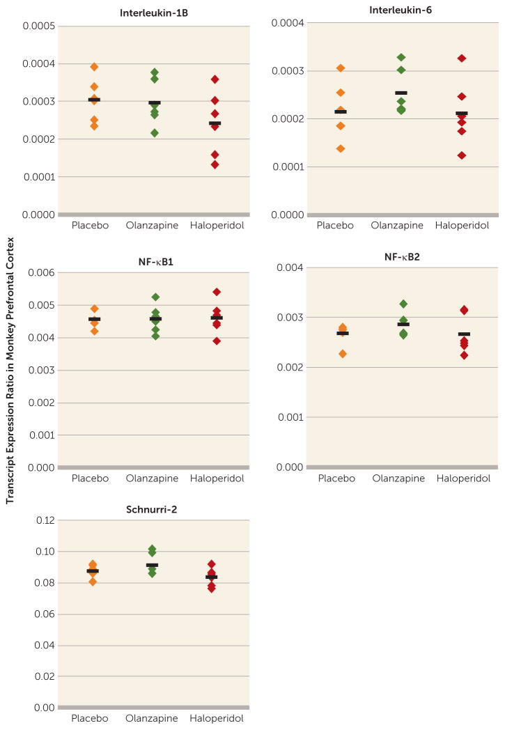
Transcript levels for IL-1B, IL-6, NF-κB1, NF-κB2, and Schnurri-2 in the prefrontal cortex did not differ between monkeys chronically exposed to haloperidol, olanzapine, or placebo (Figure 3). We also previously reported that IFITM mRNA levels were not affected by exposure to antipsychotic medications in these animals (21). Transcript levels for IL-8 and interferon-β in monkey prefrontal cortex were insufficient for quantification by quantitative PCR.
FIGURE 3. Immune System-Related Cytokines and Transcriptional Regulators in Prefrontal Cortex Area 9 of Antipsychotic-Exposed Monkeys a.
aQuantitative polymerase chain reaction analysis revealed no statistically significant differences in mRNA levels for interleukin 1B (IL-1B), IL-6, nuclear factor (NF)-κB1, NF-κB2, or Schnurri-2 in monkeys chronically exposed to either olanzapine or haloperidol compared with placebo. Mean values are shown as horizontal black bars.
Effects of Maternal Immune Activation on Immune Marker Expression in the Frontal Cortex of Young Adult Mouse Offspring
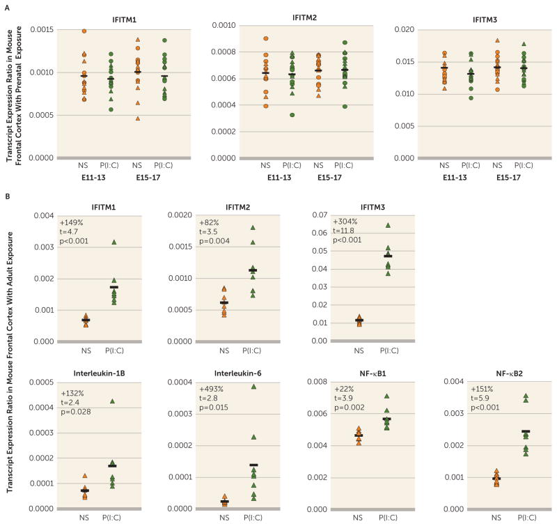
We next investigated whether maternal immune activation could lead to higher IFITM mRNA levels in the frontal cortex of adult off spring by exposing timed pregnant mice to poly(I:C) daily for 3 days in middle or late gestation. To confirm the presence of an immune response to poly(I:C), adult female nonpregnant mice were also exposed to poly(I:C) in parallel with the pregnant mice, and trunk blood was collected 3 hours after the last injection of poly(I:C). Serum IL-6 levels quantified by ELISA were massively elevated in the poly(I:C)-exposed mice (725.1 pg/mL, SD=276.6; t=7.3, df=14, p<0.0001) (see Figure S1 in the data supplement) relative to normal saline-exposed mice (8.6 pg/mL, SD=14.3), consistent with a robust immune response (25, 38). For the timed pregnant mice, exposure to poly(I:C) did not affect the mean number of offspring relative to normal saline-exposed mice (see Table S3 in the data supplement). Next, in the frontal cortex of one young adult male and/or female offspring from each poly(I:C)-injected mother (see Table S3), we quantified mRNA levels for the same immune markers that were studied in human prefrontal cortex in schizophrenia. However, exposure to daily maternal immune activation for 3 days in either middle or late gestation did not affect mRNA levels for IFITM (Figure 4A) or other immune markers in the frontal cortex of adult male or female offspring (see Table S4 in the data supplement). As in monkey prefrontal cortex, transcript levels for IL-8 and interferon-β in mouse frontal cortex were insufficient for quantification by quantitative PCR.
FIGURE 4. Transcript Levels for Immune System-Related Markers in the Frontal Cortex of Adult Mice Exposed Either Prenatally or in Adulthood to Immune Stimulationa.
aPanel A illustrates transcript levels for three variants of IFITM (IFITM1, IFITM2, IFITM3) in the frontal cortex of adult offspring (circles indicate males, triangles indicate females) of pregnant mice exposed to either normal saline (NS) or poly(I:C) [P(I:C)] daily for 3 days of gestation (embryonic day 11–13 [E11–13]: N=7 males and 7 females per condition; embryonic day 15–17 [E15–17]: N=8 males and 8 females per condition). Black bars indicate mean mRNA levels of both sexes combined for each condition. IFITM mRNA levels did not differ in adult offspring (i.e., male alone, female alone, or sexes combined) exposed prenatally to maternal immune activation in either middle (E11–13) or late (E15–17) gestation. In panel B, transcript levels for immune-related markers including different variants of IFITM, cytokines, and nuclear factor (NF)-κB were markedly higher in the frontal cortex of adult female mice exposed to poly(I:C) (N=8) relative to mice exposed to normal saline (N=8) daily for 3 days (df=14 in all analyses).
Effects of Immune Stimulation in Adulthood on Immune Marker Expression in the Frontal Cortex of Adult Mice
We also collected fresh, frozen brain tissue from the adult nonpregnant female mice that were exposed to poly(I:C) daily for 3 days, euthanized 3 hours after the last poly(I:C) injection, and confirmed to have higher serum IL-6 levels (see Figure S1 in the data supplement). Frontal cortex homogenates from these adult mice had a pattern of alterations in immune markers (Figure 4B) remarkably similar to that seen in schizophrenia, including markedly elevated mRNA levels for IFITM1 (+149%, t=4.7, df=14, p=0.0003), IFITM2 (+82%, t=3.5, df=14, p=0.004), IFITM3 (+304%, t=11.8, df=14, p<0.0001), IL-1B (+132%, t=2.4, df=14, p=0.028), IL-6 (+493%, t=2.8, df=14, p=0.015), NF-κB1 (+22%, t=3.9, df=14, p=0.002), and NF-κB2 (+151%, t=5.9, df=14, p<0.0001). However, Schnurri-2 mRNA levels were not altered in these adult mice.
DISCUSSION
In this study, we sought to determine whether higher IFITM mRNA levels and other immune-related disturbances in the prefrontal cortex of schizophrenia subjects are more consistent with being attributable to 1) the consequence of an ongoing molecular cascade contributing to immune activation or 2) a long-lasting maladaptive response to an immune-related insult that occurred during prenatal development. In the prefrontal cortex of the same schizophrenia subjects that we previously reported to have higher IFITM mRNA levels (21), we found markedly higher mRNA levels for cytokines (e.g., IL-6 and interferon-β) and transcriptional regulators (i.e., NF-κB) that induce IFITM expression, as well as lower mRNA levels for an NF-κB site-binding protein (i.e., Schnurri-2) that inhibits IFITM expression (Figure 1B). Furthermore, the within-subject correlations of IFITM mRNA with cytokine and transcription factor mRNA levels in the prefrontal cortex support the prediction that the latter could be causal of the former. This idea was further strengthened by proof-of-principle evidence that exposure to immune stimulation in adult mice produces both higher cytokine levels and higher IFITM levels in the frontal cortex in a manner similar to that seen in schizophrenia. In contrast, exposure to immune stimulation in utero (i.e., maternal immune activation) did not recapitulate schizophrenia-related immune marker expression abnormalities in the frontal cortex.
In the prefrontal cortex of schizophrenia subjects, the markedly higher mRNA levels for cytokines and transcription factors that induce IFITM expression (e.g., IL-6, interferon-β, and NF-κB), and lower mRNA levels for Schnurri-2, which suppresses IFITM expression, suggest that this combination of molecular mechanisms accounts for the elevated levels of IFITM mRNAs in the illness (Figure 1B). In addition, mRNA levels of NF-κB, a critical transcription factor that regulates the expression of many immune-related genes, including the cytokines IL-1B, IL-6, and IL-8 (41, 42), were higher in schizophrenia. Furthermore, mRNA levels for these cytokines and transcriptional regulators were significantly correlated with IFITM mRNA levels. These findings support the idea of a complex cascade of immune activation in the pre-frontal cortex in schizophrenia (Figure 1B). Consistent with this interpretation, Fillman et al. also recently reported (18) higher mRNA levels for IL-6 and IL-8 in the prefrontal cortex in schizophrenia subjects. Furthermore, higher densities of activated microglia, which produce cytokines in the brain, have been reported in the prefrontal cortex in schizophrenia (18, 44, 45). Similarly, in vivo positron emission tomography studies have also found higher [11C]-PK11195 binding, a measure of activated microglia, in the hippocampus and cerebral cortex in schizophrenia (46, 47). Taken together, these findings provide a striking convergence of evidence for a molecular cascade of immune activation in the prefrontal cortex in schizophrenia and suggest that this molecular cascade could be further explored in studies that directly quantify immune marker protein levels in the prefrontal cortex and directly determine the cell types that over express these immune markers in the disorder.
It is also possible that our findings reflect the consequences of schizophrenia that can be accompanied by life conditions (e.g., chronic institutionalization) that may carry an increased risk of exposure to infectious diseases. Although we cannot definitely exclude that possibility, several lines of evidence suggest that our findings do reflect the neurobiology of schizophrenia. Our subjects come from a community-based population (i.e., individuals with unexpected deaths and subsequent autopsies) and were not chronically institutionalized, and altered immune-related marker mRNA levels in schizophrenia did not appear to be attributable to potential confounders such as the presence of immune/inflammation-related illness or use of NSAIDs, psychotropic medications, or tobacco at time of death.
Several epidemiological studies suggest that exposure to maternal immune activation is a risk factor for schizophrenia in offspring (7–11). Furthermore, maternal immune activation has also been reported to produce epigenetic modifications that can alter gene expression postnatally (27). We therefore tested the hypothesis that maternal immune activation may contribute to higher mRNA levels for IFITM and other immune markers in the prefrontal cortex in schizophrenia. However, we found that several days of maternal immune activation in middle or late gestation in mice did not lead to a persistent elevation in mRNA levels for IFITM or several other immune system-related markers in the frontal cortex of young adult offspring such as that seen in schizophrenia. Interestingly, maternal immune activation has been reported to lead to higher cytokine levels in fetal brain (25), which may still affect the development of neural circuits (48, 49), even though cytokine mRNA levels are stable in the frontal cortex of young adult offspring with prenatal exposure to immune activation. Furthermore, some evidence suggests that maternal immune activation may interact with genetic risk factors (50) or adolescent stress (51) to produce schizophrenia-related abnormalities. These findings suggest that maternal immune activation in isolation may not be a sufficient cause of cortical immune activation in schizophrenia. However, we cannot exclude the possibility that maternal immune activation interacts with other environmental or genetic risk factors that may act in concert to disrupt brain development. In contrast, we found that exposure to immune stimulation in adult mice resulted in a pattern of elevations in cytokine, NF-κB, and IFITM mRNA levels in the frontal cortex similar to that seen in schizophrenia.
Immune activation in the prefrontal cortex may have deleterious effects on cortical circuitry in schizophrenia. For example, we previously reported deficits in GABA neuron-related markers, including the GABA synthesizing enzyme GAD67, the calcium-binding protein parvalbumin, the neuropeptide somatostatin, and the transcription factor Lhx6 in the present cohort of schizophrenia subjects (30, 33, 52–55). Furthermore, we previously reported an inverse correlation between IFITM mRNA levels and these GABA neuron-related markers (21). Fillman et al. similarly found that a subset of individuals with schizophrenia with a “high inflammatory” state had more severe deficits in GABA neuron-related mRNAs, including somatostatin, GAD67, and parvalbumin (18). In addition, we found lower Schnurri-2 mRNA levels in the prefrontal cortex in schizophrenia, and Schnurri-2 knockout mice exhibit deficits in parvalbumin and GAD67 protein levels (43). Taken together, these findings suggest that cortical immune activation may have deleterious effects on susceptible components of inhibitory cortical circuitry, including somatostatin and parvalbumin neurons. However, additional proof-of-principle studies are needed to determine the nature of the relationship between cortical immune activation and cortical GABA neuron disturbances in schizophrenia. Such studies will help determine whether immune-related markers represent attractive therapeutic targets in the disorder, as supported by preliminary evidence of clinical efficacy of anti-inflammatory agents in the treatment of schizophrenia (56), in light of our findings that elevated immune markers appear to reflect an active process rather than the remnants of a prenatal scar.
Acknowledgments
Supported by NIH grant MH100066 to Dr. Volk and grants MH043784 and MH051234 to Dr. Lewis.
Footnotes
Presented in part at the 54th annual meeting of the American College of Neuropsychopharmacology, Phoenix, Dec. 7–11, 2014.
Dr. Lewis receives investigator-initiated research support from Bristol-Myers Squibb and Pfizer and has served as a consultant for Autifony, Bristol-Myers Squibb, Concert Pharmaceuticals, and Sunovion. The other authors report no financial relationships with commercial interests.
References
- 1.Purcell SM, Wray NR, Stone JL, et al. Common polygenic variation contributes to risk of schizophrenia and bipolar disorder. Nature. 2009;460:748–752. doi: 10.1038/nature08185. [DOI] [PMC free article] [PubMed] [Google Scholar]
- 2.Shi J, Levinson DF, Duan J, et al. Common variants on chromosome 6p22. 1 are associated with schizophrenia. Nature. 2009;460:753–757. doi: 10.1038/nature08192. [DOI] [PMC free article] [PubMed] [Google Scholar]
- 3.Stefansson H, Ophoff RA, Steinberg S, et al. Common variants conferring risk of schizophrenia. Nature. 2009;460:744–747. doi: 10.1038/nature08186. [DOI] [PMC free article] [PubMed] [Google Scholar]
- 4.Jia P, Wang L, Meltzer HY, et al. Common variants conferring risk of schizophrenia: a pathway analysis of GWAS data. Schizophr Res. 2010;122:38–42. doi: 10.1016/j.schres.2010.07.001. [DOI] [PMC free article] [PubMed] [Google Scholar]
- 5.Ripke S, Sanders AR, Kendler KS, et al. Genome-wide association study identifies five new schizophrenia loci. Nat Genet. 2011;43:969–976. doi: 10.1038/ng.940. [DOI] [PMC free article] [PubMed] [Google Scholar]
- 6.Schizophrenia Working Group of the Psychiatric Genomics Consortium. Biological insights from 108 schizophrenia-associated genetic loci. Nature. 2014;511:421–427. doi: 10.1038/nature13595. [DOI] [PMC free article] [PubMed] [Google Scholar]
- 7.Brown AS, Begg MD, Gravenstein S, et al. Serologic evidence of prenatal influenza in the etiology of schizophrenia. Arch Gen Psychiatry. 2004;61:774–780. doi: 10.1001/archpsyc.61.8.774. [DOI] [PubMed] [Google Scholar]
- 8.Brown AS, Schaefer CA, Quesenberry CP, Jr, et al. Maternal exposure to toxoplasmosis and risk of schizophrenia in adult offspring. Am J Psychiatry. 2005;162:767–773. doi: 10.1176/appi.ajp.162.4.767. [DOI] [PubMed] [Google Scholar]
- 9.Brown AS, Hooton J, Schaefer CA, et al. Elevated maternal interleukin-8 levels and risk of schizophrenia in adult off spring. Am J Psychiatry. 2004;161:889–895. doi: 10.1176/appi.ajp.161.5.889. [DOI] [PubMed] [Google Scholar]
- 10.Canetta S, Sourander A, Surcel HM, et al. Elevated maternal C-reactive protein and increased risk of schizophrenia in a national birth cohort. Am J Psychiatry. 2014;171:960–968. doi: 10.1176/appi.ajp.2014.13121579. [DOI] [PMC free article] [PubMed] [Google Scholar]
- 11.Brown AS, Derkits EJ. Prenatal infection and schizophrenia: a review of epidemiologic and translational studies. Am J Psychiatry. 2010;167:261–280. doi: 10.1176/appi.ajp.2009.09030361. [DOI] [PMC free article] [PubMed] [Google Scholar]
- 12.Benros ME, Pedersen MG, Rasmussen H, et al. A nationwide study on the risk of autoimmune diseases in individuals with a personal or a family history of schizophrenia and related psychosis. Am J Psychiatry. 2014;171:218–226. doi: 10.1176/appi.ajp.2013.13010086. [DOI] [PubMed] [Google Scholar]
- 13.Benros ME, Eaton WW, Mortensen PB. The epidemiologic evidence linking autoimmune diseases and psychosis. Biol Psychiatry. 2014;75:300–306. doi: 10.1016/j.biopsych.2013.09.023. [DOI] [PMC free article] [PubMed] [Google Scholar]
- 14.Potvin S, Stip E, Sepehry AA, et al. Inflammatory cytokine alterations in schizophrenia: a systematic quantitative review. Biol Psychiatry. 2008;63:801–808. doi: 10.1016/j.biopsych.2007.09.024. [DOI] [PubMed] [Google Scholar]
- 15.Miller BJ, Buckley P, Seabolt W, et al. Meta-analysis of cytokine alterations in schizophrenia: clinical status and antipsychotic effects. Biol Psychiatry. 2011;70:663–671. doi: 10.1016/j.biopsych.2011.04.013. [DOI] [PMC free article] [PubMed] [Google Scholar]
- 16.Di Nicola M, Cattaneo A, Hepgul N, et al. Serum and gene expression profile of cytokines in first-episode psychosis. Brain Behav Immun. 2013;31:90–95. doi: 10.1016/j.bbi.2012.06.010. [DOI] [PMC free article] [PubMed] [Google Scholar]
- 17.Fillman SG, Sinclair D, Fung SJ, et al. Markers of inflammation and stress distinguish subsets of individuals with schizophrenia and bipolar disorder. Transl Psychiatr. 2014;4:e365. doi: 10.1038/tp.2014.8. [DOI] [PMC free article] [PubMed] [Google Scholar]
- 18.Fillman SG, Cloonan N, Catts VS, et al. Increased inflammatory markers identified in the dorsolateral prefrontal cortex of individuals with schizophrenia. Mol Psychiatry. 2013;18:206–214. doi: 10.1038/mp.2012.110. [DOI] [PubMed] [Google Scholar]
- 19.Arion D, Unger T, Lewis DA, et al. Molecular evidence for increased expression of genes related to immune and chaperone function in the prefrontal cortex in schizophrenia. Biol Psychiatry. 2007;62:711–721. doi: 10.1016/j.biopsych.2006.12.021. [DOI] [PMC free article] [PubMed] [Google Scholar]
- 20.Saetre P, Emilsson L, Axelsson E, et al. Inflammation-related genes up-regulated in schizophrenia brains. BMC Psychiatry. 2007;7:46. doi: 10.1186/1471-244X-7-46. [DOI] [PMC free article] [PubMed] [Google Scholar]
- 21.Siegel BI, Sengupta EJ, Edelson JR, et al. Elevated viral restriction factor levels in cortical blood vessels in schizophrenia. Biol Psychiatry. 2014;76:160–167. doi: 10.1016/j.biopsych.2013.09.019. [DOI] [PMC free article] [PubMed] [Google Scholar]
- 22.Huang IC, Bailey CC, Weyer JL, et al. Distinct patterns of IFITM-mediated restriction of filoviruses, SARS coronavirus, and influenza A virus. PLoS Pathog. 2011;7:e1001258. doi: 10.1371/journal.ppat.1001258. [DOI] [PMC free article] [PubMed] [Google Scholar]
- 23.Bailey CC, Huang IC, Kam C, et al. IFITM3 limits the severity of acute influenza in mice. PLoS Pathog. 2012;8:e1002909. doi: 10.1371/journal.ppat.1002909. [DOI] [PMC free article] [PubMed] [Google Scholar]
- 24.Diamond MS, Farzan M. The broad-spectrum antiviral functions of IFIT and IFITM proteins. Nat Rev Immunol. 2013;13:46–57. doi: 10.1038/nri3344. [DOI] [PMC free article] [PubMed] [Google Scholar]
- 25.Meyer U, Nyffeler M, Engler A, et al. The time of prenatal immune challenge determines the specificity of inflammation-mediated brain and behavioral pathology. J Neurosci. 2006;26:4752–4762. doi: 10.1523/JNEUROSCI.0099-06.2006. [DOI] [PMC free article] [PubMed] [Google Scholar]
- 26.Garay PA, Hsiao EY, Patterson PH, et al. Maternal immune activation causes age- and region-specific changes in brain cytokines in offspring throughout development. Brain Behav Immun. 2013;31:54–68. doi: 10.1016/j.bbi.2012.07.008. [DOI] [PMC free article] [PubMed] [Google Scholar]
- 27.Tang B, Jia H, Kast RJ, et al. Epigenetic changes at gene promoters in response to immune activation in utero. Brain Behav Immun. 2013;30:168–175. doi: 10.1016/j.bbi.2013.01.086. [DOI] [PubMed] [Google Scholar]
- 28.Volk DW, Radchenkova PV, Walker EM, et al. Cortical opioid markers in schizophrenia and across postnatal development. Cereb Cortex. 2012;22:1215–1223. doi: 10.1093/cercor/bhr202. [DOI] [PMC free article] [PubMed] [Google Scholar]
- 29.Volk DW, Chitrapu A, Edelson JR, et al. Chemokine receptors and cortical interneuron dysfunction in schizophrenia. Schizophr Res. doi: 10.1016/j.schres.2014.10.031. (Epub ahead of print, Nov 11, 2014) [DOI] [PMC free article] [PubMed] [Google Scholar]
- 30.Volk DW, Austin MC, Pierri JN, et al. Decreased glutamic acid decarboxylase 67 messenger RNA expression in a subset of prefrontal cortical gamma-aminobutyric acid neurons in subjects with schizophrenia. Arch Gen Psychiatry. 2000;57:237–245. doi: 10.1001/archpsyc.57.3.237. [DOI] [PubMed] [Google Scholar]
- 31.Volk DW, Eggan SM, Lewis DA. Alterations in metabotropic glutamate receptor 1α and regulator of G protein signaling 4 in the prefrontal cortex in schizophrenia. Am J Psychiatry. 2010;167:1489–1498. doi: 10.1176/appi.ajp.2010.10030318. [DOI] [PMC free article] [PubMed] [Google Scholar]
- 32.Volk DW, Siegel BI, Verrico CD, et al. Endocannabinoid metabolism in the prefrontal cortex in schizophrenia. Schizophr Res. 2013;147:53–57. doi: 10.1016/j.schres.2013.02.038. [DOI] [PMC free article] [PubMed] [Google Scholar]
- 33.Volk DW, Edelson JR, Lewis DA. Cortical inhibitory neuron disturbances in schizophrenia: role of the ontogenetic transcription factor Lhx6. Schizophr Bull. 2014;40:1053–1061. doi: 10.1093/schbul/sbu068. [DOI] [PMC free article] [PubMed] [Google Scholar]
- 34.Hashimoto T, Bazmi HH, Mirnics K, et al. Conserved regional patterns of GABA-related transcript expression in the neocortex of subjects with schizophrenia. Am J Psychiatry. 2008;165:479–489. doi: 10.1176/appi.ajp.2007.07081223. [DOI] [PMC free article] [PubMed] [Google Scholar]
- 35.Vandesompele J, De Preter K, Pattyn F, et al. Accurate normalization of real-time quantitative RT-PCR data by geometric averaging of multiple internal control genes. Genome Biol. 2002;3(7):0034.1–0034.12. doi: 10.1186/gb-2002-3-7-research0034. [DOI] [PMC free article] [PubMed] [Google Scholar]
- 36.Smith SE, Li J, Garbett K, et al. Maternal immune activation alters fetal brain development through interleukin-6. J Neurosci. 2007;27:10695–10702. doi: 10.1523/JNEUROSCI.2178-07.2007. [DOI] [PMC free article] [PubMed] [Google Scholar]
- 37.Hsiao EY, Patterson PH. Activation of the maternal immune system induces endocrine changes in the placenta via IL-6. Brain Behav Immun. 2011;25:604–615. doi: 10.1016/j.bbi.2010.12.017. [DOI] [PMC free article] [PubMed] [Google Scholar]
- 38.Cunningham C, Campion S, Teeling J, et al. The sickness behaviour and CNS inflammatory mediator profile induced by systemic challenge of mice with synthetic double-stranded RNA (poly I:C) Brain Behav Immun. 2007;21:490–502. doi: 10.1016/j.bbi.2006.12.007. [DOI] [PubMed] [Google Scholar]
- 39.Paxinos G, Franklin K. The Mouse Brain in Stereotaxic Coordinates. San Diego: Academic Press; 2001. [Google Scholar]
- 40.Dorph-Petersen KA, Pierri JN, Perel JM, et al. The influence of chronic exposure to antipsychotic medications on brain size before and after tissue fixation: a comparison of haloperidol and olanzapine in macaque monkeys. Neuropsychopharmacology. 2005;30:1649–1661. doi: 10.1038/sj.npp.1300710. [DOI] [PubMed] [Google Scholar]
- 41.Tornatore L, Thotakura AK, Bennett J, et al. The nuclear factor kappa B signaling pathway: integrating metabolism with inflammation. Trends Cell Biol. 2012;22:557–566. doi: 10.1016/j.tcb.2012.08.001. [DOI] [PubMed] [Google Scholar]
- 42.Hoesel B, Schmid JA. The complexity of NF-κB signaling in inflammation and cancer. Mol Cancer. 2013;12:86. doi: 10.1186/1476-4598-12-86. [DOI] [PMC free article] [PubMed] [Google Scholar]
- 43.Takao K, Kobayashi K, Hagihara H, et al. Deficiency of schnurri-2, an MHC enhancer binding protein, induces mild chronic inflammation in the brain and confers molecular, neuronal, and behavioral phenotypes related to schizophrenia. Neuropsychopharmacology. 2013;38:1409–1425. doi: 10.1038/npp.2013.38. [DOI] [PMC free article] [PubMed] [Google Scholar]
- 44.Radewicz K, Garey LJ, Gentleman SM, et al. Increase in HLA-DR immunoreactive microglia in frontal and temporal cortex of chronic schizophrenics. J Neuropathol Exp Neurol. 2000;59:137–150. doi: 10.1093/jnen/59.2.137. [DOI] [PubMed] [Google Scholar]
- 45.Wierzba-Bobrowicz T, Lewandowska E, Lechowicz W, et al. Quantitative analysis of activated microglia, ramified and damage of processes in the frontal and temporal lobes of chronic schizophrenics. Folia Neuropathol. 2005;43:81–89. [PubMed] [Google Scholar]
- 46.van Berckel BN, Bossong MG, Boellaard R, et al. Microglia activation in recent-onset schizophrenia: a quantitative (R)-[11C]PK11195 positron emission tomography study. Biol Psychiatry. 2008;64:820–822. doi: 10.1016/j.biopsych.2008.04.025. [DOI] [PubMed] [Google Scholar]
- 47.Doorduin J, de Vries EF, Willemsen AT, et al. Neuroinflammation in schizophrenia-related psychosis: a PET study. J Nucl Med. 2009;50:1801–1807. doi: 10.2967/jnumed.109.066647. [DOI] [PubMed] [Google Scholar]
- 48.Meyer U, Nyffeler M, Yee BK, et al. Adult brain and behavioral pathological markers of prenatal immune challenge during early/middle and late fetal development in mice. Brain Behav Immun. 2008;22:469–486. doi: 10.1016/j.bbi.2007.09.012. [DOI] [PubMed] [Google Scholar]
- 49.Richetto J, Calabrese F, Riva MA, et al. Prenatal immune activation induces maturation-dependent alterations in the prefrontal GABAergic transcriptome. Schizophr Bull. 2014;40:351–361. doi: 10.1093/schbul/sbs195. [DOI] [PMC free article] [PubMed] [Google Scholar]
- 50.Lipina TV, Zai C, Hlousek D, et al. Maternal immune activation during gestation interacts with Disc 1 point mutation to exacerbate schizophrenia-related behaviors in mice. J Neurosci. 2013;33:7654–7666. doi: 10.1523/JNEUROSCI.0091-13.2013. [DOI] [PMC free article] [PubMed] [Google Scholar]
- 51.Giovanoli S, Engler H, Engler A, et al. Stress in puberty unmasks latent neuropathological consequences of prenatal immune activation in mice. Science. 2013;339:1095–1099. doi: 10.1126/science.1228261. [DOI] [PubMed] [Google Scholar]
- 52.Hashimoto T, Volk DW, Eggan SM, et al. Gene expression deficits in a subclass of GABA neurons in the prefrontal cortex of subjects with schizophrenia. J Neurosci. 2003;23:6315–6326. doi: 10.1523/JNEUROSCI.23-15-06315.2003. [DOI] [PMC free article] [PubMed] [Google Scholar]
- 53.Hashimoto T, Arion D, Unger T, et al. Alterations in GABA-related transcriptome in the dorsolateral prefrontal cortex of subjects with schizophrenia. Mol Psychiatry. 2008;13:147–161. doi: 10.1038/sj.mp.4002011. [DOI] [PMC free article] [PubMed] [Google Scholar]
- 54.Curley AA, Arion D, Volk DW, et al. Cortical deficits of glutamic acid decarboxylase 67 expression in schizophrenia: clinical, protein, and cell type-specific features. Am J Psychiatry. 2011;168:921–929. doi: 10.1176/appi.ajp.2011.11010052. [DOI] [PMC free article] [PubMed] [Google Scholar]
- 55.Volk DW, Matsubara T, Li S, et al. Deficits in transcriptional regulators of cortical parvalbumin neurons in schizophrenia. Am J Psychiatry. 2012;169:1082–1091. doi: 10.1176/appi.ajp.2012.12030305. [DOI] [PMC free article] [PubMed] [Google Scholar]
- 56.Keller WR, Kum LM, Wehring HJ, et al. A review of anti-inflammatory agents for symptoms of schizophrenia. J Psychopharmacol. 2013;27:337–342. doi: 10.1177/0269881112467089. [DOI] [PMC free article] [PubMed] [Google Scholar]


