Abstract
Background
Nosema ceranae infection not only damages honey bee (Apis melifera) intestines, but we believe it may also affect intestinal yeast development and its seasonal pattern. In order to check our hypothesis, infection intensity versus intestinal yeast colony forming units (CFU) both in field and cage experiments were studied.
Methods/Findings
Field tests were carried out from March to October in 2014 and 2015. N. ceranae infection intensity decreased more than 100 times from 7.6 x 10⁸ in March to 5.8 x 10⁶ in October 2014. A similar tendency was observed in 2015. Therefore, in the European eastern limit of its range, N. ceranae infection intensity showed seasonality (spring peak and subsequent decline in the summer and fall), however, with an additional mid-summer peak that had not been recorded in other studies. Due to seasonal changes in the N. ceranae infection intensity observed in honey bee colonies, we recommend performing studies on new therapeutics during two consecutive years, including colony overwintering. A natural decrease in N. ceranae spore numbers observed from March to October might be misinterpreted as an effect of Nosema spp. treatment with new compounds. A similar seasonal pattern was observed for intestinal yeast population size in field experiments. Furthermore, cage experiments confirmed the size of intestinal yeast population to increase markedly together with the increase in the N. ceranae infection intensity. Yeast CFUs amounted to respectively 2,025 (CV = 13.04) and 11,150 (CV = 14.06) in uninfected and N. ceranae-infected workers at the end of cage experiments. Therefore, honey bee infection with N. ceranae supported additional opportunistic yeast infections, which may have resulted in faster colony depopulations.
Introduction
A significant decrease in the number and biodiversity of pollinators has been observed over the last 50 years. It entails huge economic losses and has even been called a “pollination crisis” [1]. A decline in honey bee populations is caused by combined stress from parasites, pesticides, and bees’ diet being inadequate due to reduced diversity of wildflower resources, which are replaced by monocultures [2]. Consequently, weakened honey bees are more easily attacked by many diseases, inter alia those caused by fungi such as Nosema apis and Nosema ceranae belonging to the Microsporidia phylum, Balbiani 1882 [3].
Although these two fungal species inhabit the same host i.e. Apis mellifera, and have similar life cycles, a phylogenetic analysis of multiple sequence data sets indicated that they are not closely related. The analysis revealed that N. ceranae is a sister species to N. bombi, whereas the weight of evidence is consistent that N. apis is a basal member of the Nosema clade [4, 5]. Competition between these two parasites within the same host leads to conflicts that may influence parasitic virulence evolution as can be seen in the differences in seasonal variations and gross colony infection intensity symptoms described for N. apis and N. ceranae [5, 6, 7, 8]. Infection of A. mellifera with N. apis was well described over 100 years ago [9, 10] whereas it was not until 2005 that the first infection of A. mellifera with N. ceranae was recorded in Taiwan [11]. Soon after that N. ceranae spread to Europe and both Americas, where it has become prevalent [6]. Therefore, A. mellifera and N. apis make a mutually co-adapted endemic host-pathogen pair that is subject to invasion of a new pathogen, i.e. N. ceranae in the temperate climate zone. This is well reflected in the way the honey bee immune system responds to these parasites, since N. apis infection increases immune defense while N. ceranae leads to immunosuppression in A. mellifera [12]. Presently, a wide spread of N. ceranae is responsible for numerous honey bee infections in Europe [13, 14] and is believed to be displacing N. apis all over the world [5, 15, 16, 17, 18]. We hypothesized that such an invasion by a new pathogen developing in apian intestines [19] may affect development of other intestinal microbiota. We initially confirmed these suspicions during our preliminary research into honey bee intestinal yeast populations [20]. In previous cage experiments performed throughout two summer months, an interdependence between N. ceranae infection intensity and intestinal yeast development was observed, albeit merely in four measurements, each completed at the end of a single experiment [20]. Cage data may differ from those obtained in fully functional colonies [21, 22, 23] and seasonal changes are crucial for understanding interrelations between N. ceranae and yeasts within apian intestine microenvironment. Hence, there is a need for field research into the impact of N. ceranae on N. apis and honey bees as well as honey bee intestinal yeast populations. Not only is it significant for the protection of honey bees, but also for broadening the knowledge concerning inter-pathogen relations occurring during new pathogen invasions.
N. ceranae, just like N. apis, completes its life cycle in honey bee intestines. Even a medium Nosema spp. infection causes a complete overlay of the gut with spores, which disturbs absorption of nutrients [24]. Moreover, Nosema spp. infection affects natural honey bee intestinal microbiota, which include about: 70% of Gram-negative bacteria mainly from the Enterobacteriaceae, Alcaligenaceae and Pseudomonadaceae families, 27% of Gram-positive bacteria, primarily from the genus Bacillus and less than 1% of yeasts and other fungi, inter alia Saccharomyces rouxi, S. mellis, S. bisporus, S. roesi, S. bailli, S. heterogenicus, Aspergillus sp., Alternaria sp., Cladosporium sp., Penicillium sp., Pichia (Hansenula) anomala, Rhizopus arrhizus, Torulopsis sp. [25, 26, 27, 28, 29, 30, 31, 32]. However, it must be highlighted that yeasts can be treated as honey bee stress indicators, because healthy honey bees do not have intestinal yeasts or have them very little [20, 25, 26, 27]. Furthermore, adding yeasts to honey bee diet caused damage to the midgut epithelial layer, which drastically disturbed the absorption of nutrients and significantly shortened bees’ lifespans [33, 34, 35, 36, 37]. Therefore, yeasts are most likely honey bee opportunistic pathogens whose population expands during disruption of homeostasis in their host. Our preliminary study on caged honey bees [20] suggested that intensity of Nosema infection influenced honey bee intestinal yeast population size in two ways. A slight infection increased the yeast number, whereas a heavy infection reduced it. We wondered, however, if such interrelationships would be observed on a colony level as well, and whether they would vary reflecting seasonal changes of Nosema spp. population sizes. An infection intensity increase was recorded in the spring, which was followed by a marked decline in the summer and fall (inter alia: [38, 39, 40, 41]). On the other hand, N. ceranae, which develops in the honey bee intestine, ought to compete for attachment sites and nutrients with the remaining pathogenic fungi, including N. apis and yeasts. Therefore, the question arises whether a significant increase in the intestinal yeast population could finally suppress Nosema spp. infection intensity, and vice versa on the colony level. Moreover, we wondered if seasonal changes in Nosema intensity could be detected through cage tests performed parallel to field studies.
The aim of this research was to study how the seasonal pattern of the N. ceranae infection intensity influenced development of intestinal yeast population in honey bees from March to October through two consecutive years, based on both cage and field assays.
Material and Methods
This research was supported by an Individual Research Grant from the Vice-rector for Research and International Relations of UMCS (Lublin, Poland) for Aneta A. Ptaszyńska. The funder had no role in the study design, data collection and analysis, decision to publish, or preparation of the manuscript.
Our research had been planned in a way that reduced the number of honeybees to the minimum necessary for the proper execution of these experiments and had been accepted by the head of the Department of Experimental and Environmental Biology, University of Life Sciences in Lublin (Poland). Afterwards, honey bees, Apis mellifera carnica, were collected from the university apiary of the University of Life Sciences in Lublin.
Field and cage tests were carried out at the apiary of the University of Life Sciences, Lublin, Poland (51°13'32.2"N 22°38'08.3"E) and the laboratory of the Maria Curie-Skłodowska University, Lublin, Poland (51°14'35.2"N 22°32'33.3"E), respectively.
2.1. The field test protocol
Hive bottom-drawers were placed individually in 82 A. mellifera carnica colonies at the beginning of the overwintering period 2013–2014 in order to select colonies for field tests. Dead worker-bees were sampled from winter falls. They were collected separately from each colony (drawer) in early February 2014. Next, 50 workers from each colony sample were homogenized and examined for the presence of Nosema spp. spores (section 2.4). Colonies in which no spores were found were considered uninfected. Whenever Nosema spp. spores were found, the colony was classified as infected and put in a secluded place. No nosemosis treatment was administered. Further DNA analysis (section 2.3) revealed which colony was infected with N. apis, which with N. ceranae, and which with both these pathogens (mixed infection). Consequently, a colony infection intensity, defined as the number of N. ceranae spores observed in a single host intestinal environment [42], was assessed through determining the number of spores in the 50-worker colony homogenate, whereas the infection type was specified by means of a DNA analysis.
At the end of February 2014, three colonies infected solely with N. ceranae at a similar intensity and three colonies considered uninfected were qualified to obtain biological material for studying the seasonality of the N. ceranae infection course and its impact on the intestinal yeast CFU number. Then, in late February 2015, colonies that had been classified as infected in 2014 were checked again to determine the intensity and type of infection. Two of them, which showed similar intensity of infection with N. ceranae and also two uninfected colonies were qualified for a repetition of the study.
100 live worker-bees were sampled from each selected colony in both 2014 (6 colonies) and 2015 (4 colonies) during the first five days of each month from March to October. Each 100-bee colony sample was divided into units of 10 worker-bees to check the Nosema infection type on the basis of DNA analysis (section 2.3), to establish N. ceranae intensity (in triplicate, section 2.4), and to determine yeast CFU number (in triplicate, section 2.5). The remaining 30 workers from every 100-bee colony sample were treated as a reserve. Therefore, the database to establish the N. ceranae intensity was: 3 pooled samples of 10 bees taken on a monthly basis from each of the 6 (2014) or 4 (2015) colonies during 8 months, i.e. 144 samples of 10 bees in 2014 and 96 samples of 10 bees in 2015. The same number of samples was taken for intestinal yeast CFU analysis.
2.2. The cage test protocol
Two consecutive cage experiments (repetitions) were additionally conducted, one in June and one in July, both in 2014 and 2015 (four experiments in total) applying the following protocol: the sealed-brood combs on the 20th day of the brood development were taken out from a Nosema-free source colony and incubated in an air-conditioned chamber (35°C, RH = 60%). Next, freshly emerged workers were placed in 80 wooden cages, 40 workers per cage, and kept under laboratory conditions (darkness; 30°C; RH = 65%). The first worker sample, comprising 40 workers (2 x 10 workers for DNA analysis and 2 x 10 workers for yeast cultures) was randomly taken from all the cages one day after the workers’ caging. Molecular analysis (section 2.3) proved there was no N. apis and N. ceranae DNA in the sampled workers. Intestinal yeasts were cultured as described in section 2.5. On the third day after worker emergence, the cages were divided into two groups, 40 cages in each. In the first group, workers were infected with N. ceranae, according to the methodology described by Forsgren and Fries [43], whereas in the second, they were uninfected and maintained as a control. The cages with control (uninfected) and N. ceranae infected honey bee workers were kept in separate chambers (darkness; 30°C; RH = 65%) to prevent spore contamination.
Then, beginning from the second day after the infection of workers (4-day-old workers), the samples of 40 workers, were taken every day for 18 days (2 x 10 workers for N. ceranae spore counting, section 2.4 and 2 x 10 workers for yeast cultures, section 2.5).
Summing up, 2 groups x 18 samplings = 36 samples of 10 workers to count N. ceranae spores and the same number of samples to culture intestinal yeasts, were taken both in 2014 and 2015.
2.3. Total DNA extraction and detection of N. apis and N. ceranae using duplex PCR
DNA was extracted from each pooled sample of 10 honey bee workers, which were ground in sterile water. 100 μL of each homogenate was added to 180 μL of lysis buffer and 20 μL of proteinase K, and the total DNA was isolated with the DNeasy Blood and Tissue Kit (Qiagen) according to the producer’s instruction. Every isolate was used as a template for detection of N. apis and N. ceranae specific 16S rDNA by PCR with Nosema-specific primers: 321-APIS for N. apis and 218-MITOC for N. ceranae as described in Martin-Hernandez et al. [44].
2.4. Nosema spp. infection intensity
Abdomens were individually dissected from the workers and homogenated by grinding in a ratio of 1 abdomen for 1 mL of sterile water. 50 workers in case of field tests (section 2.1), and 10 workers in case of the cage test (section 2.2) were used to prepare one homogenate. After that, the homogenates were smeared on a microscope slide for examination. For each spore suspension, averages of 2 intensity estimates were used. The number of Nosema spp. spores per honey bee worker was estimated using Olympus BX 61 light microscope and a haemocytometer [6, 45].
2.5. Estimation of yeast colony forming units (CFUs)
Ten workers from each pooled sample, both from the field (section 2.1) and from the cage (section 2.2) experiments were individually surface sterilized in 70% ethanol and delicately (aseptic conditions) pressed to discharge feces into one sterile Eppendorf tube, 10 workers per one tube. The feces collected in each tube were mixed to obtain homogenized solution. After that, 150 μL of each homogenate was suspended in 150 μL of sterile 0.6% NaCl invertebrate saline to obtain 300 μL of the feces suspension. Then, two samples, per 100 μL of the suspension, were immediately spread in duplicate on Petri dishes containing Sabouraud dextrose agar with chloramphenicol and gentamycin. Furthermore, 100 μL of 10−2- and 10−4-fold dilutions of the preliminary feces solution were also cultivated in duplicate on Petri dishes. Each Petri dish, was incubated for 5 days after the feces spreading at 30°C. The addition of antibiotics inhibited bacteria growth and improved the cultivation of microfungi. The API® strips-Yeasts (bio Mérieux Clinical Diagnostics) were used to differentiate fungi isolated from honey bee workers’ intestinal tracts and found on the Petri dishes.
A separate procedure was performed in order to estimate the number of yeast colony forming units (CFU) per one honey bee worker. 50 honey bee workers were individually weighed and then pressed to discharge all feces into separate small tubes, one tube per worker. The volume and weight of feces discharged per worker were measured. The average honey bee worker weight was 155.7 mg, while the average weight of feces discharged per one worker was 29 mg. The average volume of single-bee feces was 23 μL. Therefore, it was assumed that 150 μL of the feces-homogenate, and consequently 300 μL of the feces solution in 0.6% NaCl saline originated on average from about 6.5 worker honey bees. Consequently, it is assumed that feces from 2.17 workers were spread on one Petri dish, when 100 μL of the feces solution was used.
2.6. Statistical analysis
All statistical analyses were performed using SAS software [46]. One-way ANOVA (N. ceranae-infected versus uninfected), in which a group effect was the experimental factor, and Student's t test were applied to compare differences between groups.
To estimate dependency between yeast CFU number and N. ceranae spore number, Model I linear regression for field tests and Spearman’s correlation for cage tests were used. Linear regression, however, does not evaluate significances of deviations from the general trend (discrepancy between the real data and regression line), i.e. does not answer whether a given peak (e.g. Fig 1) is a statistical (random) artifact or a significant deviation from the general trend. Therefore, to assess a relationship between the numbers of spores (infection intensity) along subsequent months (seasonality) the regression model with dummy variables (month indicators) was also applied (Model II). Model parameters were estimated using the Gauss-Newton least squares method [47].
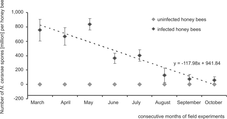
Fig 1. Field tests.
Seasonality in the N. ceranae infection intensity observed from March to October in naturally infected honey bee colonies. Data obtained from measurements conducted on a monthly basis for the first five days of each month from March to October in 2014 and 2015. The dotted line represents a linear regression. Diamond shapes (♦) represent means while error bars indicate standard deviation.
Results
3.1. Field tests
An analysis of the winter debris from 82 colonies, which was performed in 2014, revealed that 11 of them were Nosema infected (Table 1). Only one of these 11 colonies was infected solely with N. apis while as many as six were infected solely with N. ceranae. Intensity and infection type were similar in 2014 and 2015. However, the colony infected solely with N. apis in 2014 was found in 2015 to be infected both with N. apis and N. ceranae. Due to the lack of nosemosis treatment, an increase in the N. ceranae infection intensity was clearly observed in 8 out of 11 colonies during the second year of our studies. No live bees were found in the two most intensively infected colonies. On the other hand, one colony infected at a very low intensity in 2014 was found uninfected next year.
Table 1. Infection types and intensities in the Nosema spp. infected colonies.
| Colony no. | 1 | 2 | 3 | 4 | 5 | 6 | 7 | 8 | 9 | 10 | 11 | |
|---|---|---|---|---|---|---|---|---|---|---|---|---|
| II 2014 | N. apis | + | + | + | + | + | - | - | - | - | - | - |
| N. ceranae | - | + | + | + | + | + | + | + | + | + | + | |
| number of spores [million] | 244 | 1,160 | 940 | 624 | 488 | 1,800 | 828 | 792 | 664 | 320 | 0.8 | |
| II 2015 | N. apis | + | + | + | + | + | - | - | - | - | - | - |
| N. ceranae | + | + | + | + | + | + | + | + | + | + | + | |
| number of spores [million] | 328 | Θ1,480 | 820 | 780 | 560 | Θ1,920 | 984 | 920 | 592 | 640 | 0.0 | |
(+ or -); a given infection type was found or not found. The colonies analyzed for the monthly N. ceranae number of spores and yeast CFU number are in bold. The colonies depopulated in 2015 are marked with Θ.
Monitoring of the N. ceranae-infected colonies for 8 months showed that average number of spores decreased more than 100 times from May to October both in 2014 and 2015 (Fig 1). In 2014, the decrease was from approximately 7.6 x 10⁸ per honey bee worker in March to 5.8 x 10⁶ in October, whereas in 2015 the spore number decreased from 9.5 x 10⁸ to 7.1 x 10⁶, respectively. Despite this general trend to reduce the number of spores from spring to fall, which was significant (p < 0.05) in both regression Model I and Model II, two peaks were observed. Both peaks were considered to be temporary rises in Nosema infection intensity, one in May and the other in July. Corrections for the linear trend of May and July (Model II) were significant (p < 0.05). Additional calculations for Model II indicators were performed taking into account only periods when biggest increases in the number of spores were observed. In both such periods discrepancies from the linear trend were significant (p < 0.05). This indicates that the peaks were not statistical artefacts.
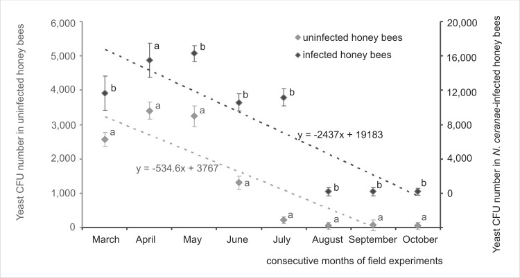
Fungi belonging to Candida and Saccharomyces genera were detected both in N. ceranae-infected and uninfected colonies. The number of intestinal yeast CFUs isolated both from uninfected and the N. ceranae-infected colonies decreased from May to October in 2014 and 2015 but CFU number was always markedly higher in infected colonies (Fig 2) (one-way ANOVA p < 0.05 was performed separately for each month). General seasonal patterns of changes in intestinal yeast CFU, showing one peak in May and later a decrease were similar in infected and uninfected colonies. However, N. ceranae-infected colonies had an additional summer peak in July. Both CFU peaks, i.e. those observed in May and July, corresponded with N. ceranae infection intensity peaks (compare Figs 1 and 2). Generally, the yeast CFU number was positively correlated with the N. ceranae infection intensity (Table 2), particularly in spring and early summer.
Fig 2. Field tests.
Changes in the intestinal yeast CFU number per honey bee worker observed from March to October in uninfected and N. ceranae-infected honey bee colonies. Data obtained from measurements conducted on a monthly basis for the first five days of each month from March to October in 2014 and 2015. Different lower case letters indicate significant differences (p < 0.05) between uninfected and N. ceranae-infected colonies (one-way ANOVA was performed separately for each month). Dotted lines represent regression lines. Diamond shapes (♦) represent means while error bars indicate standard deviation.
Table 2. Field tests.
Spearman’s correlation coefficients between yeast CFU and N. ceranae number of spores. Significant correlations (p < 0.05) are indicated with an asterisk.
| Nosema ceranae number of spores | ||
|---|---|---|
| Yeast CFU number | March | 0.6686* |
| p = 0.025 | ||
| April | 0.6393* | |
| p = 0.034 | ||
| May | 0.3624 | |
| p = 0.273 | ||
| June | 0.7114* | |
| p = 0.014 | ||
| July | 0.2312 | |
| p = 0.494 | ||
| August | -0.4737 | |
| p = 0.141 | ||
| September | -0.5528 | |
| p = 0.078 | ||
| October | 0.3007 | |
| p = 0.369 | ||
3.2. Cage experiments
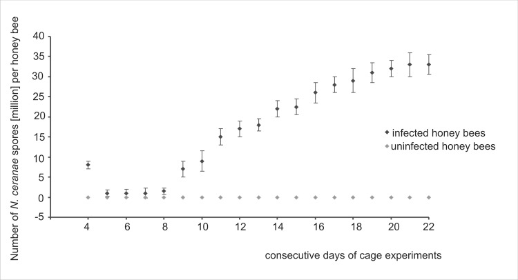
Honey bee workers were artificially infected with N. ceranae on the third day after their emergence. Nosema spores swallowed by honey bee workers were observed in microscopic samples shortly after the infection. On the second day and on the third day after the infection, spore numbers dramatically dropped (Fig 3). There were so few spores in some samples that their number was indeterminable. After that, a slight increase in the N. ceranae infection intensity was observed, and from the 8th day it became markedly intensive and permanent.
Fig 3. Cage tests.
The average N. ceranae infection intensity observed during consecutive days of cage experiments conducted in 2014 and 2015. Data obtained from daily sampling from the second day after the infection (4-day-old workers) to the end of each experiment in 2014 and 2015. Diamond shapes (♦) represent means while error bars indicate standard deviation.
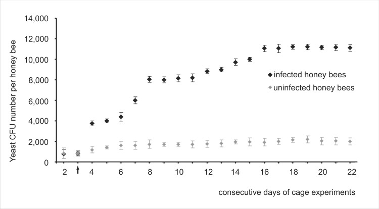
Similarly to field tests, fungi belonging to Candida and Saccharomyces genera were detected in feces of honey bees kept in cages. In uninfected honey bee workers, intestinal yeast CFU number grew slightly during the experiments (Fig 4), from 845 CFUs (CV = 24.32) detected on the second day, to 2,025 CFUs (CV = 13.04) at the end of the investigations (Fig 4). In N. ceranae-infected workers, the growth was markedly higher and two peaks were observed; the first on the 4th and the second on the 8th day of experiments. Both peaks were connected with the N. ceranae infection intensity. The first was observed a day after the bees had been infected and the second was connected with a substantial increase in N. ceranae spore number. Therefore, cage experiments confirmed that the size of the honey bee intestinal yeast population markedly increased after bees were infected with N. ceranae and was connected with the infection intensity (Fig 4). It was also confirmed with Spearman’s correlation between numbers of yeast CFUs and N. ceranae spores, whereas the correlation coefficient estimated in cage experiments was 0.82 (p = 0.0121) in 2014 and 0.79 (p = 0.0119) in 2015.
Fig 4. Cage tests.
The average yeast CFU number observed during consecutive days of cage experiments conducted in 2014 and 2015. Data obtained from daily sampling from the second day of workers emerging to the end of each experiment in 2014 and 2015. The arrow (↑) indicates the day of N. ceranae infection. Diamond shapes (♦) represent means while error bars indicate standard deviation.
Generally, it was in the N. ceranae-infected honey bee workers that the intestinal yeast CFU number reached the highest values both during field and cage tests. A comparison of cage and field tests indicated that more intestinal yeast CFUs were found in bees sampled from colonies (field experiments). Yeast CFU amounted to 11,200 (CV = 13.44) on the 19th day of the experiment in N. ceranae-infected caged workers whereas 16,280 CFU (CV = 14.16) were detected in May in N. ceranae-infected colony workers.
Discussion
Only one colony was found to be infected solely with N. apis out of 11 Nosema spp. infected colonies that were detected from the pool of 82 honey bee colonies monitored in 2014. Next year, however, the colony was found already as mix-infected, i.e. infected both with N. apis and N. ceranae (Table 1). Therefore, the N. ceranae infections were markedly prevalent. Probably, it was a weak immune response of A. mellifera to the N. ceranae infection that was the reason for this situation, since N. ceranae caused immunosuppression in honey bees [12]. Surprisingly, we noticed that one of the colonies infected with N. ceranae at a low intensity (0.8 million spores per honey bee) in 2014 was found self-cured (uninfected) next year, despite the fact that nosemosis was not treated. This may indicate that there are some defense mechanisms against N. ceranae in honey bees. If this phenomenon is confirmed, new possibilities for resistant honey bee breeding may arise.
Until recently, N. ceranae dominated in warmer climates showing non-seasonal nosemosis course [18, 44, 48]. At present, N. ceranae infections spread through temperate climate zones and were detected even in the severe climate of Finland [17, 43, 44], nonetheless having a seasonal course there. In our studies, a pattern of Nosema ceranae infections was similar to that described for temperate zones of USA, Canada, and Germany [38, 39, 40, 41], showing a spring peak and subsequent decline in the summer and fall (Fig 1). However, in our study, the second peak was observed in the middle of the summer in two consecutive years. It is believed that the summer/fall decline in N. ceranae intensity is due to a decrease in daily temperature in these seasons [16, 49]. Moreover, more potent fat body development and an increase in the immune resistance before colony wintering in mid-latitudes [50] could also lead to a reduction in the N. ceranae infection intensity in the fall. This corresponds with the seasonal, permanent decrease in N. ceranae infection intensity presented in Fig 1. A dominant impact of temperature on the N. ceranae disease course was indirectly confirmed by a non-fluctuating pattern of the N. ceranae infection course in our cage tests carried out at the constant temperature of 30°C in which, after the initial decrease in N. ceranae spore intensity, their continuous rise was observed (Fig 3). This initial decrease resulted from the fact that N. ceranae needed about 3–4 days to complete its life cycle inside honey bee intestine cells [41, 51]. After that, newly formed spores were released into the midgut lumen and could be excreted with feces or they could invade next cells of the same host. Rapid acceleration in the N. ceranae infection intensity observed from the 8th day of cage experiments (Fig 3) could be connected with a breakdown of honey bees’ immune defense and the beginning of N. ceranae invasion.
The European eastern range limit of N. ceranae was found in Ukraine [52], but there are no data about seasonal changes in the infection from this location. Therefore, data presented here show seasonality (with the additional mid-summer peak) of the N. ceranae infection intensity at the European eastern limit of its range for the first time.
A growth in the intestine yeast CFU number was observed when the intensity of the N. ceranae infection increased both in field and in cage tests (Table 2, Figs 3 and 4). This suggests that the N. ceranae infection created favorable conditions for the intestinal yeast growth. Intestinal yeast CFU numbers detected in cage tests were generally lower than those detected in field tests both in N. ceranae-infected and uninfected workers (Figs 1 and 4). These findings contradict studies by Gilliam et al. [26, 28, 29], in which caged honey bees had always more yeast than hive bees. In our research, we focused on the fungi belonging only to intestinal yeasts, such as Candida and Saccharomyces genera, whereas Gilliam et al. [26, 27, 28, 29] studied also other yeast genera. This might be the reason for this discrepancy suggesting that different intestinal yeast genera might respond to caged bees’ environment in different ways.
Generally, honey bees kept in fully functional colonies withstand a higher Nosema infection and pressure from greater yeast populations without any visible symptoms of colony weakening than the caged honey bees. Probably, a more varied diet, ability to fly, a stay in a fully functional colony, including presence of a queen bee, positively affected colony honey bees to deal with parasites and to gain appropriate overall fitness.
In mid-latitudes, foragers live under great pressure from May to the end of July when they have to collect supplies for the colony development and their whole energy demands are connected with flying and foraging. From August in the northern hemisphere, a honey bee colony switches to the overwintering preparation and workers consume more energy for maintaining immune defense than during spring or early summer [50]. Consequently, during late summer and fall, bees are well prepared to combat any microbial attack including fungal pathogens such as Nosema spp. or the opportunistic yeast pathogens such as Candida or Saccharomyces. This corresponds with our observation that the number of N. ceranae spores and intestinal yeast CFU number decreased in August. Two peaks in May and July were detected both for N. ceranae infection intensity and intestinal yeast CFU numbers (Figs 1 and 2). This additionally indicated that the reason for these rises lay probably in the seasonal differences in honey bee immune defense. The July peak may have been connected with the colony preparing to replace short-living summer honey bees with overwintering bees. Nevertheless, these data need further studies.
This natural seasonality in nosemosis development may be a cause of misunderstandings during experiments on new therapeutics against N. ceranae. A natural decrease in the number of N. ceranae spores during such experiments can be confounded with the effect of the Nosema spp. treatment with these new compounds. Therefore, we recommend performing all preliminary studies on new therapeutics in cages. Our investigation also showed that field studies on anti-Nosema drugs should be performed during two consecutive years, including colony overwintering, and only the heavily infected colonies threatened with extinction ought to be used. Colonies infected with N. ceranae at low intensity can be found naturally self-cured the following year.
Disturbances caused by N. ceranae infection lead to malabsorption of nutrients, which is inter alia manifested by the presence of large amounts of undigested sugars in feces of infected honey bees [53, 54]. This excess of sugar creates conditions favorable to rapid intestine yeast development. Consequently, sugar metabolism products in the growing yeast population increase acidity of the intestinal microenvironment. This, in turn, creates conditions favorable to N. ceranae development [55, 56, 57, 58] and a self-reinforcing circle begins. So, particularly in its initial stages, nosemosis may stimulate growth of intestinal yeast population, whereas an increase in the number of intestinal yeast, through feedback, may facilitate a development of nosemosis. However, during severe N. ceranae infection, a competition for nutrients and attachment sites begins [20]. When a honey bee’s intestine is filled with Nosema spores [24], yeast cells probably cannot convert into their hypha form and embed themselves into epithelial cells of the midgut. Moreover, Nosema-infected honey bees show decreased foraging abilities [59]. This is probably the cause of nutrient shortages, which are not suitable for yeast development. Consequently, N. ceranae, just like all intercellular parasites, draw energy directly from their host cells and may win the competition [20] with yeasts. At the end of our cage experiments, we observed a decrease in the yeast number (Fig 4), which suggests the onset of such competition. Consequently, during low and medium Nosema spp. infection intensity N. ceranae and yeasts positively affected their mutual development, however, at a high Nosema infection intensity, a suppressing effect of Nosema on yeast development was observed.
Conclusion
In the European eastern limit of its range, N. ceranae infection intensity showed seasonality and was observed as a spring peak and subsequent decline in the summer and fall. However, we also observed an additional mid-summer peak that has not been noticed in other studies. Intestinal yeast population revealed a very similar seasonal pattern to that observed in N. ceranae. There is mutual interdependence between intestine yeast and N. ceranae. At a low and medium Nosema spp. intensity, both parasites positively affected their mutual development but at high infection intensity, a suppressive effect of Nosema on yeast development can be considered.
Due to seasonal changes in the N. ceranae infection intensity in honey bee colonies, we recommend performing studies on new anti-Nosema drugs during two consecutive years, including colony overwintering. A natural decrease in the number of N. ceranae spores during a season can be misinterpreted as an effect of a Nosema spp. treatment with new compounds.
Moreover, honey bee infection with N. ceranae supports additional opportunistic yeast infections, which may result in a faster colony depopulation.
Data Availability
All relevant data are within the paper.
Funding Statement
This research was supported by the Individual Research Grant of Vice-rector for Research and International Relations of UMCS (Lublin, Poland). The funder had no role in study design, data collection and analysis, decision to publish, or preparation of the manuscript PONE-D-16-10261.
References
- 1.Levy S. The pollinator crisis: What's best for bees. Nature 2011; 10.1038/479164a [DOI] [PubMed] [Google Scholar]
- 2.Goulson D, Nicholls E, Botías C, Rotheray EL. Bee declines driven by combined stress from parasites, pesticides, and lack of flowers. Science. 2015; 10.1126/science.1255957 [DOI] [PubMed] [Google Scholar]
- 3.Adl SM, Simpson AGB, Lane CE, Lukeš J, Bass D, Bowser SS, et al. The revised classification of Eukaryotes. J Eukaryot Microbiol. 2012; 59: 429–514. 10.1111/j.1550-7408.2012.00644.x [DOI] [PMC free article] [PubMed] [Google Scholar]
- 4.Shafer ABA, Williams GR, Shutler D, Rogers REL, Stewart DT. Cophylogeny of Nosema (Microsporidia: Nosematidae) and bees (Hymenoptera: Apidae) suggests both cospeciation and a host-switch. J Parasitol. 2009; 95: 198–203. 10.1645/GE-1724.1 [DOI] [PubMed] [Google Scholar]
- 5.Fries I. Nosema ceranae in European honey bees (Apis mellifera). J Invertebr Pathol. 2010; 103: S73–S79. 10.1016/j.jip.2009.06.017 [DOI] [PubMed] [Google Scholar]
- 6.Fries I, Chauzat M-P, Chen Y-P, Doublet V, Genersch E, Gisder S, et al. Standard methods for nosema research. In: Dietemann V, Ellis JD, Neumann P., editors. The Coloss Beebook: Volume II: Standard methods for Apis mellifera pest and pathogen research. J Apicult Res. 2013; 10.3896/IBRA.1.52.1.14 [DOI]
- 7.Williams GR, Shutler D, Burgher-MacLellan KL, Rogers REL. Infra-population and -community dynamics of the parasites Nosema apis and Nosema ceranae, and consequences for honey bee (Apis mellifera) Hosts. PLoS ONE. 2014; 10.1371/journal.pone.0099465 [DOI] [PMC free article] [PubMed] [Google Scholar]
- 8.Grzybek M, Bajer A, Bednarska M, Al-Sarraf M, Behnke-Borowczyk J, Harris PD, et al. Long-term spatiotemporal stability and dynamic changes in helminth infracommunities of bank voles (Myodes glareolus) in NE Poland. Parasitology 2015; 10.1017/S0031182015001225 [DOI] [PubMed] [Google Scholar]
- 9.Zander E. Tierische Parasiten als Krankenheitserreger bei der Biene. Münchener Bienenzeitung. 1909; 31: 196–204. [Google Scholar]
- 10.Fries I. Infectivity and multiplication of Nosema apis Z. in the ventriculus of the honey bee. Apidologie. 1988; 19:319–328. [Google Scholar]
- 11.Huang WF, Jiang JH, Wang CH. Nosema ceranae infection in Apis mellifera. 38th Annual Meeting of Society for Invertebrate Pathology, Anchorage, Alaska. 2005.
- 12.Antúnez K, Martin-Hernandez R, Prieto L, Meana A, Zunino P, Higes M. Immune suppression in the honey bee (Apis mellifera) following infection by Nosema ceranae (Microsporidia). Environ Microbiol. 2009; 10.1111/j.1462-2920.2009.01953x [DOI] [PubMed] [Google Scholar]
- 13.Martin HR, Higes M, Garido ME, Meana A. Reliability of Diagnostic methods to detect Nosema spp. spores in honey bees: Molecular identification versus visual observation. Proc. Second Europ. Conf. Apidology, Prague 10–14 September 2006, p. 34.
- 14.Chauzat MP, Higes M, Martin R, Meana A, Faucon JP. Nosema ceranae and Nosema apis in France: co−infections in honey bee colonies. Proceedings of the Second European Conference of Apidology 10–14 September 2006; 30.
- 15.Klee J, Besana AM, Genersch E, Gisder S, Nanetti A, Tam DQ, et al. Widespread dispersal of the microsporidian Nosema ceranae, an emergent pathogen of the western honey bee, Apis mellifera. J Invertebr Pathol. 2007; 96: 1–10. 10.1016/j.jip.2007.02.014 [DOI] [PubMed] [Google Scholar]
- 16.Martin-Hernandez R, Meana A, Garcia-Palencia P, Marin P, Botias C, Garrido-Bailon E, et al. Effect of temperature on the biotic potential of honeybee microsporidia. Appl Environ Microbiol. 2009; 75: 2554–2557. 10.1128/AEM.02908-08 [DOI] [PMC free article] [PubMed] [Google Scholar]
- 17.Paxton RJ, Klee J, Korpela S, Fries I. Nosema ceranae has infected Apis mellifera in Europe since at least 1998 and may be more virulent than Nosema apis. Apidologie. 2007; 38: 558–565. [Google Scholar]
- 18.Tapaszti Z, Forgách P, Kövágó C, Békési L, Bakonyi T, Rusvai M. First detection and dominance of Nosema ceranae in Hungarian honeybee colonies. Acta Vet Hung. 2009; 57: 383–388. 10.1556/AVet.57.2009.3.4 [DOI] [PubMed] [Google Scholar]
- 19.Higes M, Garcia-Palencia P, Martin-Hernandez R, Aranzazu M. Experimental infection of Apis mellifera honeybees with Nosema ceranae (Microsporidia). J Invertebr Pathol. 2007; 94: 211–223. 10.1016/j.jip.2006.11.001 [DOI] [PubMed] [Google Scholar]
- 20.Borsuk G, Ptaszyńska AA, Olszewski K, Paleolog J. Impact of N. ceranae infection on the intestinal yeast flora of honey bee. Med Weter. 2013; 69: 726–730. [Google Scholar]
- 21.Milne CP Jr. The Need for using laboratory tests in breeding honeybees for improved honey production. J Apicult Res. 1985; 10.1080/00218839.1985.11100679 [DOI] [Google Scholar]
- 22.Borsuk G, Strachecka A Olszewski K, Paleolog J. The interaction of worker bees with increased genotype variance, Part 1. field tests of sugar syrup collection and storage. J Apicult Sci. 2011; 55: 53–58. [Google Scholar]
- 23.Borsuk G, Strachecka A, Olszewski K, Paleolog J. The interaction of worker bees which have increased genotype variance, Part 2. cage tests of sugar syrup collecting and mortality. J Apicult Sci. 2011, 55: 59–65. [Google Scholar]
- 24.Ptaszyńska AA, Borsuk G, Mułenko W, Demetraki-Paleolog J. Differentiation of Nosema apis and Nosema ceranae spores under Scanning Electron Microscopy (SEM). J Apicult Res. 2014; 53: 537–544, 10.3896/IBRA.1.53.5.02 [DOI] [Google Scholar]
- 25.Gilliam M. Are yeasts present in adult worker honey bees as a consequence of stress? Ann Entomol Soc Am. 1973; 66: 1176. [Google Scholar]
- 26.Gilliam M, Prest DB. Fungi isolated from the intestinal contents of foraging worker honey bees, Apis mellifera. J Invertebr Pathol. 1972; 20: 101–103. [Google Scholar]
- 27.Gilliam M, Taber S. Diseases, pests, and normal microflora of honeybees Apis mellifera, from feral colonies. J Invertebr Pathol. 1991; 58: 286–289. [Google Scholar]
- 28.Gilliam M, Valentine DK. Enterobacteriaceae isolated from foraging worker honey bees, Apis mellifera. J Invertebr Pathol. 1974; 23: 38–41. [Google Scholar]
- 29.Gilliam M, Valentine DK. Bacteria isolated from the intestinal contents of foraging worker honey bees, Apis mellifera: the genus Bacillus. J Invertebr Pathol. 1976; 28: 275–276. [Google Scholar]
- 30.Gilliam M, Wickerham LJ, Morton HL, Martin RD. Yeasts isolated from honey bees, Apis mellifera, fed 2,4-D and antibiotics. J Invertebr Pathol. 1974; 24: 349–356. [DOI] [PubMed] [Google Scholar]
- 31.Tysset C, De Rantline de la Roy Durand C. Contribution to the Study of Intestinal Microbial Infection of Healthy Honeybees: Inventory of Bacterial Population by Negative Organisms. Philadelphia, Pa, USA: Department of Agriculture, SEA-AR, Eastern Region Research Centers; 1991. [Google Scholar]
- 32.El-leithy M, El-sibael K. Role of microorganisms isolated from bees, its ripening and fermentation of honey. Egypt J Microbiol.1992; 75: 679–681. [Google Scholar]
- 33.Bradley TJ. Active transport in insect recta. J Exp Biol. 2008; 211: 835–836. 10.1242/jeb.009589 [DOI] [PubMed] [Google Scholar]
- 34.Mirjanic G, Tlak-Gajger I, Mladenovic M, Kozaric Z. 2013. Impact of different feed on intestine health of honey bees. XXXXIII International Apicultural Congress, Apimondia, Kyiv, Ukraine, 29.09–04.10 2013, p.113.
- 35.Strachecka A, Krauze M, Olszewski K, Borsuk G, Paleolog J, Merska M, Chobotow J, Bajda M, Grzywnowicz K. Unexpectedly strong effect of caffeine on the vitality of western honeybees (Apis mellifera). Biochem (Mosc). 2014; 10.1134/S0006297914110066 [DOI] [PubMed] [Google Scholar]
- 36.Strachecka A, Olszewski K, Paleolog J. Curcumin stimulates biochemical mechanisms of Apis mellifera resistance and extends the apian life-span. J Apicult Sci. 2015; 10.1515/jas-2015-0014 [DOI] [Google Scholar]
- 37.Strachecka AJ; Paleolog J, Grzywnowicz K. The surface proteolytic activity in Apis mellifera. J Apicult Sci. 2008; 52: 49–56. [Google Scholar]
- 38.Traver BE, Williams MR, Fell RD. Comparison of within hive sampling and seasonal activity of Nosema ceranae in honey bee colonies. J Invertebr Pathol. 2012; 109: 187–193. 10.1016/j.jip.2011.11.001 [DOI] [PubMed] [Google Scholar]
- 39.Mulholland GE, Traver BE, Johnson NG, Fell RD. Individual variability of Nosema ceranae infections in Apis mellifera colonies. Insects. 2012; 3:1143–1155. 10.3390/insects3041143 [DOI] [PMC free article] [PubMed] [Google Scholar]
- 40.Copley TR, Jabaji SH. Honeybee glands as possible infection reservoirs of Nosema ceranae and Nosema apis in naturally infected forager bees. J Appl Microbiol. 2012, 112: 15–24. 10.1111/j.1365-2672.2011.05192.x [DOI] [PubMed] [Google Scholar]
- 41.Gisder S, Hedtke K, Mockel N, Frielitz MC, Linde A, Genersch E. Five-year cohort study of Nosema spp. in Germany: Does climate shape virulence and assertiveness of Nosema ceranae? Appl Environ Microbiol. 2010; 76: 3032–3038. 10.1128/AEM.03097-09 [DOI] [PMC free article] [PubMed] [Google Scholar]
- 42.Bush AO, Lafferty KD, Lotz JM, Shostak AW. Parasitology meets ecology on its own terms: Margolis et al. revisited. J Parasitol. 1997; 83: 575–583. [PubMed] [Google Scholar]
- 43.Forsgren E, Fries I. Comparative virulence of Nosema ceranae and Nosema apis in individual European honey bees. Vet Parasitol. 2010; 170: 212–217. 10.1016/j.vetpar.2010.02.010 [DOI] [PubMed] [Google Scholar]
- 44.Martin-Hernandez R, Meana A, Prieto L, Salvador AM, Garrido-Bailon E, Higes M. Outcome of the colonization of Apis mellifera by Nosema ceranae. Appl Environ Microbiol. 2007; 73: 6331–6338. 10.1128/AEM.00270-07 [DOI] [PMC free article] [PubMed] [Google Scholar]
- 45.Hornitzky M. Nosema Disease–Literature review and three surveys of beekeepers–Part 2. Rural industries research and development corporation. Pub. No. 08/006, 2008.
- 46.SAS/STAT. Users Guide release 9.13, Cary, NC, Statistical Analysis System Institute; 2014. [Google Scholar]
- 47.Greene W.H. Econometric analysis, 5th edition New Jersey: Prentice Hall; 2003. [Google Scholar]
- 48.Higes M, Martín-Hernández R, Meana A. Nosema ceranae in Europe: an emergent type C N. ceranae infection. Apidologie 2010; 41: 375–392. [Google Scholar]
- 49.Martín-Hernández R, Botías C, Bailón EG, Martínez-Salvador A, Prieto L, Meana A, Higes M. Microsporidia infecting Apis mellifera: coexistence or competition. Is Nosema ceranae replacing Nosema apis? Environ Microbiol. 2012; 14: 2127–2138. 10.1111/j.1462-2920.2011.02645.x [DOI] [PubMed] [Google Scholar]
- 50.Gätschenberger H, Azzami K, Tautz J, Beier H. Antibacterial immune competence of honey bees (Apis mellifera) is adapted to different life stages and environmental risks. PLoS ONE. 2013; 10.1371/journal.pone.0066415 [DOI] [PMC free article] [PubMed] [Google Scholar]
- 51.Chen YP, Evans JD, Murphy C, Gutell R, Zuker M, Gundensen-Rindal D, Pettis JS. Morphological, molecular, and phylogenetic characterization of Nosema ceranae, a microsporidian parasite isolated from the European honey bee, Apis mellifera. J Eukaryot Microbiol. 2009; 10.1111/j.1550-7408.2008.00374.x [DOI] [PMC free article] [PubMed] [Google Scholar]
- 52.Yefimenko ТМ, Odnosum HV, Tokarev YS, Ignatieva АN. Nosema ceranae Fries et al., 1996 (Microspora, Nosematidae)–a honey bee parasite in Ukraine. Екологія. 2014; 2: 70–75. [Google Scholar]
- 53.Tlak Gajger I, Petrinec Z, Pinte Lj, Kozarić Z. Experimental treatment of nosema disease with “Nozevit” phytopharmacological preparation. Am Bee J. 2009; 149: 485–490. [Google Scholar]
- 54.Somerville D. Nosema disease in bees. Agnote DAI/124. NSW Agriculture; 2005.
- 55.Dade HA. Anatomy and dissection of the honey bee (revised Ed.) International Bee Research Association; Cardiff, UK; 2009. [Google Scholar]
- 56.Ptaszyńska AA, Borsuk G, Mułenko W, Olszewski K. Impact of ethanol on Nosema spp. infected bees. Med Weter. 2013; 69: 736–741. [Google Scholar]
- 57.Ptaszyńska AA, Borsuk G, Zdybicka-Barabas A, Cytryńska M, Małek W. Are commercial probiotics and prebiotics effective in the treatment and prevention of honeybee nosemosis C? Parasitol Res. 2016; 10.1007/s00436-015-4761-z [DOI] [PMC free article] [PubMed] [Google Scholar]
- 58.Ptaszyńska AA, Borsuk G, Mułenko W, Wilk J. 2016. Impact of vertebrate probiotics on honeybee yeast microbiota and on the course of nosemosis. Med Weter. 2016; 72: 430–434 10.21521/mw.5534 [DOI] [Google Scholar]
- 59.Lach L, Kratz M, Baer B. Parasitized honey bees are less likely to forage and carry less pollen. J Invertebr Pathol. 2015; 130:64–71. 10.1016/j.jip.2015.06.003 [DOI] [PubMed] [Google Scholar]
Associated Data
This section collects any data citations, data availability statements, or supplementary materials included in this article.
Data Availability Statement
All relevant data are within the paper.


