Abstract
As a crucial component of brain innate immunity, neuroinflammation initially contributes to neuronal tissue repair and maintenance. However, chronic inflammatory processes within the brain and associated blood-brain barrier impairment often cause neurotoxicity and hyperexcitability. Mounting evidence points to a mutual facilitation between inflammation and epilepsy, suggesting that blocking the undesired inflammatory signaling within the brain might provide novel strategies to treat seizures and epilepsy. Neuroinflammation is primarily characterized by the upregulation of proinflammatory mediators in epileptogenic foci, among which, cyclooxygenase-2/prostaglandin E2, interleukin-1β, transforming growth factor-β, toll-like receptor 4, high-mobility group box 1, and tumor necrosis factor-α have been extensively studied. Small molecules that specifically target these key proinflammatory perpetrators have been evaluated for antiepileptic and antiepileptogenic effects in animal models. These important preclinical studies provide new insights into the regulation of inflammation in epileptic brains and guide drug discovery efforts aimed at developing novel anti-inflammatory therapies for seizures and epilepsy.
Keywords: Epilepsy, Epileptogenesis, Interleukin, Neuroinflammation, Prostaglandin, Seizure
Neuroinflammation and Epilepsy
Inflammation is a sequence of biological responses to pathogenic challenges, and in the periphery, it is often symptomized by local redness, fever, swelling, pain and loss of function. The inflammatory processes can be temporally classified as either acute or chronic. During the acute phase of inflammation, plasma proteins and leukocytes from the blood vessels rapidly extravasate to the injury sites to initiate a series of biochemical and cellular events, involving the immune system, vasculature and local cells. As a key component of innate immunity, acute inflammation represents an attempt to resolve the noxious stimuli and initiate the healing process [1]. Acute inflammation typically subsides in days; however, under certain circumstances inflammation persists to become chronic. Although tissue healing continues at the chronic stage of inflammation, the excessive inflammatory processes promote a complex network of molecular and cellular signaling pathways that are linked to debilitating conditions such as rheumatoid arthritis, pain, cancer, vascular diseases, skin inflammation, diabetes, heart diseases and pulmonary diseases [2]. In contrast to our historical understanding that the central nervous system (CNS) is immune-privileged due to tight constriction of the blood-brain barrier (BBB), inflammation within the brain – or neuroinflammation – has emerged as a salient feature in virtually all neurological conditions (Box 1), and been proposed to facilitate disease progression in the CNS (Box 2). Over the past decade, an increasing body of clinical and preclinical evidence points to the involvement of inflammation in the pathophysiology of acute brain insults and subsequent epileptogenesis, a process that a normal brain becomes epileptic (Box 2) [3–6], and anti-inflammatory therapeutics targeting crucial inflammatory components have been proposed to treat seizures and epilepsy [7–13]. In this review, we highlight our current understanding of neuroinflammation in epilepsy by focusing on recent preclinical efforts to develop anti-inflammatory therapies that block acute seizures and/or the development of epilepsy via selective small molecules.
Box 1. Does seizure cause neuroinflammation?
Neuroinflammation is characterized by the activation of microglia, astrocytes, endothelial cells of the BBB, infiltration of plasma proteins and immune cells – granulocytes (mainly neutrophils) in the beginning, followed by monocytes within a day and lymphocytes days later, and the upregulation of an array of pro- or anti-inflammatory mediators [5, 163–165]. The best-known members of these inflammatory molecules can be classified into proinflammatory enzymes including COX-2, inducible NOS (iNOS) and NOX; cytokines such as IL-1β, IL-6, TNF- α; and growth factors like TGF-β and BDNF. These cellular and biochemical alterations within the brain are commonly observed in brain specimens from epilepsy patients and in brain tissues from animals with experimental epilepsy [3, 4]. In addition, recent studies show that seizures also can increase the BBB permeability, which can intensify and perpetuate neuroinflammation via extravasation of leukocytes and inflammatory molecules from blood vessels into the brain parenchyma [5, 164, 166]. In general, initial prolonged seizures can trigger acute immune and inflammatory responses within the brain, while the subsequent spontaneous recurrent seizures sustain the chronic neuroinflammation.
Box 2. Does neuroinflammation cause seizure?
Neuroinflammation appears to affect seizure severity and recurrence, which is evident by extensive clinical and experimental data. Patients with autoimmune diseases and encephalitis accompanied by severe and long-lasting neuroinflammation often have high frequency of seizures [167, 168]; infections in the CNS – caused by viruses, bacteria and parasites – often lead to severe inflammatory reactions and are a major risk factor for epilepsy, as about 6.8 - 8.3 % of survivors of CNS infections have unprovoked seizures in developed countries and the rates are much higher in the developing world [169]. These clinical observations clearly suggest that the immune and inflammatory processes are involved in some forms of epilepsy. It now becomes clear that proinflammatory mediators such as COX-2, PGE2, IL-1β, IL-6, HMGB1, TLR4, TNF-α, TGF-β and NOX2 play important roles in seizure generation and exacerbation [3, 13, 74, 132]. For instance, systemic inflammatory processes induced by fever in children can cause seizures and the proinflammatory cytokines such as IL-1β are believed to play crucial roles [170]. Furthermore, systemic administration of LPS in rodents can change the seizure threshold [171] and enhances epileptogenesis [82, 172], also involving cytokines IL-1β and TNF-α, and COX-2 activation in the brain. Thus, as a consequence of epilepsy, neuroinflammation in turn can cause or facilitate epilepsy.
Small Molecules as Anti-Inflammatory Therapeutics
Small molecules are synthetic chemicals with a low molecular weight (< 900 Da) and a size on the order of a nanometer. These organic compounds constitute the vast majority of current drugs in the market. Due to the small size, they are widely distributed throughout the body after administration and can cross biological membranes, thereby modulating a broad range of intracellular and extracellular molecular targets. Compared to biological drugs like recombinant proteins, peptides, monoclonal antibodies, etc., small molecule-based drugs provide many manufacture and delivery advantages: they are typically low-cost, stable, non-immunogenic, easy to characterize and modify, orally available, and capable of penetrating tissues [14]. The future of small-molecule paradigm is still promising particularly in the CNS field in that biological drugs generally have poor brain penetration. Up to date, a number of small-molecule compounds that selectively modify the key inflammatory signaling pathways have been evaluated for therapeutic benefits in animal models of seizures and epilepsy, with more under development or to be tested. Most of these compounds have sufficient pharmacodynamic and pharmacokinetic properties – i.e., potency, selectivity, bioavailability, in vivo half-life, brain penetration – and are considerably safe with appropriate dosage and dosing paradigm, and thus have enormous translational potential, as some compounds have advanced to clinical studies.
COX Inhibitors
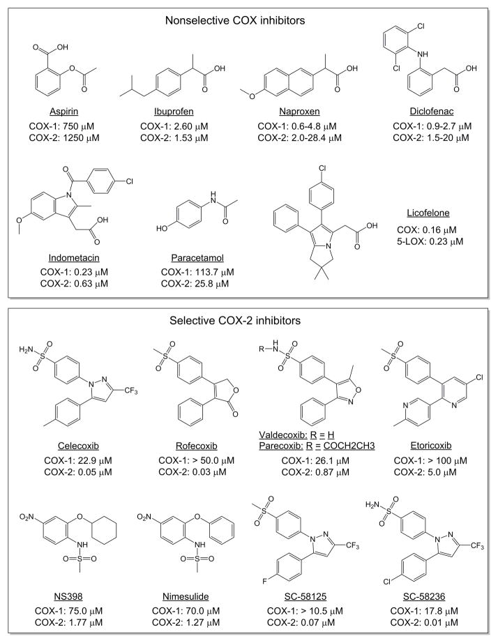
Cyclooxygenase (COX) is the rate-limiting enzyme in the synthesis of prostanoids that comprise prostaglandin D2 (PGD2), PGE2, PGF2α and PGI2, and thromboxane A2 (TXA2). COX has two isoforms: COX-1 is constitutively expressed throughout the body to maintain homeostatic prostaglandins, which are important for many normal physiological functions; COX-2 is usually undetectable in most normal tissues but strongly induced by infection, fever, inflammation and other stimuli such as growth factors and excessive neuronal activity, and is generally considered as a major proinflammatory mediator. COX-2 is rapidly and robustly induced within the brain following seizures in both human patients and experimental animals [15–17]. Chronic upregulation of COX-2 perpetuates and aggravates neuroinflammation and, thus contributes to the pathophysiology of acute and chronic seizures. The first insight into the pathogenic role for COX-2 in seizures was derived from experimental evidence that neuronal overexpression of COX-2 facilitates kainate-provoked convulsions and increases seizure-associated mortality in mice [18]. Additional evidence from a genetic strategy came from COX-2 knockout mice that show reduction of recurrent hippocampal seizures in the kindling model of status epilepticus (SE) [19], and resistance to neuronal death after kainate treatment [20]. More recently, in the mouse pilocarpine model, ablation of COX-2 from a restricted population of forebrain neurons reduced neuroinflammation and secondary neurodegeneration [16], and subtly improved retrograde memory performance [21]. Multiple COX-2-selective and nonselective inhibitors including aspirin, celecoxib, etoricoxib, indomethacin, nimesulide, NS398, parecoxib (valdecoxib), rofecoxib, SC58236, SC58125 (Figure 1), have been evaluated for antiepileptic and antiepileptogenic effects, neuroprotection, and improvements in behavioral and cognitive abnormalities in chemoconvulsant or electrical models of acute seizures and epilepsy (Table 1) [9, 19, 20, 22–37].
Figure 1.
Chemical structures of small molecules that inhibit COX and have been tested in animal models for antiepileptic and antiepileptogenic effects. The IC50s on COX-1 and COX-2 of each compound are indicated. Note that licofelone is a dual COX/LOX inhibitor and parecoxib is a pro-drug of valdecoxib.
Table 1.
Effects of COX inhibitors on neuronal loss, acute and chronic seizures, and behavior in animal models of epilepsy.
| Seizure induced by | Initial seizure duration | Species, strain, gender and age | Dosage and dose paradigm | Start-time of treatment | Major therapeutic outcomes | Reference | |
|---|---|---|---|---|---|---|---|
| Kainate (50 mg/kg, i.p.) | Not terminated | Adult male mice | 10 mg/kg, i.p. | 30 min before kainate injection | Increased mortality | [22] | |
| Lithium chloride (3 mEq/kg, i.p.) 18– 20 hr prior to pilocarpine (30 mg/kg, i.p.) | 70 min (terminated by diazepam) | Adult male Sprague-Dawley rats |
20 mg/kg, i.p., once daily for 20 days | 0 hr, 3 hr, or 24 hr after termination of SE | Reduction in frequency and duration of SRSs, hippocampal neuronal loss, aberrant migration of newborn granule cells, formation of hilar basal dendrites, and mossy fiber sprouting | [23] | |
| Pilocarpine (280 mg/kg, i.p.) | 2 hr (terminated by diazepam) | Male C57BL/6 mice (8 weeks) | 15 or 150 mg/kg, i.p. | 3 days before pilocarpine injection for 10 days | Shorter latency to SE, higher mortality, no effect on neuronal loss and glial responses | [24] | |
| Kainate (50 mg/kg, i.p.) | Not terminated | Adult male mice | 10 mg/kg, i.p. | 30 min before kainate injection | Increase of seizure severity, mortality, and neuronal loss in the hippocampus | [22] | |
| Electrical stimulation of amygdala | Not terminated | Adult male NMRI mice |
10 mg/kg, i.p. | 30 min before kindling | No effect on the generation, spread and termination of seizure activity | [25] | |
| Kainate (12 or 6 mg/kg, i.p.) | Not terminated | Adult male Wistar rats | 6 mg/kg, i.p., once daily for 5 days or once | 2 hr after kainate injection | Attenuation of BDNF level in hippocampus and reduction of the learning and object exploration deficits in MWM task, but no neuroprotection | [26] | |
| Kainate (12 or 6 mg/kg, i.p.) | Not terminated | Adult male Wistar rats |
6 mg/kg, i.p., once daily for 5 days or once | 2 hr before kainate injection | No effect on BDNF induction, cognitive deficits or neuronal loss | [26] | |
| Lithium (127 mg/kg, i.p.) 24 h prior to pilocarpine (30 mg/kg, i.p.) | 60 min (terminated by diazepam) | Male Sprague- Dawley rats (12 weeks) |
20 mg/kg, p.o., once daily for 14 days | 24 hr after SE | Reduction of SRSs and microglia activation; neuroprotection in the hippocampus | [27] | |
| Pentylenetetrazol (60 mg/kg, i.p.) | Not terminated | Adult male Wistar rats |
2 mg/kg, p.o., once | 1 hr before pentylenetetrazol injection | Anticonvulsant effect | [28] | |
| Pentylenetetrazol (60 mg/kg, i.p.) | Not terminated | Adult male Wistar rats |
0.2 or 20 mg/kg, p.o., once | 1 hr before pentylenetetrazol injection | No anticonvulsant effect | [28] | |
| Animal model of absence epilepsy and epileptogenesis | - | Male WAG/Rij rats (45 days) | 10 mg/kg daily, p.o. | 17 consecutive weeks starting from 45 days postnatal age | Anti-epileptogenic effect | [29] | |
| Animal model of absence epilepsy and epileptogenesis | - | Male WAG/Rij rats (6 months) | 10 and 20 mg/kg, i.p., once | - | Anti-absence seizure effect | [29] | |
| Indomethacin | Kainate (50 mg/kg, i.p.) | Not terminated | Adult male mice | 5 mg/kg, i.p. | 30 min before kainate injection | Increase of seizure severity, mortality, and neuronal loss in the hippocampus | [22] |
| Kainate (10 mg/kg, i.p.) | Not terminated, 6–8 hr | Adult male Sprague-Dawley rats |
10 mg/kg, i.p. | 1 hr before, then 8 hr after kainate injection, and twice daily for 2 days | Augmented seizure activities and increased mortality | [30] | |
| Kainate (10 mg/kg, i.p.) | Not terminated, 6–8 hr | Adult male Sprague-Dawley rats |
10 mg/kg, i.p. | 8 hr after kainate injection and twice daily for 2 days | No neuroprotection | [30] | |
| Electrical stimulation of hippocampus | Not terminated | C57BL/6J mice (9–12 weeks) | 10 mg/kg, p.o., once | 30 min before SE- induction | Reduction of PGE2 level in the forebrain; decrease of recurrence of electrographic and behavioral seizures | [19] | |
| Kainate (50 mg/kg, i.p.) | Not terminated | Adult male mice | 10 mg/kg, i.p. | 30 min before kainate injection | Increase of seizure severity, mortality, and neuronal loss in the hippocampus | [22] | |
| Electrical stimulation of amygdala | Not terminated | Adult male NMRI mice |
10 mg/kg, i.p. | 30 min before kindling | No effect on the generation, spread and termination of seizure activity | [25] | |
| Kainate (1 μg, intrahippocampal injection) | Not terminated | Adult male Wistar rats (9–12 weeks) |
10 mg/kg, i.p., twice daily for 3 days | 5 hr after kainate injection | Reduction of PGE2 level and seizure- induced neuronal loss in hippocampus | [20] | |
| Kainate (1 μg, intrahippocampal injection) | Not terminated | Adult male Wistar rats (9–12 weeks) |
5 mg/kg, i.p., once | 30 min before kainate injection | Transient reduction of PGE2 level and no neuroprotection in hippocampus | [20] | |
| Lithium chloride (127 mg/kg, i.p.) 18 hr prior to pilocarpine (50 mg/kg, i.p.) | 30 min (treated by diazepam) | Adult male Sprague-Dawley rats |
10 mg/kg, i.p., twice | 30 min after SE onset, and another injection 6 hr later | Reduction in neuronal damage in the hippocampus | [31] | |
| Lithium chloride (127 mg/kg, i.p.) 12–17 hr prior to pilocarpine (20–50 mg/kg, i.p.) | 90 min (terminated by diazepam) | Female Sprague-Dawley rats |
10 mg/kg, i.p., twice daily for 17 days | 1.5 hr after SE onset | Reduction of PGE2 levels in hippocampus, frontal cortex, amygdala and piriform cortex; reduction of the severity, but not the frequency and duration of SRSs; neuroprotection in hippocampus and piriform cortex; moderate reduction of learning impairment and prevention of locomotor hyperactivity in the MWM | [32] | |
| Kainate (10 mg/kg, i.p.) | Not terminated | Adult male Sprague-Dawley rats |
10 mg/kg, i.p., twice daily for 3 days | 6 or 8 hr after kainate injection | Neuroprotection in hippocampus, but not in thalamus, amygdala and piriform cortex | [33] | |
| Pentylenetetrazol (i.v.) | - | Adult male Albino mice |
2 or 4 mg/kg, i.p., once |
45 min before pentylenetetrazol injection | Increase of seizure threshold | [34] | |
| Kainate (10 mg/kg, s.c.) | Not terminated | Adult male Sprague-Dawley rats |
3 mg/kg, p.o., once | 1 hr before kainate injection | Attenuation of PGE2 level in hippocampus | [35] | |
| Kainate (0.6 μg, i.c.v.) | Not terminated | Adult male Sprague-Dawley rats |
3 mg/kg, p.o., 3 times | 1 hr before, 24 and 48 hr after the kainate injection | Neuroprotection in hippocampus | [35] | |
| Electrical stimulation of hippocampus | Not terminated, 9–10 hr | Adult male Sprague-Dawley rats |
10 mg/kg, p.o., once daily for 7 days | 4 hr after SE | Reduction of PGE2 level in hippocampus; no anti-epileptic or anti-epileptogenic effect; no neuroprotection | [36] | |
| Electrical stimulation of hippocampus | Not terminated, 9–10 hr | Adult male Sprague-Dawley rats |
10 mg/kg, p.o., once daily for 3 days or for 14 days | 1 day before SE- induction | No obvious anti-epileptic or anti- epileptogenic effect; increased mortality | [37] |
Abbreviations: BDNF, brain-derived neurotrophic factor; hr, hour; i.c.v., intracerebroventricular; i.p., intraperitoneal; i.v., intravenous; p.o., per os; min, minute; MWM, Morris water maze; PGE2, prostaglandin E2; s.c., subcutaneous; SE, status epilepticus; SRSs, spontaneous recurrent seizures.
Adapted from [9].
Interestingly, it appears that treatment with these COX-2 inhibitors beginning after – but not before – SE onset usually shows beneficial effects including reductions in neuronal damage, recurrent seizures and behavior abnormalities (Table 1). The inadequate therapeutic outcomes from preadministration of COX-2 inhibitors in animal SE models have been known for nearly two decades [22]. For instance, treatment by NS398, the first documented small molecule that selectively inhibits COX-2, beginning five hours after but not 30 minutes before kainate injection is neuroprotective in rats [20]. Similarly, celecoxib, the first COX-2 selective inhibitor to reach market, when administered once daily (6 mg/kg, i.p.) for five days after but not before kainate injection in rats, significantly attenuates brain-derived neurotrophic factor (BDNF) induction in hippocampus and reduces the learning and object exploration deficits in Morris water maze task [26]. In another study, oral administration of celecoxib (20 mg/kg) one day after a one-hour episode of lithium/pilocarpine-induced SE in rats reduces spontaneous recurrent seizures, and prevents neuronal death and microglia activation in the hippocampus [27]. Furthermore, nonselective COX inhibitor aspirin, when administered commencing hours after pilocarpine-induced SE, reduces neuronal damage in hippocampus and recurrent seizures in rats [23], whereas aspirin pre-treatment beginning three days before pilocarpine injection does not show such beneficial effects but increases the seizure susceptibility in mice [24].
These seemingly conflicting outcomes by pre- and post-treatment of COX-2 inhibitors suggest a possible dual consequence of COX-2 induction – early neuroprotection and later neurotoxicity involving persistent inflammation following brain insults. This is also evident by a trend of aggregating hippocampal injury detected one day after pilocarpine-induced SE in mice with COX-2 deficiency in forebrain neurons, but a substantial neuroprotection measured three days later [16]. In addition, the dosage, dosing interval, dosing duration, as well as the pharmacodynamics and pharmacokinetics of test compounds such as off-target activities and in vivo half-life could also potentially influence the therapeutic outcomes. Whether antiepileptic drugs (AEDs) are used to terminate seizures along with COX-2 inhibition in these animal studies is another potential contributory factor to the incongruent results [31, 32, 36, 37], in that long duration of SE – up to 10 hours in some cases – might impede the outcomes of COX-2 inhibition (Table 1). Nonetheless, the dichotomous actions of seizure-promoted COX-2 induction suggest that identifying the therapeutic time window and optimizing the treatment paradigm might help resolve the inconsistent efficacy of COX-2 inhibition in these animal models [17, 38].
Other than the desired inhibition on the synthesis of PGE2, the prostaglandin type that is largely responsible for COX-2 cascade-governed inflammation, COX-2 inhibition also causes collateral suppression of endothelial COX-2-derived PGI2, imposing vascular risks that discourage the clinical use of COX-2 inhibitors [39]. As such, rofecoxib (Vioxx®) and valdecoxib (Bextra®) were withdrawn from the USA market in 2004 and 2005, respectively, while celecoxib (Celebrex® or Onsenal®) that is less COX-2 selective is still available to treat chronic conditions such as arthritis, pain and polyps, but with a black box warning for cardiovascular hazard. However, short-term and low-dose medication with selective COX-2 inhibitors is overall safe, evident by their negligible effects in control animals, and could be considered to treat seizure-promoted neuronal injury [40]. Nonetheless, the safety concerns raised on COX-2 inhibitors inspire that targeting a specific downstream prostaglandin synthase or receptor might offer an alternative strategy to control neuroinflammation and neuronal injury associated with seizures and epilepsy.
Prostaglandin Receptor Antagonists
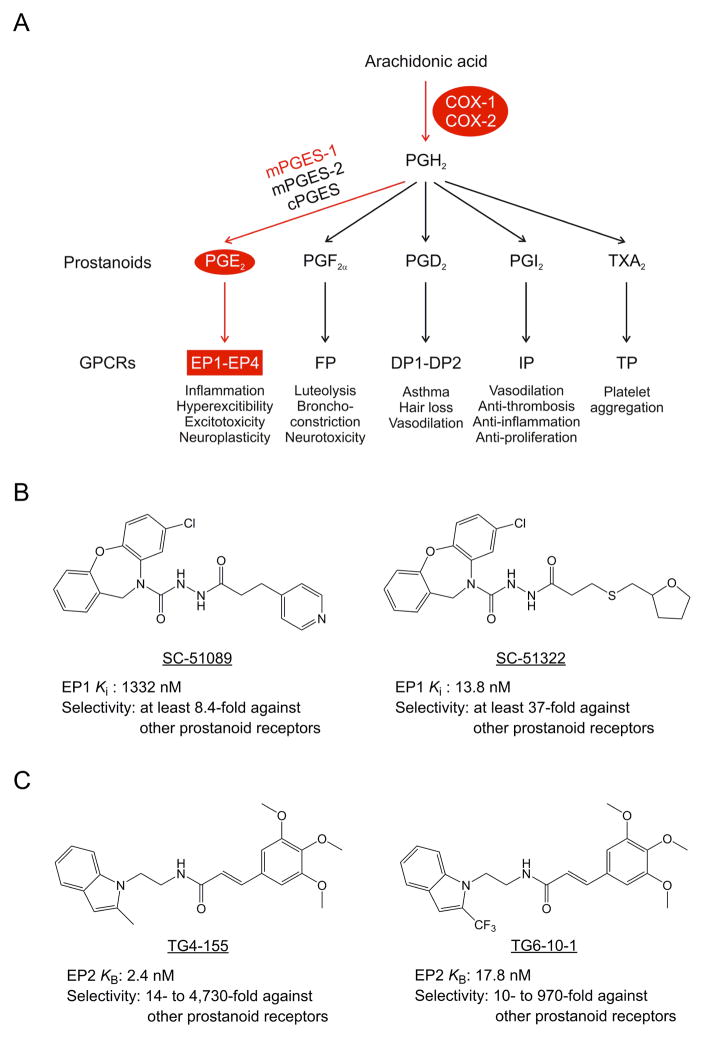
As a major COX-2 product in the CNS, PGE2 is now widely recognized for its crucial roles in neuroinflammation, neuronal hyperexcitability and excitotoxicity, via promoting local vasodilation, infiltration of immune cells, and upregulation of many proinflammatory mediators in the inflamed brain regions (Figure 2A) [40, 41]. Interestingly, the anticonvulsant action of COX-2 inhibitor celecoxib in pentylenetetrazol-induced seizures can be reversed by intracerebroventricular administration of PGE2 [28], suggesting that COX-2 facilitates seizures through PGE2. Conversely, another prostaglandin type PGD2 has been demonstrated to show antiepileptic effect via the DP1 receptor after pentylenetetrazol administration in both rats and mice [42, 43]. Likewise, intracisternal administration of PGF2α reduces seizure intensity and mortality after kainate injection in mice [44]. Therefore, targeting specific PGE2 receptors instead of COX-2 enzyme itself could avoid compromising the PGD2- or PGF2α-mediated anti-seizure effects.
Figure 2.
COX-2 and inflammatory prostaglandin signaling pathways. (A) COX cascade mediates a variety of physiological and pathological events via its prostanoid products. In response to disparate stimuli, the membrane-bound arachidonic acid is freed and converted to unstable intermediate PGH2 by COX, which has two forms: constitutive COX-1 and inducible COX-2. Short-lived PGH2 is then quickly converted to five prostanoids, consisting of prostaglandin PGD2, PGE2, PGF2α, prostacyclin PGI2, and thromboxane TXA2, by tissue-specific prostanoid synthases. Prostanoids exert their functions via activating a suite of GPCRs. Four receptors – EP1, EP2, EP3 and EP4 – are activated by PGE2, and two by PGE2 (DP1 and DP2), whereas each of the other three prostanoids activates a single receptor (FP, IP, TP). The inflammatory prostaglandin signaling is mainly mediated by PGE2 and its receptors. Only the major pathways are shown. (B) Chemical structures of example small molecules that selectively block prostaglandin PGE2 receptor subtype EP2. The potency (Schild KB) and selectivity of each compound are indicated. (C) Chemical structures of small molecules that selectively block EP1 receptor. The potency (Ki) and selectivity of each compound are indicated. Abbreviations: COX, cyclooxygenase; GPCRs, G protein-coupled receptors; PG, prostaglandin; PGES, prostaglandin E synthase.
PGE2 acts on four G protein-coupled receptors (GPCRs), namely EP1, EP2, EP3 and EP4 (Figure 2A). Over the past decade, studies have demonstrated that genetic ablation or pharmacological inhibition of Gαq-coupled EP1 receptor is neuroprotective following experimental ischemic injuries [45–48]. These results from ischemia models suggest that the EP1 receptor might contribute to PGE2-mediated neurotoxicity. In addition, EP1 receptor activation plays an important role in the BBB impairment following ischemic strokes [49]. A recent study revealed that global ablation of EP1 receptor reduces seizure threshold, neuronal damage and proinflammatory responses in hippocampus after kainate injection in mice [50]. Furthermore, the EP1-selective antagonist SC-51089 (Figure 2B) can abolish the seizure-induced upregulation of the BBB efflux transporter P-glycoprotein following pilocarpine-induced SE in rats, suggesting that EP1 receptor antagonism by small molecules might facilitate the access and efficacy of therapeutic agents such as phenytoin via restoring the BBB integrity [51]. Systemic pre-treatment with SC-51089 only moderately decreases the seizure severity without affecting the seizure threshold in amygdala-kindled mice [25], indicating that the EP1 antagonist alone may not afford sufficient anti-seizure effects.
PGE2 via the Gαs-coupled EP2 receptor at basal level mediates some normal physiological function; however, mounting evidence provides strong support for a link between the EP2 receptor activation and the secondary neurotoxicity in models of chronic inflammation and neurodegeneration (Figure 2A) [41, 52–54]. Thus, selective EP2 antagonism by small molecules has been proposed to prevent COX-2 cascade-governed pathogenesis in the CNS. A number of potent and selective antagonists for EP2 receptor have been developed in the past few years (Figure 2C) [52, 55]. For instance, compound TG4-155, an EP2-competitive antagonist identified by high-throughput screening (HTS), shows a Schild KB of 2.4 nM on human EP2 receptor and good selectivity against other prostanoid receptors with a plasma half-life of 0.6 hour and a brain-to-plasma ratio of 0.3 in mice [56, 57]. TG4-155 attenuates the induction of COX-2 and other proinflammatory mediators caused by EP2 receptor activation in rat microglia [53, 56]. Intraperitoneal administration of TG4-155 (5 mg/kg) in mice one hour and again 12 hours after termination of pilocarpine-induced SE reduces neuronal injury in the hippocampus [56]. Another EP2 antagonist, TG6-10-1, was created by introducing a trifluoromethyl group in the methylindol ring of TG4-155, aiming to improve its pharmacokinetics (Figure 2C) [58]. This chemical modification indeed substantially improves its plasma half-life to 1.6 hours and the brain-to-plasma ratio to 1.6 in mice without markedly compromising the potency and selectivity [56, 57, 59], enabling TG6-10-1 suitable for more extensive in vivo testing. Systemic administration of TG6-10-1 (5 mg/kg, i.p.) beginning two to four hours, but not one or 21 hours, after pilocarpine SE onset in mice reduces delayed mortality, accelerates recovery from weight loss and functional loss, prevents the BBB impairment, and reduces neuroinflammation and neuronal injury in the hippocampus [17, 59]. These studies also uncover a therapeutic time window for using TG6-10-1 to suppress seizure provoked-neuroinflammation that coincides with the time-course of COX-2 induction, taking into account the compound pharmacokinetics [38]. Furthermore, administration of TG6-10-1 is also neuroprotective and accelerates functional recovery in rats following SE induced by acute exposure to diisopropyl fluorophosphate (DFP), an analog of nerve agent sarin [60]. Intriguingly, treatment with TG6-10-1 doesn’t modify seizures acutely [59, 60], suggesting that these benefits from EP2 inhibition after SE are not caused from a direct anticonvulsant effect, rather likely derive from an anti-inflammatory action. To move these EP2 antagonists toward clinical studies, future efforts in medicinal chemistry and drug formulation are required to further improve their in vivo half-time without affecting their desirable potency, selectivity and brain-to-plasma ratio [61–63].
Whether these EP1 and EP2 receptor antagonists have effect on chronic epilepsy or cognitive deficit after SE awaits further investigation with long-term electroencephalogram (EEG) recording. Nonetheless, these preclinical studies suggest that PGE2 signaling pathways via EP1 and EP2 receptors are critically involved in neuroinflammation and neurodegeneration after seizures, and point to EP1 and/or EP2 antagonism as a possible adjunctive therapeutics – for the well-documented neuroprotection – to treat SE, along with the current first-line AED therapies [11]. Other prostaglandin receptors such as EP3, EP4 and FP also contribute to inflammation in both the periphery and the brain (Figure 2A) [64–66]. Whether the activation of these receptors is proconvulsant as well remains unknown.
IL-1β Synthesis Inhibitors and Receptor Antagonists
Proinflammatory cytokines such as interleukin-1β (IL-1β), IL-6 and tumor necrosis factor-α (TNF-α) normally express at low basal levels in the brain and are rapidly upregulated in response to acute brain insults like seizures. Elevated expression of IL-1β, its receptor type 1 (IL-1R1), as well as its biosynthetic enzyme – caspase-1/interleukin converting enzyme (ICE) have been reported in both glia and neurons in human epilepsy foci from pharmacoresistant forms of symptomatic epilepsies and in experimental models of seizures and epilepsy [67–70]. Pharmacological and genetic interventions in animal models showed that this cytokine contributes to pathophysiology of epilepsy in multiple ways: i). increases neuronal hyperexcitability via blocking astrocyte-mediated reuptake of glutamate from the synaptic space [71]; ii). enhances NMDA receptor function through activation of Src tyrosine kinases and subsequent NR2A/B subunit phosphorylation [72]; iii). alters GABAergic neurotransmission [73]; iv). modulates voltage-gated ion channels and contributes to channelopathies [74]. In addition, Caspase-1 activation has been recently demonstrated to promote the secretion of high-mobility group box 1 (HMGB1) [75], a proinflammatory molecule also implicated in seizure generation [76–78]. Intrahippocampal administration of IL-1 receptor antagonist (IL-1RA) or its overexpression in astrocytes inhibits behavioral and electrographic seizures triggered by injection of kainate or bicuculline, or electrical stimulation of the hippocampus in mice [79, 80]. In addition, intravenous administration of IL-1RA reduces SE onset and the BBB disruption in rat lithium/pilocarpine model of SE [81]. Moreover, IL-1RA also reduces the augmentation of epileptogenesis enhanced by lipopolysaccharide (LPS) – a classical inducer of immune and inflammatory responses [82], suggesting that blocking IL-1β signaling might prevent SE and chronic epilepsy.
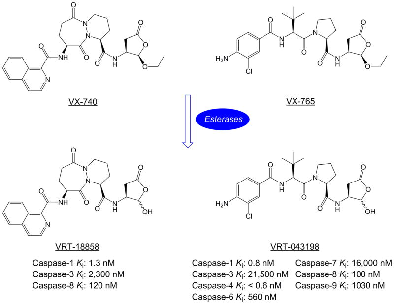
HMR 3480/VRT-18858 and VRT-043198 are selective inhibitors of caspase-1/ICE that cleaves IL-1β precursor (pro-IL-1β) into active mature IL-1β (Figure 3) [83]. In the periphery, VX-740 (pralnacasan) – a pro-drug of HMR 3480/VRT-18858 – can reduce joint damage in two mouse models of osteoarthritis [84], and attenuate dextran sulfate sodium-induced colitis and T helper 1 T-Cell activation in mice [85]. Pro-drug VX-765 can be efficiently converted to VRT-043198 when administered systemically to mice and inhibits LPS-induced IL-1β secretion. VX-765 also reduces disease severity of rheumatoid arthritis in mice and cytokine secretion in a mouse model of skin inflammation [86]. These studies on peripheral inflammation in mice suggest that inhibition of IL-1β synthesis is sufficient to block excessive inflammatory processes. In the CNS, treatment with VX-765 (50 mg/kg, i.p.) decreases chronic stress-induced depressive symptom in mice with reduced serum and hippocampal IL-1β levels [87]. Interestingly, administration of VX-740 (50 μg, i.c.v.) or VX-765 (25-200 mg/kg, i.p.) decreases seizure-induced IL-1β production in the hippocampus, delays SE onset, and reduces seizure duration in rats with intrahippocampal injection of kainate. These beneficial effects are associated with caspase-1 inhibition and blockade of IL-1β biosynthesis in the brain, and recapitulated in mice that lack caspase-1 [88]. In addition, caspase-1 inhibition by VX-765 (200 mg/kg, i.p.) suppresses epileptogenesis in rat hippocampal kindling model by blocking astrocytic IL-1β production [89]. Furthermore, systemic administration of VX-765 shows powerful anticonvulsant activity on acute seizures and in chronic epileptic mice that are refractory to conventional AEDs in a dose-dependent manner [90]. These results taken together strongly support that pharmacological inhibition of caspase-1 by selective small molecules represents a possible therapeutic strategy to suppress acute seizures and the development of epilepsy [13].
Figure 3.
Chemical structures of caspase-1 inhibitors and pro-drugs. VX-740 and VX-765 are rapidly converted to VRT-18858 and VRT-043198, respectively, by plasma and liver esterases in vivo. The Kis of each compound on caspase-1 and other caspases are indicated.
TGF-β Signaling Inhibitors
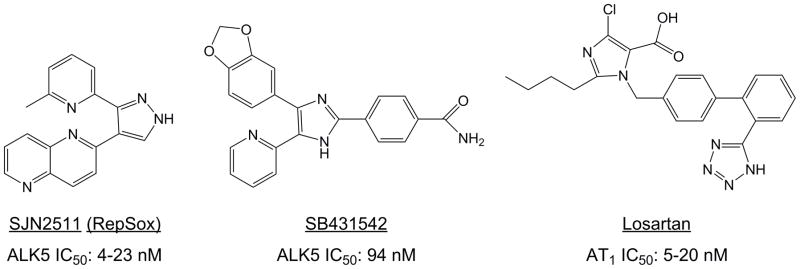
Transforming growth factor-β (TGF-β) is a multifunctional cytokine that mediates essential roles in cell proliferation and differentiation, apoptosis, embryogenesis and inflammatory responses [91]. TGF-β dimers first activate a type II receptor (TβRII), which in turn phosphorylates a type I receptor (TβRI) or activin-like kinase 5 (ALK5). The phosphorylated ALK5 then activates the intracellular SMAD protein complexes and multiple mitogen-activated protein kinase (MAPK) pathways to regulate a wide range of downstream signaling pathways [91, 92]. In addition, ALK5-dependent TGF-β signaling can regulate the late stages of adult hippocampal neurogenesis, suggestive of its involvement in neurogenesis during aging and disease [93]. Recent studies provide evidence that TGF-β signaling is involved in epileptogenesis after brain injury associated with the BBB destruction [94]. Shortly after the BBB function is compromised, albumin begins to enter the brain’s extracellular space [95], and induces activation of TGF-β signaling in astrocytes, causing astrocytic activation, impaired potassium buffering and glutamate metabolism, upregulation of proinflammatory cytokines, and synaptogenesis that eventually result in increased neuronal excitability and spontaneous seizures [96–98]. Moreover, activation of the astrocytic TGF-β/ALK5 pathway by the intracerebroventricular infusion of albumin induces excitatory – but not inhibitory –synaptogenesis prior to the appearance of spontaneous seizures. Interestingly, the albumin-initiated synaptogenesis and seizures can be abolished by SJN2511 (RepSox) (i.c.v.), a potent and selective TGF-β/ALK5 inhibitor (Figure 4) [99]. In addition, albumin-induced TGF-β upregulation and associated SMAD2/3 phosphorylation are blocked by the ALK5 antagonists –SJN2511 or SB431542 (Figure 4) [100]. Furthermore, losartan (Figure 4), a USA FDA-approved small-molecule antagonist for angiotensin II receptor type 1 (AT1) that has been reported to block the peripheral TGF-β signaling, can effectively suppress albumin-induced TGF-β activation in the brain and prevent the subsequent spontaneous seizures [100]. These results reinforce the notion that targeting TGF-β signaling is a feasible strategy for disease modification and prevention of epilepsy.
Figure 4.
Chemical structures of small molecules that suppress TGF-β1/ALK5 signaling. SJN2511 and SB431542 are selective ALK5 inhibitors. Losartan, a selective antagonist for AT1 can downregulate TGF-β/ALK5 signaling in both the periphery and the CNS. Abbreviations: ALK5, activin-like kinase 5; AT1, angiotensin II receptor type 1.
NOX2 Inhibitors
Oxidative stress in the brain is caused by the imbalance between the generation and detoxification of reactive oxygen and nitrogen species (ROS/RNS) that attack brain cells and thus play important roles in brain inflammation, aging and degeneration [101]. Activated NADPH oxidases (NOXs) – a family including NOX1, NOX2, NOX3, NOX4, NOX5, dual oxidase 1 (DUOX1) and DUOX2 – are a primary source of ROS via transporting electrons from intracellular NADPH, across biological membrane, and then to extracellular oxygen, generating superoxide [102, 103]. Among these seven isozymes, NOX2 is the prototype form and plays central roles in neuroinflammation, neurodegeneration and associated functional deficits in neurological conditions such as spinal cord injury [104], Parkinson’s disease [105, 106], Alzheimer’s disease (AD) [107], amyotrophic lateral sclerosis [108], multiple sclerosis [109], and epilepsy [110].
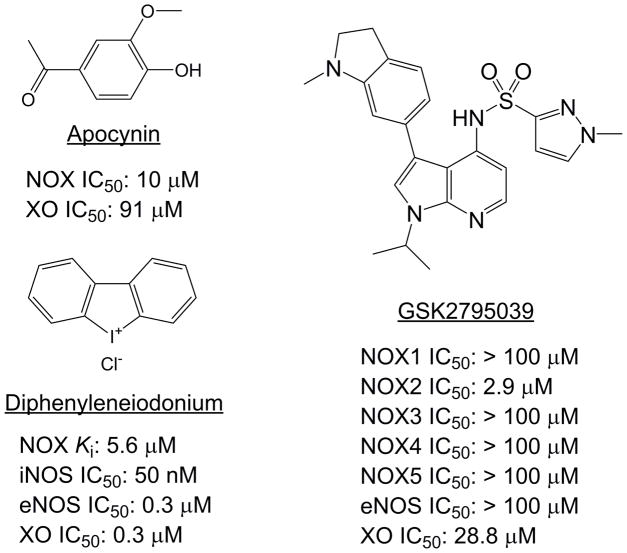
Evidence suggests that seizure-induced neuronal damage in the hippocampus is associated with ROS induction after pilocarpine injection in rats, characterized by a decrease in reduced glutathione level and increases in nitrite content and lipid peroxidation [111]. Likewise, kainate-induced SE in rats results in a time-dependent translocation of NOX subunits (gp91phox, p47phox, p67phox and rac-1) from hippocampal cytosol to membrane where active NOX complex is assembled. Interestingly, the seizure-induced NOX activation coincides with microglial activation in the hippocampus [112]. Furthermore, ROS generated by NOX contributes to neurodegeneration in pilocarpine-treated rats, which is substantially suppressed by apocynin –a selective NOX2 inhibitor (Figure 5) [113]. In another study, apocynin – when systemically administered at two and 24 hours after lithium/pilocarpine-induced SE in rats – reduced seizure-induced ROS production, lipid peroxidation, BBB disruption, neutrophil infiltration, microglial activation and neuronal death [114]. There thus is a growing interest in NOX2 as therapeutic target for seizures and epilepsy. However, apocynin and other historical NOX2 inhibitors including diphenyleneiodonium (DPI) are often challenged for their off-target activities (Figure 5). For instance, apocynin shows marked ROS scavenging activity and inhibition on rho kinases; DPI is a general inhibitor of flavoproteins, e.g., xanthine oxidase and endothelial nitric oxide synthase (eNOS) [115]. Owing to recent advances in chemical biology and HTS, several new classes of small-molecule NOX inhibitors with improved specificity have been identified [115–117]. However, whether these novel NOX2 inhibitors would show any antiepileptic or antiepileptogenic effect in animal models requires investigation.
Figure 5.
Chemical structures of classical NOX inhibitors apocynin and diphenyleneiodonium. Note that both compounds have considerable off-target inhibitions. Recently developed compounds like GSK2795039 with improved selectivity await to be tested in animal models of seizures and epilepsy. Abbreviations: eNOS, endothelial nitric oxide synthase (NOS); iNOS, inducible NOS; XO, xanthine oxidase.
Other Potential Anti-Inflammatory Targets for Small Molecules
Lipoxygenase (LOX)
Similar to prostaglandins, leukotrienes produced by LOX from arachidonic acids are also proinflammatory and increase microvascular permeability, therefore drugs that are able to inhibit both COX and LOX are proposed to treat inflammation-associated conditions due to their dual actions [118]. For instance, natural product flavonoids containing baicalin and catechin show neuroprotection during kainate-induced excitotoxicity via reducing both COX-2 and 5-LOX activities [119]. Likewise, licofelone (Figure 1), a dual COX/LOX small-molecule inhibitor, which recently was approved as an effective treatment for osteoarthritis, shows anticonvulsant effect in a mouse pentylenetetrazol seizure model [120].
Microsomal prostaglandin E synthase-1 (mPGES-1)
PGES is the enzyme that produces PGE2 from PGH2, a precursor molecule directly synthesized by COX, and has three isozymes: mPGES-1 (or PTGES), mPGES-2 (or PTGES2) and cytosolic PGES (cPGES or PTGES3) (Figure 2A). Among these, mPGES-1 is inducible, membrane-bound, and functionally linked to COX-2 in preference to COX-1 [40]. mPGES-1 promotes inflammatory and pain reactions via synthesis of PGE2 in a disease model of human rheumatoid arthritis [121]. In the CNS, mPGES-1 is upregulated in pyramidal neurons of AD human patients and transgenic mice [122–124], and contributes to pathogenesis of AD [123, 124]. In addition, mPGES-1 exacerbates inflammation and demyelination via disrupting the local vessel structure in a model of multiple sclerosis [125]. mPGES-1 is also robustly induced in mice following pilocarpine-induced seizures and aggravates neuronal loss induced by kainate, suggesting that mPGES-1 is involved in seizure-triggered pathogenesis [17, 126, 127]. mPGES-1 also has been shown to exacerbate epileptic seizures and hippocampal gliosis in mouse pentylenetetrazol seizure model [128]. The first clinical trial with the mPGES-1 inhibitor LY3023703 (structure not disclosed) by Eli Lilly and Company (https://clinicaltrials.gov/show/NCT01872910) reveals more potent inhibition of PGE2 synthesis than COX-2 inhibitor celecoxib without decreasing PGI2 levels in the blood [129]. Thus, blockade of mPGES-1 by small molecules should be explored as an anti-inflammatory therapy for epilepsy in the future [128, 130].
Toll-like receptor 4 (TLR4)
Toll-like receptors (TLRs) have significant homology in the cytosolic region to the IL-1R family and, thus shares partly overlapping signaling molecules with IL-1β/IL-1R [131]. Among TLR family members, TLR4 is the LPS sensing receptor and a crucial component of innate immunity. TLR4 can be activated by HMGB1, an endogenous danger signal protein released by immune cells or by neurons and glia in the CNS in response to cell damage or neuronal hyperexcitability. Interestingly, both HMGB1 and TLR4 are upregulated in brain specimens from epilepsy patients and in brain tissues from animals with chronic seizures [3, 132]. HMGB1, particularly the active disulfide form, enhances NMDA receptor function and excitotoxicity, as well as exacerbates kainate-induced seizures by activating TLR4 in hippocampal neurons [76, 78]. On the other hand, HMGB1 also activates the receptor for advanced glycation end products (RAGE), contributing to hyperexcitability and proictogenic effects that are TLR4-independent [77]. Antagonism of HMGB1/TLR4 signaling by pseudo-peptide BOX A or LPS-RS – an inactive LPS from photosynthetic bacterium Rhodobacter sphaeroides – reduces initial and chronic seizures. Moreover, TLR4 knockout mice show resistance to kainate-induced seizures [76]. Intriguingly, TLR4 has been reported to upregulate COX-2 expression via activation of the immune and inflammatory transcription factor – nuclear factor-κB (NF-κB) in several models of inflammation and injury [133–135]. Therefore, the HMGB1/TLR4 signaling has been proposed as antiepileptic and antiepileptogenic target [13, 136]. However, LPS-RS has poor pharmacokinetics, e.g., low brain penetration. Another LPS-mimicking antagonist eritoran recently failed in a multinational Phase III clinical trial for severe sepsis (https://clinicaltrials.gov/show/NCT00334828). As an alternative strategy, small-molecule inhibitors (e.g., TAK-242) that target TLR4 have been developed. However, test of these novel TLR4 antagonists in animal models of acute seizures or epilepsy has not been reported to date.
TNF-α
TNF-α is a crucial proinflammatory cytokine that belongs to the TNF superfamily of ligands, which are involved in the activation, differentiation, proliferation and infiltration of immune cells into the CNS during systemic inflammation [137]. Systemic administration of TNF-α 24 hours after amygdala kindling in rats facilitates the behavioral seizures and increases the epileptiform discharges, while the electrical stimulation in turn significantly enhances TNF-α levels in both blood and the brain, suggesting a mutual facilitative interaction between seizure and proinflammatory cytokines [138]. Intriguingly, it becomes clear that TNF-α plays a dichotomous role in the pathophysiology of seizures and epilepsy – pro-convulsive effect through TNF receptor type 1 (TNFR1 or p55) and anti-convulsive effect via receptor 2 (TNFR2 or p75), depending on engaged cellular context [139–142]. Therefore, inhibition of TNFR1 and/or activation of TNFR2 represents a novel strategy to treat neurological conditions including epilepsy [11, 13]. In fact, a PEGylated TNFR1-selective antagonistic TNF mutant (PEG-R1antTNF) suppresses arterial inflammation [143], whereas a specific TNFR2 agonist – TNC-scTNF(R2) can protect human dopaminergic neurons from oxidative stress-induced cell death [144]. However, small molecules that specifically modulate TNFR1 or TNFR2 with sufficient pharmacodynamics and pharmacokinetics are not available.
IL-6
IL-6 regulates inflammatory and immune responses through its receptor (IL6R or CD126) that is coupled to gp130 (CD130). Like other prototypical proinflammatory cytokines such as IL-1β and TNF-α, IL-6 signaling also can induce COX-2 to synthesize PGE2 via activating NF-κB transcriptional signaling [145–147]. IL-6 induction is widely observed in epilepsy patients and in animal models of experimental epilepsy [3, 148]. Recent evidence in the clinical setting and experimental models suggests active roles of IL-6 in seizure generation and exacerbation [149]. For example, intranasal administration of recombinant IL-6 one hour before systemic injection of pentylenetetrazol exacerbates the severity of seizures in rats [150]. The proconvulsant action of IL-6 is also supported by extensive studies on IL-6 deficient mice [151, 152] and IL-6 overexpressing mice [153], and is presumably associated with a constitutive loss of inhibitory interneuron function [153]. Future efforts should be directed toward developing potent and selective small molecules that target the synthesis of IL-6 or its downstream receptor IL6R, and evaluating their antiepileptic and antiepileptogenic effects in animal models.
Concluding Remarks
Current anti-inflammatory therapies in epilepsy are limited to immunosuppressants such as adrenocorticotropic hormone, immunoglobulin, plasmapheresis, monoclonal antibodies and corticotropic steroid hormones [154, 155]. However these seldom-used treatments are restrictively effective for some specific types of epilepsy, e.g., those associated with severe encephalitis and autoimmune conditions, and their mechanism of action remains largely unknown [3, 154, 155]. Identifying key proinflammatory mediators involved in epilepsy and developing potent and brain-permeable small molecules that specifically modify their inflammatory functions might lead to novel antiepileptic and antiepileptogenic therapies. These efforts brought on a number of studies on pharmacological inhibition of several well-recognized proinflammatory mediators by small molecules in animal models of acute seizures and epilepsy with many on the way. Particularly, the involvement of several prominent proinflammatory mediators – COX-2, PGE2, IL-1β, IL6, TNF-α, TGF-β, NOX2, HMGB1 and TLR4 – in initial seizures and chronic epilepsy has been extensively studied, and their pathogenic roles in the epileptic brain are becoming clear. However, more studies are imperative to determine whether any of small molecules targeting these proinflammatory mediators could successfully evolve into clinical innovations (see Outstanding Questions).
OUTSTANDING QUESTIONS.
Does inflammation in the brain cause epileptic seizures and vice versa?
What are the limitations of current immunosuppressants for epilepsy treatment?
Why do current AEDs merely provide symptomatic relief rather than disease prevention or modification?
What are the feasible anti-inflammatory targets for treatment of seizures and epilepsy?
What are the current status and challenges for moving the experimental discoveries about small molecule-mediated anti-inflammatory therapeutics into a clinical setting?
What are the advantages of small molecules over the biological drugs for anti-inflammatory therapeutics in epilepsy treatment?
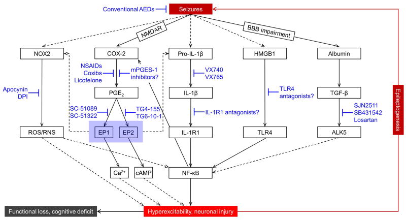
Activation of the COX-2 and prostaglandin cascade induces proinflammatory cytokines including IL-1β, IL-6 and TNF-α, which in turn enhance COX-2 transcription engaging the NF-κB pathway and, thus perpetuate inflammatory reactions in both periphery and brain [145–147, 156]. COX-2 inhibition by small molecules could break this self-reinforcing circle, thereby reducing chronic inflammation and subsequent sequelae (Figure 6). The studies on several COX-2 inhibitors in rodent models of seizures and epilepsy yielded some positive outcomes but discrepancy as well. Some COX-2 inhibitors, if not all, appear to confer anticonvulsant effects and reduce neuronal injury developing after SE when administered properly, although their moderate effects on behavioral and cognitive alterations need to be further characterized. A Phase IV clinical trial had been conducted to evaluate the antipyretic medication with a combination of selective and nonselective COX-2 inhibitors – diclofenac, paracetamol and ibuprofen – for preventing the recurrence of febrile seizures from 1997 to 2005 (https://clinicaltrials.gov/show/NCT00568217). This combined therapy failed to effectively prevent the recurrence of febrile seizures, nor did it reduce the body temperature in patients that had a febrile episode prior to a recurrent seizure [157]. However, the highest recommended doses with the rectal route of administration used in this study might have compromised the therapeutic outcomes because the high doses could increase the drug off-target activities and the rapid effects from rectal administration might impede the early benefits from COX-2 induction.
Figure 6.
Strategies of targeting inflammation to treat seizures and epilepsy. Inflammatory signaling pathways are upregulated by initial brain insults such as prolonged seizures and are proposed to contribute to the development of epilepsy. Only the major signaling pathways are indicated. AEDs alone cannot terminate the vicious circle of chronic inflammatory events that may lead to epileptogenesis. Small-molecule compounds that target the key proinflammatory mediators or their downstream signaling molecules might represent novel adjunctive strategies, along with AEDs and general anesthetics, to treat acute seizures and chronic epilepsy. Similar to IL-1β, cytokines IL-6 and TNF-α also can induce COX-2 via NF-κB and facilitate excitotoxicity; however, small molecules that target these two proinflammatory mediators or their receptors are either not available or have not been tested in animal models for antiepileptic or antiepileptogenic effect. Abbreviations: AEDs, antiepileptic drugs; ALK5, activin-like kinase 5; BBB, blood-brain barrier; cAMP, cyclic AMP; COX, cyclooxygenase; Coxibs, COX-2 inhibitors; DPI, diphenyleneiodonium; HMGB1, high-mobility group box 1 protein; IL-1β, interleukin-1β; IL-1R, IL-1 receptor type 1; IL-6, interleukin-6; mPGES-1, microsomal prostaglandin E synthase-1; NF-κB, nuclear factor κB; NMDAR, N-methyl-D-aspartate (NMDA) receptor; NOX2, NADPH oxidase 2; NSAIDs, nonsteroidal anti-inflammatory drugs; ROS/RNS, reactive oxygen and nitrogen species; TGF-β1, transforming growth factor-β1; TLR4, toll-like receptor 4; TNF-α, tumor necrosis factor-α.
As a canonical marker of neuroinflammation, IL-1β is upregulated by seizures and initiates NF-κB-dependent transcription of many proinflammatory genes including COX-2 and, therefore contributes to subsequent chronic epilepsy. VX-765, a small molecule that selectively blocks IL-1β synthesis, shows promising therapeutic effects in several rodent models of chronic epilepsy. To evaluate its safety, tolerability and proof-of-concept clinical activity, Vertex Pharmaceuticals initiated a randomized, placebo-controlled, double-blind study of VX-765 that involved 60 people with refractory partial onset epilepsy in 2010 (https://clinicaltrials.gov/show/NCT01048255). The results from this Phase II trial indicate a beneficial effect from a four-week treatment with VX-765 that occurred at the end of treatment period and continued during the beginning of the observation period, suggestive of a delayed mechanism of action from this compound [158]. This potential efficacy signal encouraged a longer-duration study to evaluate any more robust or additional clinical activity of VX-765. A larger Phase IIb trial therefore was launched in 2011 to further characterize VX-765 for a longer treatment period (https://clinicaltrials.gov/show/NCT01501383). Unfortunately, the trial was discontinued for administrative reasons. Nonetheless, VX-765 is generally tolerable and safe, and future study is necessitated to determine its efficacy in patients with refractory focal epilepsy [159]. As an alternative approach to the inhibition of IL-1β synthesis, blocking IL-1R1 –the effector for the inflammatory actions of IL-1 – might represent another anti-inflammatory strategy. For instance, a combined therapy with anakinra – a recombinant IL-1RA – and VX-765 shows marked neuroprotection in both rat lithium/pilocarpine and electrical SE models [160]. Likewise, anti-inflammatory drug cocktail containing anakinra, COX-2 inhibitor CAY10404 and caspase-1 expression inhibitor minocycline shows neuroprotective and antiepileptogenic effects in rat pups following lithium/pilocarpine SE [161]. However, current available therapies that directly inhibit IL-1β/IL-1R1 signaling are limited to neutralizing antibodies or peptides, as small-molecule antagonists targeting IL-1R1 complex are not available at present [162].
In sum, accumulating evidence from preclinical and clinical studies suggests that targeting inflammatory signaling pathways represent a complementary approach for the current symptomatic therapies – AEDs – to control initial and recurrent seizures, particularly in epilepsy forms that are refractory to conventional treatments (Figure 6). The therapeutic strategy by resolving the brain inflammation could increase the seizure threshold and reduce the likelihood of spontaneous recurrent seizures, thereby providing disease prevention or modification rather than only symptomatic relief [8, 11, 13]. However, translating the anti-inflammatory strategies to clinical uses is challenging and necessitates extra caution partially due to the complexity of inflammatory networks that are concurrently regulated by an array of components and signals that often reinforce each other. Combined therapies targeting multiple key proinflammatory molecules provide comprehensive suppression of inflammatory networks and thus might lead to more consistent and promising therapeutic outcomes (Figure 6) [119, 120, 160, 161]. Compared to biological drugs, brain-permeable small molecules are more realistic candidate agents for this therapeutic strategy due to their advantages in production, delivery and pharmacokinetics, and represent a future direction of developing novel therapeutics for epilepsy. As these small-molecule compounds continue growing, so too will our current understanding on chronic inflammatory processes in the epileptic brain, which will help to identify common pathophysiological mechanisms in different forms of epilepsy and ultimately lead to clinical innovations.
TRENDS.
Unresolved inflammation in the brain is a common feature in epilepsy, and is primarily characterized by elevated proinflammatory mediators in epileptogenic foci.
Blocking excessive inflammatory processes in the brain increases the seizure threshold and reduces the likelihood of recurrent seizures, thereby providing disease prevention or modification.
Anti-inflammatory therapeutics represent a complementary strategy to the current symptomatic treatments.
Small molecules that specifically target the proinflammatory mediators are realistic candidate agents for anti-inflammatory therapeutics owing to their advantages in production, delivery and pharmacokinetics.
Acknowledgments
We are very grateful to Annamaria Vezzani, Ph.D. and Michael Privitera, M.D. for insightful comments and constructive criticisms on an early draft of the manuscript. This work was supported by National Institutes of Health (NIH)/National Institute of Neurological Disorders and Stroke (NINDS) grant R00NS082379 (J.J.) and NARSAD Young Investigator Grant 20940 (J.J.) from the Brain & Behavior Research Foundation.
Footnotes
Publisher's Disclaimer: This is a PDF file of an unedited manuscript that has been accepted for publication. As a service to our customers we are providing this early version of the manuscript. The manuscript will undergo copyediting, typesetting, and review of the resulting proof before it is published in its final citable form. Please note that during the production process errors may be discovered which could affect the content, and all legal disclaimers that apply to the journal pertain.
References
- 1.Abbas AK, et al. Basic Immunology: Functions and Disorders of the Immune System. Elsevier/Saunders; 2012. Chapter 2 - Innate Immunity; pp. 23–48. [Google Scholar]
- 2.Aoki T, Narumiya S. Prostaglandins and chronic inflammation. Trends in pharmacological sciences. 2012;33:304–311. doi: 10.1016/j.tips.2012.02.004. [DOI] [PubMed] [Google Scholar]
- 3.Vezzani A, et al. The role of inflammation in epilepsy. Nature reviews Neurology. 2011;7:31–40. doi: 10.1038/nrneurol.2010.178. [DOI] [PMC free article] [PubMed] [Google Scholar]
- 4.Aronica E, Crino PB. Inflammation in epilepsy: clinical observations. Epilepsia. 2011;52(Suppl 3):26–32. doi: 10.1111/j.1528-1167.2011.03033.x. [DOI] [PubMed] [Google Scholar]
- 5.Marchi N, et al. Inflammatory pathways of seizure disorders. Trends in neurosciences. 2014;37:55–65. doi: 10.1016/j.tins.2013.11.002. [DOI] [PMC free article] [PubMed] [Google Scholar]
- 6.Wilcox KS, Vezzani A. Does brain inflammation mediate pathological outcomes in epilepsy? Advances in experimental medicine and biology. 2014;813:169–183. doi: 10.1007/978-94-017-8914-1_14. [DOI] [PMC free article] [PubMed] [Google Scholar]
- 7.D'Ambrosio R, et al. Novel frontiers in epilepsy treatments: preventing epileptogenesis by targeting inflammation. Expert review of neurotherapeutics. 2013;13:615–625. doi: 10.1586/ern.13.54. [DOI] [PMC free article] [PubMed] [Google Scholar]
- 8.Loscher W, et al. New avenues for anti-epileptic drug discovery and development. Nat Rev Drug Discov. 2013;12:757–776. doi: 10.1038/nrd4126. [DOI] [PubMed] [Google Scholar]
- 9.Rojas A, et al. Cyclooxygenase-2 in epilepsy. Epilepsia. 2014;55:17–25. doi: 10.1111/epi.12461. [DOI] [PMC free article] [PubMed] [Google Scholar]
- 10.Vezzani A. Anti-inflammatory drugs in epilepsy: does it impact epileptogenesis? Expert opinion on drug safety. 2015;14:583–592. doi: 10.1517/14740338.2015.1010508. [DOI] [PubMed] [Google Scholar]
- 11.Varvel NH, et al. Candidate drug targets for prevention or modification of epilepsy. Annual review of pharmacology and toxicology. 2015;55:229–247. doi: 10.1146/annurev-pharmtox-010814-124607. [DOI] [PMC free article] [PubMed] [Google Scholar]
- 12.Vezzani A, et al. Immunity and inflammation in status epilepticus and its sequelae: possibilities for therapeutic application. Expert review of neurotherapeutics. 2015;15:1081–1092. doi: 10.1586/14737175.2015.1079130. [DOI] [PMC free article] [PubMed] [Google Scholar]
- 13.Iori V, et al. Modulation of neuronal excitability by immune mediators in epilepsy. Curr Opin Pharmacol. 2016;26:118–123. doi: 10.1016/j.coph.2015.11.002. [DOI] [PMC free article] [PubMed] [Google Scholar]
- 14.Samanen J. Chapter 5 - Similarities and differences in the discovery and use of biopharmaceuticals and small-molecule chemotherapeutics. In: Ganellin R, Jefferis SR, editors. Introduction to Biological and Small Molecule Drug Research and Development. Elsevier; 2013. pp. 161–203. [Google Scholar]
- 15.Desjardins P, et al. Induction of astrocytic cyclooxygenase-2 in epileptic patients with hippocampal sclerosis. Neurochemistry international. 2003;42:299–303. doi: 10.1016/s0197-0186(02)00101-8. [DOI] [PubMed] [Google Scholar]
- 16.Serrano GE, et al. Ablation of cyclooxygenase-2 in forebrain neurons is neuroprotective and dampens brain inflammation after status epilepticus. The Journal of neuroscience : the official journal of the Society for Neuroscience. 2011;31:14850–14860. doi: 10.1523/JNEUROSCI.3922-11.2011. [DOI] [PMC free article] [PubMed] [Google Scholar]
- 17.Jiang J, et al. Therapeutic window for cyclooxygenase-2 related anti-inflammatory therapy after status epilepticus. Neurobiol Dis. 2015;76:126–136. doi: 10.1016/j.nbd.2014.12.032. [DOI] [PMC free article] [PubMed] [Google Scholar]
- 18.Kelley KA, et al. Potentiation of excitotoxicity in transgenic mice overexpressing neuronal cyclooxygenase-2. The American journal of pathology. 1999;155:995–1004. doi: 10.1016/S0002-9440(10)65199-1. [DOI] [PMC free article] [PubMed] [Google Scholar]
- 19.Takemiya T, et al. Inducible brain COX-2 facilitates the recurrence of hippocampal seizures in mouse rapid kindling. Prostaglandins & other lipid mediators. 2003;71:205–216. doi: 10.1016/s1098-8823(03)00040-6. [DOI] [PubMed] [Google Scholar]
- 20.Takemiya T, et al. Prostaglandin E2 produced by late induced COX-2 stimulates hippocampal neuron loss after seizure in the CA3 region. Neuroscience research. 2006;56:103–110. doi: 10.1016/j.neures.2006.06.003. [DOI] [PubMed] [Google Scholar]
- 21.Levin JR, et al. Reduction in delayed mortality and subtle improvement in retrograde memory performance in pilocarpine-treated mice with conditional neuronal deletion of cyclooxygenase-2 gene. Epilepsia. 2012;53:1411–1420. doi: 10.1111/j.1528-1167.2012.03584.x. [DOI] [PMC free article] [PubMed] [Google Scholar]
- 22.Baik EJ, et al. Cyclooxygenase-2 selective inhibitors aggravate kainic acid induced seizure and neuronal cell death in the hippocampus. Brain research. 1999;843:118–129. doi: 10.1016/s0006-8993(99)01797-7. [DOI] [PubMed] [Google Scholar]
- 23.Ma L, et al. Aspirin attenuates spontaneous recurrent seizures and inhibits hippocampal neuronal loss, mossy fiber sprouting and aberrant neurogenesis following pilocarpine-induced status epilepticus in rats. Brain research. 2012;1469:103–113. doi: 10.1016/j.brainres.2012.05.058. [DOI] [PubMed] [Google Scholar]
- 24.Jeong KH, et al. Influence of aspirin on pilocarpine-induced epilepsy in mice. The Korean journal of physiology & pharmacology : official journal of the Korean Physiological Society and the Korean Society of Pharmacology. 2013;17:15–21. doi: 10.4196/kjpp.2013.17.1.15. [DOI] [PMC free article] [PubMed] [Google Scholar]
- 25.Fischborn SV, et al. Targeting the prostaglandin E2 EP1 receptor and cyclooxygenase-2 in the amygdala kindling model in mice. Epilepsy Res. 2010;91:57–65. doi: 10.1016/j.eplepsyres.2010.06.012. [DOI] [PubMed] [Google Scholar]
- 26.Gobbo OL, O'Mara SM. Post-treatment, but not pre-treatment, with the selective cyclooxygenase-2 inhibitor celecoxib markedly enhances functional recovery from kainic acid-induced neurodegeneration. Neuroscience. 2004;125:317–327. doi: 10.1016/j.neuroscience.2004.01.045. [DOI] [PubMed] [Google Scholar]
- 27.Jung KH, et al. Cyclooxygenase-2 inhibitor, celecoxib, inhibits the altered hippocampal neurogenesis with attenuation of spontaneous recurrent seizures following pilocarpine-induced status epilepticus. Neurobiol Dis. 2006;23:237–246. doi: 10.1016/j.nbd.2006.02.016. [DOI] [PubMed] [Google Scholar]
- 28.Oliveira MS, et al. Cyclooxygenase-2/PGE2 pathway facilitates pentylenetetrazol-induced seizures. Epilepsy Res. 2008;79:14–21. doi: 10.1016/j.eplepsyres.2007.12.008. [DOI] [PubMed] [Google Scholar]
- 29.Citraro R, et al. Antiepileptogenic effects of the selective COX-2 inhibitor etoricoxib, on the development of spontaneous absence seizures in WAG/Rij rats. Brain research bulletin. 2015;113:1–7. doi: 10.1016/j.brainresbull.2015.02.004. [DOI] [PubMed] [Google Scholar]
- 30.Kunz T, Oliw EH. Nimesulide aggravates kainic acid-induced seizures in the rat. Pharmacology & toxicology. 2001;88:271–276. doi: 10.1034/j.1600-0773.2001.d01-116.x. [DOI] [PubMed] [Google Scholar]
- 31.Trandafir CC, et al. Co-administration of subtherapeutic diazepam enhances neuroprotective effect of COX-2 inhibitor, NS-398, after lithium pilocarpine-induced status epilepticus. Neuroscience. 2015;284:601–610. doi: 10.1016/j.neuroscience.2014.10.021. [DOI] [PMC free article] [PubMed] [Google Scholar]
- 32.Polascheck N, et al. The COX-2 inhibitor parecoxib is neuroprotective but not antiepileptogenic in the pilocarpine model of temporal lobe epilepsy. Experimental neurology. 2010;224:219–233. doi: 10.1016/j.expneurol.2010.03.014. [DOI] [PubMed] [Google Scholar]
- 33.Kunz T, Oliw EH. The selective cyclooxygenase-2 inhibitor rofecoxib reduces kainate-induced cell death in the rat hippocampus. The European journal of neuroscience. 2001;13:569–575. doi: 10.1046/j.1460-9568.2001.01420.x. [DOI] [PubMed] [Google Scholar]
- 34.Akula KK, et al. Rofecoxib, a selective cyclooxygenase-2 (COX-2) inhibitor increases pentylenetetrazol seizure threshold in mice: possible involvement of adenosinergic mechanism. Epilepsy Res. 2008;78:60–70. doi: 10.1016/j.eplepsyres.2007.10.008. [DOI] [PubMed] [Google Scholar]
- 35.Kawaguchi K, et al. Cyclooxygenase-2 expression is induced in rat brain after kainate-induced seizures and promotes neuronal death in CA3 hippocampus. Brain research. 2005;1050:130–137. doi: 10.1016/j.brainres.2005.05.038. [DOI] [PubMed] [Google Scholar]
- 36.Holtman L, et al. Effects of SC58236, a selective COX-2 inhibitor, on epileptogenesis and spontaneous seizures in a rat model for temporal lobe epilepsy. Epilepsy Res. 2009;84:56–66. doi: 10.1016/j.eplepsyres.2008.12.006. [DOI] [PubMed] [Google Scholar]
- 37.Holtman L, et al. Cox-2 inhibition can lead to adverse effects in a rat model for temporal lobe epilepsy. Epilepsy Res. 2010;91:49–56. doi: 10.1016/j.eplepsyres.2010.06.011. [DOI] [PubMed] [Google Scholar]
- 38.Du Y, et al. Defining the therapeutic time window for suppressing the inflammatory prostaglandin E2 signaling after status epilepticus. Expert review of neurotherapeutics. 2016;16:123–130. doi: 10.1586/14737175.2016.1134322. [DOI] [PMC free article] [PubMed] [Google Scholar]
- 39.Funk CD, FitzGerald GA. COX-2 inhibitors and cardiovascular risk. J Cardiovasc Pharmacol. 2007;50:470–479. doi: 10.1097/FJC.0b013e318157f72d. [DOI] [PubMed] [Google Scholar]
- 40.Takemiya T, et al. Roles of prostaglandin synthesis in excitotoxic brain diseases. Neurochemistry international. 2007;51:112–120. doi: 10.1016/j.neuint.2007.05.009. [DOI] [PubMed] [Google Scholar]
- 41.Andreasson K. Emerging roles of PGE2 receptors in models of neurological disease. Prostaglandins & other lipid mediators. 2010;91:104–112. doi: 10.1016/j.prostaglandins.2009.04.003. [DOI] [PMC free article] [PubMed] [Google Scholar]
- 42.Kaushik MK, et al. Prostaglandin D(2) is crucial for seizure suppression and postictal sleep. Experimental neurology. 2014;253:82–90. doi: 10.1016/j.expneurol.2013.12.002. [DOI] [PubMed] [Google Scholar]
- 43.Akarsu ES, et al. Inhibition of pentylenetetrazol-induced seizures in rats by prostaglandin D2. Epilepsy Res. 1998;30:63–68. doi: 10.1016/s0920-1211(97)00092-2. [DOI] [PubMed] [Google Scholar]
- 44.Chung JI, et al. Seizure susceptibility in immature brain due to lack of COX-2-induced PGF2alpha. Experimental neurology. 2013;249:95–103. doi: 10.1016/j.expneurol.2013.08.014. [DOI] [PubMed] [Google Scholar]
- 45.Shimamura M, et al. Prostaglandin E2 type 1 receptors contribute to neuronal apoptosis after transient forebrain ischemia. Journal of cerebral blood flow and metabolism : official journal of the International Society of Cerebral Blood Flow and Metabolism. 2013;33:1207–1214. doi: 10.1038/jcbfm.2013.69. [DOI] [PMC free article] [PubMed] [Google Scholar]
- 46.Zhou P, et al. Neuroprotection by PGE2 receptor EP1 inhibition involves the PTEN/AKT pathway. Neurobiol Dis. 2008;29:543–551. doi: 10.1016/j.nbd.2007.11.010. [DOI] [PMC free article] [PubMed] [Google Scholar]
- 47.Kawano T, et al. Prostaglandin E2 EP1 receptors: downstream effectors of COX-2 neurotoxicity. Nature medicine. 2006;12:225–229. doi: 10.1038/nm1362. [DOI] [PubMed] [Google Scholar]
- 48.Zhen G, et al. PGE2 EP1 receptor exacerbated neurotoxicity in a mouse model of cerebral ischemia and Alzheimer's disease. Neurobiol Aging. 2012;33:2215–2219. doi: 10.1016/j.neurobiolaging.2011.09.017. [DOI] [PMC free article] [PubMed] [Google Scholar]
- 49.Frankowski JC, et al. Detrimental role of the EP1 prostanoid receptor in blood-brain barrier damage following experimental ischemic stroke. Scientific reports. 2015;5:17956. doi: 10.1038/srep17956. [DOI] [PMC free article] [PubMed] [Google Scholar]
- 50.Rojas A, et al. The prostaglandin EP1 receptor potentiates kainate receptor activation via a protein kinase C pathway and exacerbates status epilepticus. Neurobiol Dis. 2014;70:74–89. doi: 10.1016/j.nbd.2014.06.004. [DOI] [PMC free article] [PubMed] [Google Scholar]
- 51.Pekcec A, et al. Targeting prostaglandin E2 EP1 receptors prevents seizure-associated P-glycoprotein up-regulation. The Journal of pharmacology and experimental therapeutics. 2009;330:939–947. doi: 10.1124/jpet.109.152520. [DOI] [PubMed] [Google Scholar]
- 52.Jiang J, Dingledine R. Prostaglandin receptor EP2 in the crosshairs of anti-inflammation, anti-cancer, and neuroprotection. Trends in pharmacological sciences. 2013;34:413–423. doi: 10.1016/j.tips.2013.05.003. [DOI] [PMC free article] [PubMed] [Google Scholar]
- 53.Quan Y, et al. EP2 receptor signaling pathways regulate classical activation of microglia. The Journal of biological chemistry. 2013;288:9293–9302. doi: 10.1074/jbc.M113.455816. [DOI] [PMC free article] [PubMed] [Google Scholar]
- 54.Fu Y, et al. EP2 Receptor Signaling Regulates Microglia Death. Molecular pharmacology. 2015;88:161–170. doi: 10.1124/mol.115.098202. [DOI] [PMC free article] [PubMed] [Google Scholar]
- 55.Jiang J, et al. Prostaglandin receptor EP2 antagonists, derivatives, compositions, and uses related thereto. 20140179750A1 US.
- 56.Jiang J, et al. Small molecule antagonist reveals seizure-induced mediation of neuronal injury by prostaglandin E2 receptor subtype EP2. Proceedings of the National Academy of Sciences of the United States of America. 2012;109:3149–3154. doi: 10.1073/pnas.1120195109. [DOI] [PMC free article] [PubMed] [Google Scholar]
- 57.Jiang J, Dingledine R. Role of prostaglandin receptor EP2 in the regulations of cancer cell proliferation, invasion, and inflammation. The Journal of pharmacology and experimental therapeutics. 2013;344:360–367. doi: 10.1124/jpet.112.200444. [DOI] [PMC free article] [PubMed] [Google Scholar]
- 58.Hagmann WK. The many roles for fluorine in medicinal chemistry. Journal of medicinal chemistry. 2008;51:4359–4369. doi: 10.1021/jm800219f. [DOI] [PubMed] [Google Scholar]
- 59.Jiang J, et al. Inhibition of the prostaglandin receptor EP2 following status epilepticus reduces delayed mortality and brain inflammation. Proceedings of the National Academy of Sciences of the United States of America. 2013;110:3591–3596. doi: 10.1073/pnas.1218498110. [DOI] [PMC free article] [PubMed] [Google Scholar]
- 60.Rojas A, et al. Inhibition of the prostaglandin EP2 receptor is neuroprotective and accelerates functional recovery in a rat model of organophosphorus induced status epilepticus. Neuropharmacology. 2015;93:15–27. doi: 10.1016/j.neuropharm.2015.01.017. [DOI] [PMC free article] [PubMed] [Google Scholar]
- 61.Ganesh T, et al. Discovery and characterization of carbamothioylacrylamides as EP selective antagonists. ACS medicinal chemistry letters. 2013;4:616–621. doi: 10.1021/ml400112h. [DOI] [PMC free article] [PubMed] [Google Scholar]
- 62.Ganesh T, et al. Lead optimization studies of cinnamic amide EP2 antagonists. Journal of medicinal chemistry. 2014;57:4173–4184. doi: 10.1021/jm5000672. [DOI] [PMC free article] [PubMed] [Google Scholar]
- 63.Ganesh T, et al. Development of second generation EP2 antagonists with high selectivity. European journal of medicinal chemistry. 2014;82:521–535. doi: 10.1016/j.ejmech.2014.05.076. [DOI] [PMC free article] [PubMed] [Google Scholar]
- 64.Kim YT, et al. Prostaglandin FP receptor inhibitor reduces ischemic brain damage and neurotoxicity. Neurobiol Dis. 2012;48:58–65. doi: 10.1016/j.nbd.2012.06.003. [DOI] [PMC free article] [PubMed] [Google Scholar]
- 65.Shi J, et al. Inflammatory prostaglandin E2 signaling in a mouse model of Alzheimer disease. Annals of neurology. 2012;72:788–798. doi: 10.1002/ana.23677. [DOI] [PMC free article] [PubMed] [Google Scholar]
- 66.Ni H, et al. EP3, Prostaglandin E2 Receptor Subtype 3, Associated with Neuronal Apoptosis Following Intracerebral Hemorrhage. Cell Mol Neurobiol. 2015:1–10. doi: 10.1007/s10571-015-0287-2. [DOI] [PMC free article] [PubMed] [Google Scholar]
- 67.Ravizza T, Vezzani A. Status epilepticus induces time-dependent neuronal and astrocytic expression of interleukin-1 receptor type I in the rat limbic system. Neuroscience. 2006;137:301–308. doi: 10.1016/j.neuroscience.2005.07.063. [DOI] [PubMed] [Google Scholar]
- 68.Ravizza T, et al. Innate and adaptive immunity during epileptogenesis and spontaneous seizures: evidence from experimental models and human temporal lobe epilepsy. Neurobiol Dis. 2008;29:142–160. doi: 10.1016/j.nbd.2007.08.012. [DOI] [PubMed] [Google Scholar]
- 69.Boer K, et al. Inflammatory processes in cortical tubers and subependymal giant cell tumors of tuberous sclerosis complex. Epilepsy Res. 2008;78:7–21. doi: 10.1016/j.eplepsyres.2007.10.002. [DOI] [PubMed] [Google Scholar]
- 70.Henshall DC, et al. Alterations in bcl-2 and caspase gene family protein expression in human temporal lobe epilepsy. Neurology. 2000;55:250–257. doi: 10.1212/wnl.55.2.250. [DOI] [PubMed] [Google Scholar]
- 71.Hu S, et al. Cytokine effects on glutamate uptake by human astrocytes. Neuroimmunomodulation. 2000;7:153–159. doi: 10.1159/000026433. [DOI] [PubMed] [Google Scholar]
- 72.Viviani B, et al. Interleukin-1beta enhances NMDA receptor-mediated intracellular calcium increase through activation of the Src family of kinases. The Journal of neuroscience : the official journal of the Society for Neuroscience. 2003;23:8692–8700. doi: 10.1523/JNEUROSCI.23-25-08692.2003. [DOI] [PMC free article] [PubMed] [Google Scholar]
- 73.Roseti C, et al. GABAA currents are decreased by IL-1beta in epileptogenic tissue of patients with temporal lobe epilepsy: implications for ictogenesis. Neurobiol Dis. 2015;82:311–320. doi: 10.1016/j.nbd.2015.07.003. [DOI] [PubMed] [Google Scholar]
- 74.Vezzani A, Viviani B. Neuromodulatory properties of inflammatory cytokines and their impact on neuronal excitability. Neuropharmacology. 2015;96:70–82. doi: 10.1016/j.neuropharm.2014.10.027. [DOI] [PubMed] [Google Scholar]
- 75.Lamkanfi M, et al. Inflammasome-dependent release of the alarmin HMGB1 in endotoxemia. Journal of immunology. 2010;185:4385–4392. doi: 10.4049/jimmunol.1000803. [DOI] [PMC free article] [PubMed] [Google Scholar]
- 76.Maroso M, et al. Toll-like receptor 4 and high-mobility group box-1 are involved in ictogenesis and can be targeted to reduce seizures. Nature medicine. 2010;16:413–419. doi: 10.1038/nm.2127. [DOI] [PubMed] [Google Scholar]
- 77.Iori V, et al. Receptor for Advanced Glycation Endproducts is upregulated in temporal lobe epilepsy and contributes to experimental seizures. Neurobiol Dis. 2013;58:102–114. doi: 10.1016/j.nbd.2013.03.006. [DOI] [PubMed] [Google Scholar]
- 78.Balosso S, et al. Disulfide-containing high mobility group box-1 promotes N-methyl-D-aspartate receptor function and excitotoxicity by activating Toll-like receptor 4-dependent signaling in hippocampal neurons. Antioxid Redox Signal. 2014;21:1726–1740. doi: 10.1089/ars.2013.5349. [DOI] [PubMed] [Google Scholar]
- 79.Vezzani A, et al. Powerful anticonvulsant action of IL-1 receptor antagonist on intracerebral injection and astrocytic overexpression in mice. Proceedings of the National Academy of Sciences of the United States of America. 2000;97:11534–11539. doi: 10.1073/pnas.190206797. [DOI] [PMC free article] [PubMed] [Google Scholar]
- 80.Vezzani A, et al. Functional role of inflammatory cytokines and antiinflammatory molecules in seizures and epileptogenesis. Epilepsia. 2002;43(Suppl 5):30–35. doi: 10.1046/j.1528-1157.43.s.5.14.x. [DOI] [PubMed] [Google Scholar]
- 81.Marchi N, et al. Antagonism of peripheral inflammation reduces the severity of status epilepticus. Neurobiol Dis. 2009;33:171–181. doi: 10.1016/j.nbd.2008.10.002. [DOI] [PMC free article] [PubMed] [Google Scholar]
- 82.Auvin S, et al. Inflammation induced by LPS enhances epileptogenesis in immature rat and may be partially reversed by IL1RA. Epilepsia. 2010;51(Suppl 3):34–38. doi: 10.1111/j.1528-1167.2010.02606.x. [DOI] [PMC free article] [PubMed] [Google Scholar]
- 83.Braddock M, Quinn A. Targeting IL-1 in inflammatory disease: new opportunities for therapeutic intervention. Nat Rev Drug Discov. 2004;3:330–339. doi: 10.1038/nrd1342. [DOI] [PubMed] [Google Scholar]
- 84.Rudolphi K, et al. Pralnacasan, an inhibitor of interleukin-1beta converting enzyme, reduces joint damage in two murine models of osteoarthritis. Osteoarthritis and cartilage /OARS, Osteoarthritis Research Society. 2003;11:738–746. doi: 10.1016/s1063-4584(03)00153-5. [DOI] [PubMed] [Google Scholar]
- 85.Loher F, et al. The interleukin-1 beta-converting enzyme inhibitor pralnacasan reduces dextran sulfate sodium-induced murine colitis and T helper 1 T-cell activation. The Journal of pharmacology and experimental therapeutics. 2004;308:583–590. doi: 10.1124/jpet.103.057059. [DOI] [PubMed] [Google Scholar]
- 86.Wannamaker W, et al. (S)-1-((S)-2-{[1-(4-amino-3-chloro-phenyl)-methanoyl]-amino}-3,3-dimethyl-butanoy l)-pyrrolidine-2-carboxylic acid ((2R,3S)-2-ethoxy-5-oxo-tetrahydro-furan-3-yl)-amide (VX-765), an orally available selective interleukin (IL)-converting enzyme/caspase-1 inhibitor, exhibits potent anti-inflammatory activities by inhibiting the release of IL-1beta and IL-18. The Journal of pharmacology and experimental therapeutics. 2007;321:509–516. doi: 10.1124/jpet.106.111344. [DOI] [PubMed] [Google Scholar]
- 87.Zhang Y, et al. NLRP3 Inflammasome Mediates Chronic Mild Stress-Induced Depression in Mice via Neuroinflammation. Int J Neuropsychopharmacol. 2015:18. doi: 10.1093/ijnp/pyv006. [DOI] [PMC free article] [PubMed] [Google Scholar]
- 88.Ravizza T, et al. Inactivation of caspase-1 in rodent brain: a novel anticonvulsive strategy. Epilepsia. 2006;47:1160–1168. doi: 10.1111/j.1528-1167.2006.00590.x. [DOI] [PubMed] [Google Scholar]
- 89.Ravizza T, et al. Interleukin Converting Enzyme inhibition impairs kindling epileptogenesis in rats by blocking astrocytic IL-1beta production. Neurobiol Dis. 2008;31:327–333. doi: 10.1016/j.nbd.2008.05.007. [DOI] [PubMed] [Google Scholar]
- 90.Maroso M, et al. Interleukin-1beta biosynthesis inhibition reduces acute seizures and drug resistant chronic epileptic activity in mice. Neurotherapeutics: the journal of the American Society for Experimental NeuroTherapeutics. 2011;8:304–315. doi: 10.1007/s13311-011-0039-z. [DOI] [PMC free article] [PubMed] [Google Scholar]
- 91.Yingling JM, et al. Development of TGF-beta signalling inhibitors for cancer therapy. Nat Rev Drug Discov. 2004;3:1011–1022. doi: 10.1038/nrd1580. [DOI] [PubMed] [Google Scholar]
- 92.Yu L, et al. TGF-beta receptor-activated p38 MAP kinase mediates Smad-independent TGF-beta responses. The EMBO journal. 2002;21:3749–3759. doi: 10.1093/emboj/cdf366. [DOI] [PMC free article] [PubMed] [Google Scholar]
- 93.He Y, et al. ALK5-dependent TGF-beta signaling is a major determinant of late-stage adult neurogenesis. Nature neuroscience. 2014;17:943–952. doi: 10.1038/nn.3732. [DOI] [PMC free article] [PubMed] [Google Scholar]
- 94.Cacheaux LP, et al. Transcriptome profiling reveals TGF-beta signaling involvement in epileptogenesis. The Journal of neuroscience : the official journal of the Society for Neuroscience. 2009;29:8927–8935. doi: 10.1523/JNEUROSCI.0430-09.2009. [DOI] [PMC free article] [PubMed] [Google Scholar]
- 95.van Vliet EA, et al. Blood-brain barrier leakage may lead to progression of temporal lobe epilepsy. Brain. 2007;130:521–534. doi: 10.1093/brain/awl318. [DOI] [PubMed] [Google Scholar]
- 96.Ivens S, et al. TGF-beta receptor-mediated albumin uptake into astrocytes is involved in neocortical epileptogenesis. Brain. 2007;130:535–547. doi: 10.1093/brain/awl317. [DOI] [PubMed] [Google Scholar]
- 97.Friedman A, et al. Blood-brain barrier breakdown-inducing astrocytic transformation: novel targets for the prevention of epilepsy. Epilepsy Res. 2009;85:142–149. doi: 10.1016/j.eplepsyres.2009.03.005. [DOI] [PMC free article] [PubMed] [Google Scholar]
- 98.Heinemann U, et al. Blood-brain barrier dysfunction, TGFbeta signaling, and astrocyte dysfunction in epilepsy. Glia. 2012;60:1251–1257. doi: 10.1002/glia.22311. [DOI] [PMC free article] [PubMed] [Google Scholar]
- 99.Weissberg I, et al. Albumin induces excitatory synaptogenesis through astrocytic TGF-beta/ALK5 signaling in a model of acquired epilepsy following blood-brain barrier dysfunction. Neurobiol Dis. 2015;78:115–125. doi: 10.1016/j.nbd.2015.02.029. [DOI] [PMC free article] [PubMed] [Google Scholar]
- 100.Bar-Klein G, et al. Losartan prevents acquired epilepsy via TGF-beta signaling suppression. Annals of neurology. 2014;75:864–875. doi: 10.1002/ana.24147. [DOI] [PMC free article] [PubMed] [Google Scholar]
- 101.Uttara B, et al. Oxidative stress and neurodegenerative diseases: a review of upstream and downstream antioxidant therapeutic options. Curr Neuropharmacol. 2009;7:65–74. doi: 10.2174/157015909787602823. [DOI] [PMC free article] [PubMed] [Google Scholar]
- 102.Panday A, et al. NADPH oxidases: an overview from structure to innate immunity-associated pathologies. Cell Mol Immunol. 2015;12:5–23. doi: 10.1038/cmi.2014.89. [DOI] [PMC free article] [PubMed] [Google Scholar]
- 103.Bedard K, Krause KH. The NOX family of ROS-generating NADPH oxidases: physiology and pathophysiology. Physiol Rev. 2007;87:245–313. doi: 10.1152/physrev.00044.2005. [DOI] [PubMed] [Google Scholar]
- 104.Khayrullina G, et al. Inhibition of NOX2 reduces locomotor impairment, inflammation, and oxidative stress after spinal cord injury. J Neuroinflammation. 2015;12:172. doi: 10.1186/s12974-015-0391-8. [DOI] [PMC free article] [PubMed] [Google Scholar]
- 105.Wang Q, et al. Post-treatment with an ultra-low dose of NADPH oxidase inhibitor diphenyleneiodonium attenuates disease progression in multiple Parkinson's disease models. Brain. 2015;138:1247–1262. doi: 10.1093/brain/awv034. [DOI] [PMC free article] [PubMed] [Google Scholar]
- 106.Chen SH, et al. Critical role of the Mac1/NOX2 pathway in mediating reactive microgliosis-generated chronic neuroinflammation and progressive neurodegeneration. Curr Opin Pharmacol. 2016;26:54–60. doi: 10.1016/j.coph.2015.10.001. [DOI] [PMC free article] [PubMed] [Google Scholar]
- 107.Wilkinson BL, et al. Ibuprofen attenuates oxidative damage through NOX2 inhibition in Alzheimer's disease. Neurobiol Aging. 2012;33:197 e121–132. doi: 10.1016/j.neurobiolaging.2010.06.014. [DOI] [PMC free article] [PubMed] [Google Scholar]
- 108.Apolloni S, et al. The NADPH oxidase pathway is dysregulated by the P2X7 receptor in the SOD1-G93A microglia model of amyotrophic lateral sclerosis. Journal of immunology. 2013;190:5187–5195. doi: 10.4049/jimmunol.1203262. [DOI] [PubMed] [Google Scholar]
- 109.Fischer MT, et al. NADPH oxidase expression in active multiple sclerosis lesions in relation to oxidative tissue damage and mitochondrial injury. Brain. 2012;135:886–899. doi: 10.1093/brain/aws012. [DOI] [PMC free article] [PubMed] [Google Scholar]
- 110.Puttachary S, et al. Seizure-induced oxidative stress in temporal lobe epilepsy. Biomed Res Int. 2015;2015:745613. doi: 10.1155/2015/745613. [DOI] [PMC free article] [PubMed] [Google Scholar]
- 111.Freitas RM, et al. Oxidative stress in the hippocampus after pilocarpine-induced status epilepticus in Wistar rats. FEBS J. 2005;272:1307–1312. doi: 10.1111/j.1742-4658.2004.04537.x. [DOI] [PubMed] [Google Scholar]
- 112.Patel M, et al. Activation of NADPH oxidase and extracellular superoxide production in seizure-induced hippocampal damage. Journal of neurochemistry. 2005;92:123–131. doi: 10.1111/j.1471-4159.2004.02838.x. [DOI] [PubMed] [Google Scholar]
- 113.Pestana RR, et al. Reactive oxygen species generated by NADPH oxidase are involved in neurodegeneration in the pilocarpine model of temporal lobe epilepsy. Neuroscience letters. 2010;484:187–191. doi: 10.1016/j.neulet.2010.08.049. [DOI] [PubMed] [Google Scholar]
- 114.Kim JH, et al. Post-treatment of an NADPH oxidase inhibitor prevents seizure-induced neuronal death. Brain research. 2013;1499:163–172. doi: 10.1016/j.brainres.2013.01.007. [DOI] [PubMed] [Google Scholar]
- 115.Altenhofer S, et al. Evolution of NADPH Oxidase Inhibitors: Selectivity and Mechanisms for Target Engagement. Antioxid Redox Signal. 2015;23:406–427. doi: 10.1089/ars.2013.5814. [DOI] [PMC free article] [PubMed] [Google Scholar]
- 116.Maraldi T. Natural compounds as modulators of NADPH oxidases. Oxidative medicine and cellular longevity. 2013;2013:271602. doi: 10.1155/2013/271602. [DOI] [PMC free article] [PubMed] [Google Scholar]
- 117.Hirano K, et al. Discovery of GSK2795039, a Novel Small Molecule NADPH Oxidase 2 Inhibitor. Antioxid Redox Signal. 2015;23:358–374. doi: 10.1089/ars.2014.6202. [DOI] [PMC free article] [PubMed] [Google Scholar]
- 118.Leone S, et al. Dual acting anti-inflammatory drugs. Current topics in medicinal chemistry. 2007;7:265–275. doi: 10.2174/156802607779941341. [DOI] [PubMed] [Google Scholar]
- 119.Minutoli L, et al. A dual inhibitor of cyclooxygenase and 5-lipoxygenase protects against kainic acid-induced brain injury. Neuromolecular medicine. 2015;17:192–201. doi: 10.1007/s12017-015-8351-0. [DOI] [PubMed] [Google Scholar]
- 120.Payandemehr B, et al. A COX/5-LOX Inhibitor Licofelone Revealed Anticonvulsant Properties Through iNOS Diminution in Mice. Neurochem Res. 2015;40:1819–1828. doi: 10.1007/s11064-015-1669-z. [DOI] [PubMed] [Google Scholar]
- 121.Trebino CE, et al. Impaired inflammatory and pain responses in mice lacking an inducible prostaglandin E synthase. Proceedings of the National Academy of Sciences of the United States of America. 2003;100:9044–9049. doi: 10.1073/pnas.1332766100. [DOI] [PMC free article] [PubMed] [Google Scholar]
- 122.Chaudhry UA, et al. Elevated microsomal prostaglandin-E synthase-1 in Alzheimer's disease. Alzheimer's & dementia : the journal of the Alzheimer's Association. 2008;4:6–13. doi: 10.1016/j.jalz.2007.10.015. [DOI] [PMC free article] [PubMed] [Google Scholar]
- 123.Akitake Y, et al. Microsomal prostaglandin E synthase-1 is induced in alzheimer's disease and its deletion mitigates alzheimer's disease-like pathology in a mouse model. Journal of neuroscience research. 2013;91:909–919. doi: 10.1002/jnr.23217. [DOI] [PubMed] [Google Scholar]
- 124.Koeberle A, Werz O. Perspective of microsomal prostaglandin E2 synthase-1 as drug target in inflammation-related disorders. Biochemical pharmacology. 2015;98:1–15. doi: 10.1016/j.bcp.2015.06.022. [DOI] [PubMed] [Google Scholar]
- 125.Takeuchi C, et al. Microsomal prostaglandin E synthase-1 aggravates inflammation and demyelination in a mouse model of multiple sclerosis. Neurochemistry international. 2013;62:271–280. doi: 10.1016/j.neuint.2012.12.007. [DOI] [PubMed] [Google Scholar]
- 126.Turrin NP, Rivest S. Innate immune reaction in response to seizures: implications for the neuropathology associated with epilepsy. Neurobiol Dis. 2004;16:321–334. doi: 10.1016/j.nbd.2004.03.010. [DOI] [PubMed] [Google Scholar]
- 127.Takemiya T, et al. Endothelial microsomal prostaglandin E synthase-1 exacerbates neuronal loss induced by kainate. Journal of neuroscience research. 2010;88:381–390. doi: 10.1002/jnr.22195. [DOI] [PubMed] [Google Scholar]
- 128.Shimada T, et al. Role of inflammatory mediators in the pathogenesis of epilepsy. Mediators of inflammation. 2014;2014:901902. doi: 10.1155/2014/901902. [DOI] [PMC free article] [PubMed] [Google Scholar]
- 129.Jin Y, et al. Pharmacodynamic comparison of LY3023703, a novel microsomal prostaglandin e synthase 1 inhibitor, with celecoxib. Clinical pharmacology and therapeutics. 2016;99:274–284. doi: 10.1002/cpt.260. [DOI] [PubMed] [Google Scholar]
- 130.Koeberle A, et al. Design and Development of Microsomal Prostaglandin E Synthase-1 Inhibitors: Challenges and Future Directions. Journal of medicinal chemistry. 2016 doi: 10.1021/acs.jmedchem.5b01750. [DOI] [PubMed] [Google Scholar]
- 131.Bowie A, O'Neill LA. The interleukin-1 receptor/Toll-like receptor superfamily: signal generators for pro-inflammatory interleukins and microbial products. Journal of leukocyte biology. 2000;67:508–514. doi: 10.1002/jlb.67.4.508. [DOI] [PubMed] [Google Scholar]
- 132.Walker A, et al. Proteomic profiling of epileptogenesis in a rat model: Focus on inflammation. Brain, behavior, and immunity. 2015 doi: 10.1016/j.bbi.2015.12.007. [DOI] [PubMed] [Google Scholar]
- 133.Moses T, et al. TLR4-mediated Cox-2 expression increases intestinal ischemia/reperfusion-induced damage. Journal of leukocyte biology. 2009;86:971–980. doi: 10.1189/jlb.0708396. [DOI] [PMC free article] [PubMed] [Google Scholar]
- 134.Fukata M, et al. Cox-2 is regulated by Toll-like receptor-4 (TLR4) signaling: Role in proliferation and apoptosis in the intestine. Gastroenterology. 2006;131:862–877. doi: 10.1053/j.gastro.2006.06.017. [DOI] [PMC free article] [PubMed] [Google Scholar]
- 135.Kuper C, et al. Toll-like receptor 4 activates NF-kappaB and MAP kinase pathways to regulate expression of proinflammatory COX-2 in renal medullary collecting duct cells. American journal of physiology Renal physiology. 2012;302:F38–46. doi: 10.1152/ajprenal.00590.2010. [DOI] [PubMed] [Google Scholar]
- 136.Vezzani A, et al. IL-1 receptor/Toll-like receptor signaling in infection, inflammation, stress and neurodegeneration couples hyperexcitability and seizures. Brain, behavior, and immunity. 2011;25:1281–1289. doi: 10.1016/j.bbi.2011.03.018. [DOI] [PubMed] [Google Scholar]
- 137.Sonar S, Lal G. Role of Tumor Necrosis Factor Superfamily in Neuroinflammation and Autoimmunity. Frontiers in immunology. 2015;6:364. doi: 10.3389/fimmu.2015.00364. [DOI] [PMC free article] [PubMed] [Google Scholar]
- 138.Shandra AA, et al. The role of TNF-alpha in amygdala kindled rats. Neuroscience research. 2002;42:147–153. doi: 10.1016/s0168-0102(01)00309-1. [DOI] [PubMed] [Google Scholar]
- 139.Balosso S, et al. Tumor necrosis factor-alpha inhibits seizures in mice via p75 receptors. Annals of neurology. 2005;57:804–812. doi: 10.1002/ana.20480. [DOI] [PubMed] [Google Scholar]
- 140.Weinberg MS, et al. Opposing actions of hippocampus TNFalpha receptors on limbic seizure susceptibility. Experimental neurology. 2013;247:429–437. doi: 10.1016/j.expneurol.2013.01.011. [DOI] [PMC free article] [PubMed] [Google Scholar]
- 141.Balosso S, et al. Molecular and functional interactions between tumor necrosis factor-alpha receptors and the glutamatergic system in the mouse hippocampus: implications for seizure susceptibility. Neuroscience. 2009;161:293–300. doi: 10.1016/j.neuroscience.2009.03.005. [DOI] [PubMed] [Google Scholar]
- 142.Balosso S, et al. The dual role of TNF-alpha and its receptors in seizures. Experimental neurology. 2013;247:267–271. doi: 10.1016/j.expneurol.2013.05.010. [DOI] [PubMed] [Google Scholar]
- 143.Kitagaki M, et al. Novel TNF-alpha receptor 1 antagonist treatment attenuates arterial inflammation and intimal hyperplasia in mice. Journal of atherosclerosis and thrombosis. 2012;19:36–46. doi: 10.5551/jat.9746. [DOI] [PubMed] [Google Scholar]
- 144.Fischer R, et al. A TNF receptor 2 selective agonist rescues human neurons from oxidative stress-induced cell death. PloS one. 2011;6:e27621. doi: 10.1371/journal.pone.0027621. [DOI] [PMC free article] [PubMed] [Google Scholar]
- 145.Nakao S, et al. Tumor necrosis factor alpha (TNF-alpha)-induced prostaglandin E2 release is mediated by the activation of cyclooxygenase-2 (COX-2) transcription via NFkappaB in human gingival fibroblasts. Molecular and cellular biochemistry. 2002;238:11–18. doi: 10.1023/a:1019927616000. [DOI] [PubMed] [Google Scholar]
- 146.Medeiros R, et al. The role of TNF-alpha signaling pathway on COX-2 upregulation and cognitive decline induced by beta-amyloid peptide. Behavioural brain research. 2010;209:165–173. doi: 10.1016/j.bbr.2010.01.040. [DOI] [PubMed] [Google Scholar]
- 147.Chikuma T, et al. Interleukin-6 induces prostaglandin E(2) synthesis in mouse astrocytes. Journal of molecular neuroscience : MN. 2009;39:175–184. doi: 10.1007/s12031-009-9187-6. [DOI] [PubMed] [Google Scholar]
- 148.Li G, et al. Cytokines and epilepsy. Seizure. 2011;20:249–256. doi: 10.1016/j.seizure.2010.12.005. [DOI] [PubMed] [Google Scholar]
- 149.Vezzani A, et al. The role of cytokines in the pathophysiology of epilepsy. Brain, behavior, and immunity. 2008;22:797–803. doi: 10.1016/j.bbi.2008.03.009. [DOI] [PubMed] [Google Scholar]
- 150.Kalueff AV, et al. Intranasal administration of human IL-6 increases the severity of chemically induced seizures in rats. Neuroscience letters. 2004;365:106–110. doi: 10.1016/j.neulet.2004.04.061. [DOI] [PubMed] [Google Scholar]
- 151.De Luca G, et al. Susceptibility to audiogenic seizure and neurotransmitter amino acid levels in different brain areas of IL-6-deficient mice. Pharmacology, biochemistry, and behavior. 2004;78:75–81. doi: 10.1016/j.pbb.2004.02.004. [DOI] [PubMed] [Google Scholar]
- 152.Penkowa M, et al. Interleukin-6 deficiency reduces the brain inflammatory response and increases oxidative stress and neurodegeneration after kainic acid-induced seizures. Neuroscience. 2001;102:805–818. doi: 10.1016/s0306-4522(00)00515-7. [DOI] [PubMed] [Google Scholar]
- 153.Samland H, et al. Profound increase in sensitivity to glutamatergic- but not cholinergic agonist-induced seizures in transgenic mice with astrocyte production of IL-6. Journal of neuroscience research. 2003;73:176–187. doi: 10.1002/jnr.10635. [DOI] [PubMed] [Google Scholar]
- 154.Quek AL, et al. Autoimmune epilepsy: Clinical characteristics and response to immunotherapy. Archives of neurology. 2012;69:582–593. doi: 10.1001/archneurol.2011.2985. [DOI] [PMC free article] [PubMed] [Google Scholar]
- 155.Vincent A, et al. The growing recognition of immunotherapy-responsive seizure disorders with autoantibodies to specific neuronal proteins. Current opinion in neurology. 2010;23:144–150. doi: 10.1097/WCO.0b013e32833735fe. [DOI] [PubMed] [Google Scholar]
- 156.Laflamme N, et al. An essential role of interleukin-1beta in mediating NF-kappaB activity and COX-2 transcription in cells of the blood-brain barrier in response to a systemic and localized inflammation but not during endotoxemia. The Journal of neuroscience : the official journal of the Society for Neuroscience. 1999;19:10923–10930. doi: 10.1523/JNEUROSCI.19-24-10923.1999. [DOI] [PMC free article] [PubMed] [Google Scholar]
- 157.Strengell T, et al. Antipyretic agents for preventing recurrences of febrile seizures: randomized controlled trial. Archives of Pediatrics & Adolescent Medicine. 2009;163:799–804. doi: 10.1001/archpediatrics.2009.137. [DOI] [PubMed] [Google Scholar]
- 158.Bialer M, et al. Progress report on new antiepileptic drugs: a summary of the Eleventh Eilat Conference (EILAT XI) Epilepsy Res. 2013;103:2–30. doi: 10.1016/j.eplepsyres.2012.10.001. [DOI] [PubMed] [Google Scholar]
- 159.French J, et al. AES 2013 Annual Meeting - Online Abstract Supplement. Epilepsy Currents. 2014;14:1–475. [Google Scholar]
- 160.Noe FM, et al. Pharmacological blockade of IL-1beta/IL-1 receptor type 1 axis during epileptogenesis provides neuroprotection in two rat models of temporal lobe epilepsy. Neurobiol Dis. 2013;59:183–193. doi: 10.1016/j.nbd.2013.07.015. [DOI] [PubMed] [Google Scholar]
- 161.Kwon YS, et al. Neuroprotective and antiepileptogenic effects of combination of anti-inflammatory drugs in the immature brain. J Neuroinflammation. 2013;10:30. doi: 10.1186/1742-2094-10-30. [DOI] [PMC free article] [PubMed] [Google Scholar]
- 162.Yang CY. Identification of potential small molecule allosteric modulator sites on IL-1R1 ectodomain using accelerated conformational sampling method. PloS one. 2015;10:e0118671. doi: 10.1371/journal.pone.0118671. [DOI] [PMC free article] [PubMed] [Google Scholar]
- 163.Janigro D. Are you in or out? Leukocyte, ion, and neurotransmitter permeability across the epileptic blood-brain barrier. Epilepsia. 2012;53(Suppl 1):26–34. doi: 10.1111/j.1528-1167.2012.03472.x. [DOI] [PMC free article] [PubMed] [Google Scholar]
- 164.Marchi N, et al. Seizure-promoting effect of blood-brain barrier disruption. Epilepsia. 2007;48:732–742. doi: 10.1111/j.1528-1167.2007.00988.x. [DOI] [PMC free article] [PubMed] [Google Scholar]
- 165.Zattoni M, et al. Brain infiltration of leukocytes contributes to the pathophysiology of temporal lobe epilepsy. The Journal of neuroscience : the official journal of the Society for Neuroscience. 2011;31:4037–4050. doi: 10.1523/JNEUROSCI.6210-10.2011. [DOI] [PMC free article] [PubMed] [Google Scholar]
- 166.Gorter JA, et al. Status epilepticus, blood-brain barrier disruption, inflammation, and epileptogenesis. Epilepsy & behavior : E&B. 2015;49:13–16. doi: 10.1016/j.yebeh.2015.04.047. [DOI] [PubMed] [Google Scholar]
- 167.Levite M. Autoimmune epilepsy. Nature immunology. 2002;3:500. doi: 10.1038/ni0602-500. [DOI] [PubMed] [Google Scholar]
- 168.Bien CG, et al. Limbic encephalitis as a precipitating event in adult-onset temporal lobe epilepsy. Neurology. 2007;69:1236–1244. doi: 10.1212/01.wnl.0000276946.08412.ef. [DOI] [PubMed] [Google Scholar]
- 169.Vezzani A, et al. Infections, inflammation and epilepsy. Acta neuropathologica. 2016;131:211–234. doi: 10.1007/s00401-015-1481-5. [DOI] [PMC free article] [PubMed] [Google Scholar]
- 170.Dube C, et al. Interleukin-1beta contributes to the generation of experimental febrile seizures. Annals of neurology. 2005;57:152–155. doi: 10.1002/ana.20358. [DOI] [PMC free article] [PubMed] [Google Scholar]
- 171.Kovacs Z, et al. Facilitation of spike-wave discharge activity by lipopolysaccharides in Wistar Albino Glaxo/Rijswijk rats. Neuroscience. 2006;140:731–742. doi: 10.1016/j.neuroscience.2006.02.023. [DOI] [PubMed] [Google Scholar]
- 172.Auvin S, et al. Inflammation enhances epileptogenesis in the developing rat brain. Neurobiol Dis. 2010;40:303–310. doi: 10.1016/j.nbd.2010.06.004. [DOI] [PMC free article] [PubMed] [Google Scholar]


