Abstract
Embryonic germ cell migration is a vital component of the germline lifecycle. The translocation of germ cells from the place of origin to the developing somatic gonad involves several processes including passive movements with underlying tissues, transepithelial migration, cell adhesion dynamics, the establishment of environmental guidance cues and the ability to sustain directed migration. How germ cells accomplish these feats in established model organisms will be discussed in this review, with a focus on recent discoveries and themes conserved across species.
Introduction
Embryonic development involves the complex and coordinated movement of many cell types. In many metazoans, germ cells are specified at one location in the embryo and must translocate across a large distance to find the developing somatic gonad. This translocation often involves more than one process, including moving passively with underlying somatic cells, traversing epithelial barriers and responding to environmental guidance cues during active migration. As defects in any one of these processes can compromise fertility, the migration of germ cells is a critical component of the germline lifecycle and propagation of many metazoan species. Therefore, it is not surprising that germ cell migration has been the subject of intense scientific interest for more than one hundred years [1–3]. Investigations into germ cell movements have yielded a wealth of insights into the mechanisms of cell migration in the context of dynamically developing embryos. This review will focus on recent discoveries and highlight features and strategies shared by many model organisms.
Migratory paths of germ cells
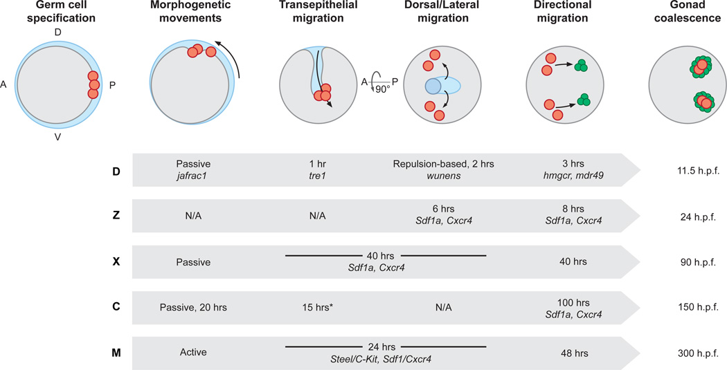
Germ cell migration is being investigated in an ever-growing number of organisms [4–7]. Established model organisms include mice, chicken, frogs, fruitflies and two teleost fish: zebrafish and medaka [8–13]. Despite divergence, features of overall path of embryonic germ cells can be remarkably similar between these species. For instance, germ cells are often specified at the posterior edge of the embryo or at the border between embryonic and extraembryonic tissues (Figure 1). Germ cells then translocate during morphogenetic movements. These movements usually occur during gastrulation and involve movements with endodermal tissue toward the center of the embryo. In Drosophila and Xenopus, the translocation with endodermal tissue is a passive process and known to require germ cell adhesion to underlying endodermal epithelium [14,15], while germ cell morphology suggests that endoderm translocation may be an active process in mice [16,17]. Germ cells that get enclosed within the developing endoderm must undergo a transepithelial migration to enter the mesoderm before migrating both dorsally and laterally to form two groups of germ cells that will occupy each somatic gonad. In Drosophila and mice, these dorsal/lateral movements occur after gut exit, while in Xenopus the dorsal/lateral movements occur before endoderm exit [10,14].
Figure 1. Shared themes in the migration path of embryonic germ cells.
Shown are highly stylized schematics of an embryo not meant to represent any one species. The ‘species-less’ embryo is shown at six key events during germ cell migration in chronological order from left to right. First, germ cells (red) are specified, often at the posterior or edge between embryonic (gray) and extra-embryonic (blue) tissue. Second, germ cells move during somatic morphogenetic movements (dashed arrow). In many species, germ cells move passively during gastrulation and often move within the developing mid or hindgut. Third, germ cells in several species undergo a transepithelial migration to exit the gut. Fourth, germ cells move dorsally and laterally to sort into two populations. Fifth, germ cells undergo a sustained, directed migration toward the developing somatic gonad (green circles). Sixth, germ and somatic gonadal cells coalesce to form the complete embryonic gonad. Shown underneath each stage of germ cell migration is a table with characteristic, key factors and length of stage noted for specific model organisms: D-Drosophila, Z- Zebrafish, X- Xenopus, C- Chicken, M- Mouse. Hpf – hours post fertilization. A- anterior, P- posterior, D- dorsal, V- ventral *Unlike other species, chicken germ cells migrate through the vascular epithelium rather than the gut epithelium.
Alternative migration paths are observed in two model organisms. In chicken embryos, germ cells translocate through the vasculature before migrating along the endoderm toward the developing somatic gonads [18]. In zebrafish, germ cells do not appear to enter the endoderm and because they are specified at four random locations, germ cells do not have to bilaterally sort in order to form two separate groups [19]. Instead, Zebrafish germ cells migrate dorsally to occupy a large zone along the dorsal midline and only a portion of germ cells migrates laterally [19,20]. Despite these unique features, all germ cells studied in depth seem to undergo an active migration guided by attractive and repulsive cues toward the genital ridges or somatic gonadal precursors of the developing gonad. Somatic gonadal cells and germ cells then coalesce to form the complete embryonic gonad. The mechanisms by which germ cells navigate several tissue types in order to reach the gonad are often similar in many organisms and will be discussed in further detail.
Transepithelial migration
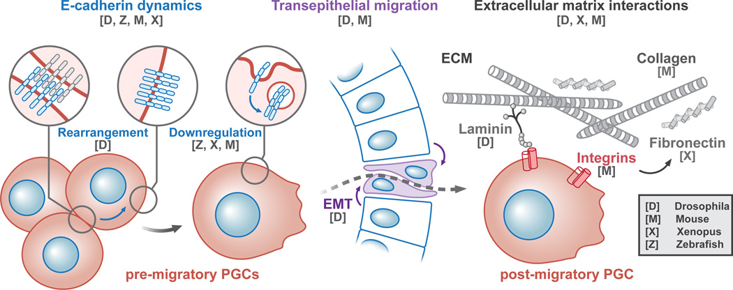
Germ cells in many species must traverse an epithelium to reach the gonad. Insights into how germ cells traverse this barrier have been made through studies in mice and Drosophila. Several signal transduction pathways have been implicated in mouse germ cell exit from the hindgut, including Fibroblast growth factor (FGF) [21], Wnt [22] and Transforming growth factor beta (TGF-β) [23,24]. Which cells produce and respond to these signals has yet to be determined, though the TGF-β responsive gene foxc1 is expressed in the mouse hindgut, suggesting a non-autonomous role germ cell exit [24]. FGF signaling facilitates germ cell exit in both mice and Drosophila. In mouse explants, addition of FGF2 causes germ cells to exit the explanted gut with increased velocity [21]. In Drosophila, FGF signaling is required for dynamic E-cadherin localization within the endodermal epithelium to prevent a midgut collapse that traps germ cells [25]. Together, these findings suggest that germ cells require a dynamic gut epithelium for a properly timed germ cell exit. Strong support for this postulate was recently found in Drosophila. By genetically manipulating the timing of endodermal remodeling, it was demonstrated that an endodermal epithelial to mesenchymal transition is both necessary and sufficient for germ cell migration out of the midgut (Figure 2) [26]. Thus, at least in some organisms, germ cells capitalize on epithelial dynamics to traverse barriers.
Figure 2. Transitions between migratory stages coincide with adhesion dynamics.
Shown is a stylized schematic of germ cell transepithelial migration that occurs during embryogenesis in many species. The transition between tissue types often accompanies changes in localization or downregulation of the cell-cell adhesion protein, E-Cadherin (blue links on red germ cells). In Drosophila, transepithelial migration requires an endodermal (blue columnar cell) epithelial to mesenchymal transition (EMT) (purple cells). Once out of the gut, germ cells in some species make contacts with select extracellular matrix (ECM) (gray crosshatch). The model organism for which specific factors and processes have been discovered is noted as a single letter with the key code in the bottom right corner.
Adhesion dynamics
Changes in adhesion are often observed in germ cells during endodermal exit or the initiation of active migration. The cell-cell adhesion protein E-Cadherin is dynamically regulated in germ cells in many organisms (Figure 2). In zebrafish, E-Cadherin downregulation is facilitated by the depletion of regulator of G–protein signaling 14a (Rgs14a) [27,28]. Similarly, recent studies of isolated Xenopus germ cells using single cell force spectroscopy have shown that isolated migratory Xenopus germ cells have less E-Cadherin-mediated adhesion capabilities compared to isolated pre-migratory Xenopus cells [28–30]. Interestingly, mouse germ cells display an increase in E-cadherin expression during exit from the hindgut, although mouse E-Cadherin does not appear to be strictly required for migration to the somatic gonad [31]. In Drosophila, E-cadherin localization is altered as germ cells dissociate from one another prior to midgut exit, a process that requires the G protein-coupled receptor (GPCR) Trapped in endoderm (Tre1) [32,33]. Together, these data suggest a conserved rearrangement in the cell-cell adhesive properties of germ cells prior to guided migration in the mesoderm.
Dynamic adhesion to the extracellular matrix (ECM) is also observed in germ cells of some species (Figure 2). For instance, isolated migrating Xenopus germ cells display less adhesion to Collagen I and fibronectin than isolated pre-migratory germ cells [29]. In contrast, isolated migrating mouse germ cells display more adhesion to fibronectin than germ cells isolated at the end of migration, while adhesion to Collagen IV and laminin remains unchanged [34]. Together these data suggest that differential ECM localization may facilitate germ cell migration. Indeed, high levels of select ECM components are found along the paths of migrating germ cells in several species [14,23,35] and mouse germ cells dynamically express a select few matrix metalloproteinases during migration to the developing gonad [36]. Consistent with differential adhesive properties and expression patterns, fibronectin in Xenopus [37], laminin and the ECM protein Shifted in Drosophila [38,39] and β1-integrin in mice have all been shown to be necessary for efficient germ cell migration [35]. However, Drosophila and zebrafish germ cells motility was not enhanced by the addition of ECM components in vitro and interestingly Drosophila germ cells do not require beta-integrins in vivo [38,40,41]. Future studies will be needed to clarify the role of adhesion to the ECM as germ cells actively migrate to gonad.
Guidance to the gonad
Germ cells ultimately undertake an active migration toward the developing somatic gonad. The high fidelity in which germ cells reach the gonad was noted over sixty year ago, leading to the postulate that this stage of germ cell migration is guided by environmental chemical gradients [2,3]. Initial support for this chemotaxis postulate came from two experimental strategies. First, in vitro studies demonstrated that mouse genital ridge explants attract isolated germ cells across a large distance [42–44]. Second, genetic screens in Drosophila solidified that germ cell migration requires both the specification and maintenance of somatic gonadal precursor fate [45,46]. In fact, such genetic studies continue to identify new factors that are required for somatic gonad fate maintenance and morphogenesis [47,48] and a wealth of studies have shown that active germ cell migration is guided by one or more attractive or repulsive cues in established model organisms.
SDF-1/CXCR4
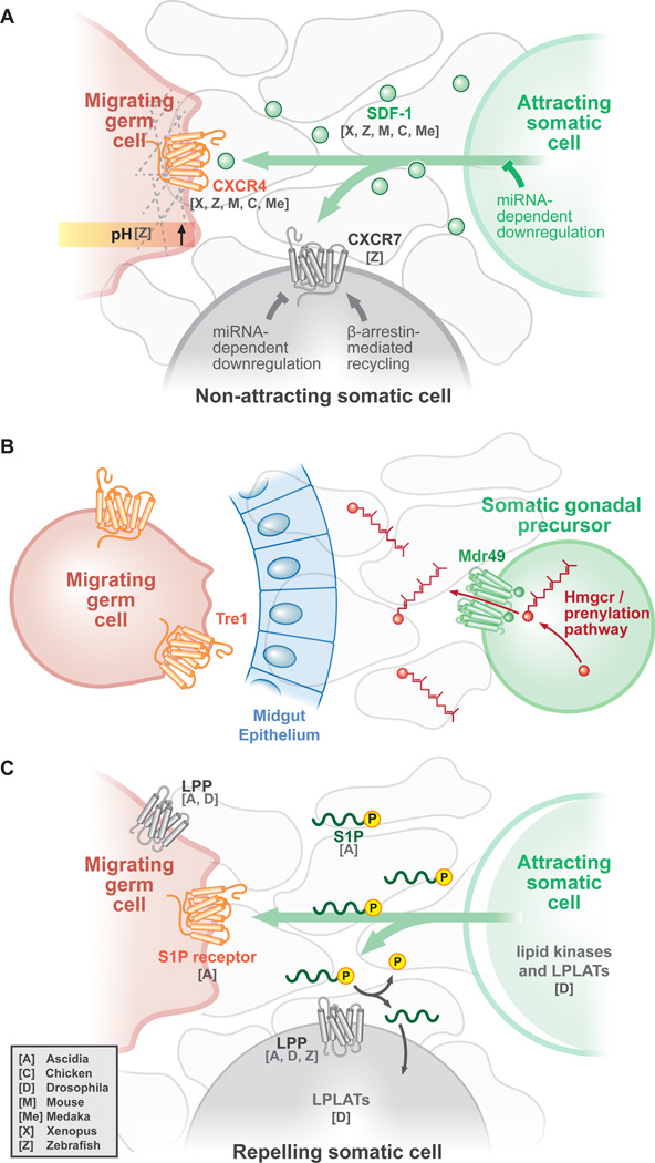
One significant germ cell guidance mechanism is mediated by a gradient of the small chemokine Stromal-cell-derived factor 1 (SDF-1 also called CXCL12). SDF-1 binds to and activates the GPCR chemokine (C-X-C motif), receptor 4 (CXCR4) expressed in migrating germ cells (Figure 3a). The SDF-1/CXCR4 axis is well-studied in cell migration, being required for lymphocyte chemotaxis, neural cell migration, growth cone guidance, stem cell homing and metastasis, reviewed in [49]. First discovered in zebrafish [50,51], the SDF-1/CXCR4 germ cell guidance mechanism was subsequently found in mouse [52–54], chicken [54] and more recently Xenopus [55], medaka [56], and other marine animals [4,6]. The importance of the SDF-1/CXCR4 axis is highlighted by a recent study uncovering a requirement for the pluripotency transcription factor Nanog in medaka germ cell migration. Interestingly, the Nanog requirement can be completely circumvented by overexpression of cxcr4, suggesting CXCR4 is the only Nanog target critical for medaka germ cell migration [57]. Thus, the SDF-1/CXCR4 axis has emerged as a central germ cell guidance mechanism in many species.
Figure 3. Germ cells are guided to the gonad by one or more environmental cues.
Shown here are three simplified schematics, each with three cell types: germ cell-attracting somatic cells (green), migratory germ cells (red) and non-attracting somatic cells (gray) A. The SDF-1/CXCR4 axis functions in many vertebrates. The attracting somatic cell secretes the chemoattractant SDF-1 (small green circles), which either binds to the CXCR4 GPCR in the migrating germ cell or is taken up by the molecular sink GPCR CXCR7 in non-attracting somatic cells. Factors recently identified as promoting or inhibiting ligand or receptor components are highlighted. B. Study of the invertebrate Drosophila model has revealed other guidance mechanisms, wherein HMGCR, additional factors required for protein prenylation, and the ABC transporter Mdr49 are required in somatic attracting cells for germ cell guidance by a secreted factor (red). Germ cells require the GPCR Tre1 for exit from the midgut and migration to the gonad. C. Extracellular lipids guide germ cell migration in highly divergent species. Phospholipids are produced by attracting somatic cells, which can bind to receptors such as the S1P receptor in migrating germ cells. To amplify the phospholipid gradient, nonattracting somatic cells express lipid phosphate phosphatases (LPP), which depletes the local environment of phospholipids. The model organism for which specific factors and processes have been discovered is noted as a single letter with the key code in the bottom left corner.
A steep chemokine gradient is crucial for effective directed germ cell migration. In fact, a recent study has shown that ectopic expression of sdf-1 in germ cells is sufficient to sterilize zebrafish [58]. Steep gradient establishment and maintenance is complicated by the fact that sdf-1 expression is not restricted to the developing somatic gonad target tissue but instead is highly dynamic, with highest levels found just in front of migrating germ cells [50,51,53,54]. With the source of SDF-1 changing as germ cells migrate, it is perhaps not surprising that several mechanisms exist to regulate the SDF-1 gradient. One important regulatory layer was discovered in 2008, during which it was discovered that depletion of a second SDF-1-binding GPCR CXCR7b yielded a migration phenotype similar to that observed upon CXCR4 loss [59]. Interestingly, CXCR7b is not expressed within germ cells and instead was found to function in the surrounding soma as molecular sink to steepen the SDF-1 gradient (Figure 3a) [59]. Two additional regulatory layers were recently discovered. First, it was shown that miRNA processing enzymes or specifically miR-430 depletion increases sdf1a and cxcr7b expression in zebrafish leading to germ cell migration defects, although observed variations in phenotypic penetrance suggests that miRNA-mediated regulation of SDF-1/CXCR7 might not be apparent at all developmental stages or required under all environmental conditions [60–62]. Interestingly, miR-430 also regulates myosin light chain kinase (mlck) and Zeb1 mRNA, suggesting that downregulation of several targets influence the timing and success of germ cell migration [63]. A second regulatory layer involves β-arrestin-mediated endosomal recycling of CXCR7b, which increases the number of times CXCR7b can function as a molecular sink [64]. Interestingly, β-arrestin does not facilitate the endosomal recycling of CXCR4, consistent with CXCR7b being the first GPCR to be identified as having an intrinsic β-arrestin bias [64,65]. Together, these discoveries highlight the importance of a robust SDF-1 gradient in zebrafish. Future investigations will be needed to determine whether similar regulatory mechanisms exists in other organisms that rely on the SDF-1/CXCR4 axis for germ cell guidance.
Additional guidance mechanisms discovered in Drosophila
SDF-1-mediated germ cell guidance is not conserved in all animals even when the overall migration path is similar. One well-established example is Drosophila, in which the newly evolved CXCR family of GPCRs does not appear to exist [66]. Instead, in vivo genetic screens have identified other mechanisms of germ cell guidance. One prominent guidance mechanism requires 3-hydroxy-3-methylglutaryl coenzyme A (Hmgcr), which like SDF-1, displays a dynamic expression pattern that initially encompasses the mesoderm before becoming restricted to the somatic gonad [67]. Also like SDF-1, ectopic expression of hmgcr, as well as other components of the mevalonate pathway, is sufficient to attract germ cells in vivo [50,67,68]. Interestingly, hmgcr-dependent attraction requires the ABC transporter Multidrug resistance 49 [69]. One biological process that requires each of these components is the biogenesis and secretion of the yeast pheromone a-mating factor, suggesting that a secreted prenylated factor may guide Drosophila germ cells (Figure 3b) [69,70]. Some studies suggest that Hedgehog is the hmgcr-dependent germ cell attractant [39,71]; however intensive study of Hedgehog lipid modifications have not revealed prenylation [72]. Furthermore, the attracting capabilities of ectopic hedgehog expression has not proven reproducible and using several methods to alter Hedgehog signaling failed to affect germ cell migration [73]. Thus, the identity of the hmgcr-dependent germ cell attractant remains elusive.
Response to guidance cues is often mediated by GPCRs or receptor tyrosine kinases (RTKs). A key receptor required for Drosophila germ cell migration is the GPCR Tre1, named for the loss of function phenotype ‘trapped in endoderm’ wherein the majority of tre1 mutant germ cells fail to exit the midgut (Figure 3b) [32,33]. Interestingly, tre1 null germ cells that circumvent the need for transepithelial migration migrate to the somatic gonad, while germ cells carrying a mutation in the conserved DRY domain of Tre1 are able to exit the midgut but not migrate to the gonad, instead scattering throughout the embryo [33,74]. These curious findings suggest that Tre1 may be required to both exit the gut and respond to guidance cues. Computational modeling suggests that mutations in the DRY domain prevents the formation of a salt bridge necessary for protein stability, however this seems inconsistent with the phenotypic differences between the hypomorphic tre1sctt (DRY domain mutation) and null mutations [75]. A detailed structure-function analysis is needed to determine whether there is indeed multiple requirements for Tre1 in germ cell migration. While the ligand for Tre1 is as yet unknown, the closest mammalian homolog, GPR84, binds to medium chain fatty acids, leaving open the prospect that Tre1 mediates germ cell response to lipid guidance cues [76].
Lipid-mediated germ cell guidance
Growing evidence suggests that extracellular lipids play an important role in germ cell guidance. Lipid-mediated germ cell guidance was first discovered in Drosophila, in which mutations in the genes encoding lipid phosphate phosphatases (LPP), wunen and wunen2, disrupt bilateral sorting and cause germ cells to scatter throughout the embryo instead of reaching the gonad [77,78]. The genes wunen and wunen2 encode six-pass transmembrane LPPs wherein the enzymatic lipid phosphatase activity faces the extracellular space and modulate the lipid environment within at least a ten-micron distance [79,80]. Interestingly, germ cells are repelled from regions of high wunen expression, suggesting that Wunens may deplete the local environment of a phospholipid germ cell attractant (Figure 3c) [77,81,82]. Consistently, several additional lipid-modifying factors have been implicated in Drosophila germ cell guidance. The simultaneous loss of two lysophospholipid acyltransferases, Oysgerdart and Nessy or two lipid kinases Dmulk and Dcerk compromise germ cell migration to the developing somatic gonad [83,84]. While the requirements for lipids in Drosophila germ cell guidance is becoming more defined, much less is known in other organisms, perhaps due to functional redundancy, which is already evident in the relatively simple Drosophila genome [83,84]. Indeed, the recent discovery that LPPs repel germ cells away from nearby somites required the simultaneous knockout of the six LPP genes encoded in the zebrafish genome [85]. In addition, it was recently shown that germ cells in the colonial ascidian Botryllus schlosseri respond to sphingosine-1-phospate (S1P) gradients and S1P receptors are required for germ cell migration to the niche (Figure 3c) [5]. These exciting findings suggest that lipid-mediated germ cell guidance may be conserved in quite divergent species.
Sustaining motility
Germ cells require both environmental guidance and the ability to initiate and sustain motility. In some species, germ cells must sustain directed migration for twenty-four to forty-eight hours (Figure 1). The study of zebrafish germ cell migration has proven fruitful for the discovery of autonomous motility factors (for a recent detailed review see [11]). One motility factor, Dead end regulates cell shape, actomyosin contractility, and cell-cell adhesion to facilitate the initiation of migration [63,86]. Recent evidence suggests that filopodia may play a role in polarized CXCR4 activation, which in turn facilitates elevated pH to polarize Rac1 activity at the leading edge in zebrafish germ cells [87,88]. In Xenopus, germ cell migration requires the kinesin KIF123B for polarized Phosphatidylinositol (3,4,5)-trisphosphate (PIP3) accumulation at bleb-like protrusions [89] and the PDZ domain-containing germ plasm protein XGRIP2, although the function of XGRIP2 is not yet known [90,91]. In mice and chicken, germ cell motility is enhanced by the somatically-expressed Stem cell factor (SCF, or Steel factor) ligand and the corresponding receptor c-Kit in germ cells [92–94]. Interestingly, the RTK Ror2 is required in mouse germ cells for SCF-mediated motility in vitro [95]. Ror2 is a receptor for Wnt5a, a ligand that is also required for germ cell migration, though not as a chemoattractant, suggesting that Wnt5a-mediated Ror2 activation may permit robust SCF-mediated c-Kit activation [22,95]. Less is known about motility factors in Drosophila germ cells due to the perdurance of maternally-provided factors in Drosophila germ cells through most stages of migration [45]. One strategy to overcome this hurdle is to deplete maternal RNAs, as was done recently by injecting interfering dsRNAs into developing embryos to identify factors potentially required autonomously for germ cell migration [96]. Development of more refined tools, such as protein-degradation systems could also be used to deplete maternally-provided factors and has the added potential to shed light on temporal requirements for germ cell migration and motility factors [97].
Post-migration fates
Germ cells that successfully reach the target tissue undergo several maturation steps to yield a functional gonad. The fate of germ cells that do not reach the gonad vary depending upon species. In Drosophila, Xenopus, and mice, germ cells that do not make it to the gonad eventually disappear, either through apoptosis or loss of germ cell fate [52,53,98,99], while in zebrafish, mis-migrated germ cells persist quite some time during development [50]. The fate for the mis-migrated cell may lie in differences in the hospitality of non-gonadal somatic tissues. Indeed, wunen deficient Drosophila germ cells die upon migration into regions of high wunen expression but are protected if they remain in the midgut, while ectopic expression of zebrafish LPPs do not lead to germ cell loss [79,85,100]. Similarly, mouse germ cells that migrate into the midline encounter low levels of SCF and undergo apoptosis [101]. Thus, Wunen in Drosophila and SCF in mice modulate both germ cell migration and survival and dynamic regulation of such mechanisms may safeguard organisms against neonatal germ cell tumors.
Conclusions
The study of germ cell migration has yielded valuable insights into how cells navigate several tissues in a dynamic environment to reach their target. Despite mechanistic differences in migrational paths and guidance cues, the themes required for germ cell migration remain remarkably conserved across model organism species. In all species, the migration of germ cells is a multi-step process involving passive and active movements. While these translocation steps might seem distinct, emerging evidence suggests that defects in an early stage can, through unknown mechanisms, affect migration at a later stage [102–104]. The development of new genetic tools and imaging capabilities will shed light on the interconnectedness of each migration step. In addition, insights into germ cell dynamics will continue to be aided by in vitro systems, including recent developments using single cell force spectroscopy and defined germ cell culturing strategies [30,105]. Thus, the study of germ cell migration is primed for additional breakthroughs in our understanding of this vital process in the germline lifecycle.
Acknowledgments
We would like to thank Alexey Soshnev for assistance with figures and members of the Lehmann lab for helpful discussions. This work was by the National Institutes of Health R37 HD49100 to R.L.. R.L. is a Howard Hughes Medical Institute Investigator. L.J.B. is a Damon Runyon Fellow supported by the Damon Runyon Cancer Research Foundation (DRG-2235-15).
Footnotes
Publisher's Disclaimer: This is a PDF file of an unedited manuscript that has been accepted for publication. As a service to our customers we are providing this early version of the manuscript. The manuscript will undergo copyediting, typesetting, and review of the resulting proof before it is published in its final citable form. Please note that during the production process errors may be discovered which could affect the content, and all legal disclaimers that apply to the journal pertain.
Contributor Information
Lacy J. Barton, Email: Lacy.Barton@med.nyu.edu.
Michelle G. LeBlanc, Email: Michelle.Leblanc@med.nyu.edu.
Ruth Lehmann, Email: Ruth.Lehmann@med.nyu.edu.
References
- 1.Rubaschkin W. On the question of the origin of the germ cells in mammalian embryos. Anat. 1908;32:222–224. [Google Scholar]
- 2.Witschi E. Migration of the germ cells of human embryos from the yolk sac to the primitive gonadal folds. Control Embryology Carnegie Institute. 1948;32:67–80. [Google Scholar]
- 3.Chiquoine AD. The identification, origin, and migration of the primordial germ cells in the mouse embryo. Anat Rec. 1954;118:135–146. doi: 10.1002/ar.1091180202. [DOI] [PubMed] [Google Scholar]
- 4.Fernandez JA, Bubner EJ, Takeuchi Y, Yoshizaki G, Wang T, Cummins SF, Elizur A. Primordial germ cell migration in the yellowtail kingfish (Seriola lalandi) and identification of stromal cell-derived factor 1. Gen Comp Endocrinol. 2015;213:16–23. doi: 10.1016/j.ygcen.2015.02.007. [DOI] [PubMed] [Google Scholar]
- 5. Kassmer SH, Rodriguez D, Langenbacher AD, Bui C, De Tomaso AW. Migration of germline progenitor cells is directed by sphingosine-1-phosphate signalling in a basal chordate. Nat Commun. 2015;6:8565. doi: 10.1038/ncomms9565. •• This study uncovered a role for lipid signaling in ascidian germ cell migration. Previously only implicated in Drosophila, this discovery suggests lipid signaling guidance may be conserved across phyla.
- 6.Li M, Tan X, Jiao S, Wang Q, Wu Z, You F, Zou Y. A new pattern of primordial germ cell migration in olive flounder (Paralichthys olivaceus) identified using nanos3. Dev Genes Evol. 2015;225:195–206. doi: 10.1007/s00427-015-0503-6. [DOI] [PubMed] [Google Scholar]
- 7.Saito T, Psenicka M, Goto R, Adachi S, Inoue K, Arai K, Yamaha E. The origin and migration of primordial germ cells in sturgeons. PLoS One. 2014;9:e86861. doi: 10.1371/journal.pone.0086861. [DOI] [PMC free article] [PubMed] [Google Scholar]
- 8.Nakamura Y, Kagami H, Tagami T. Development, differentiation and manipulation of chicken germ cells. Dev Growth Differ. 2013;55:20–40. doi: 10.1111/dgd.12026. [DOI] [PubMed] [Google Scholar]
- 9. Kang KS, Lee HC, Kim HJ, Lee HG, Kim YM, Lee HJ, Park YH, Yang SY, Rengaraj D, Park TS, et al. Spatial and temporal action of chicken primordial germ cells during initial migration. Reproduction. 2015;149:179–187. doi: 10.1530/REP-14-0433. • Researchers in this study used a combination of transplanation and live imaging assays to defined the stages of passive and active migration during avian germ cell migration.
- 10.Richardson BE, Lehmann R. Mechanisms guiding primordial germ cell migration: strategies from different organisms. Nat Rev Mol Cell Biol. 2010;11:37–49. doi: 10.1038/nrm2815. [DOI] [PMC free article] [PubMed] [Google Scholar]
- 11.Paksa A, Raz E. Zebrafish germ cells: motility and guided migration. Curr Opin Cell Biol. 2015;36:80–85. doi: 10.1016/j.ceb.2015.07.007. [DOI] [PubMed] [Google Scholar]
- 12.Shinomiya A, Tanaka M, Kobayashi T, Nagahama Y, Hamaguchi S. The vasa-like gene, olvas, identifies the migration path of primordial germ cells during embryonic body formation stage in the medaka, Oryzias latipes. Dev Growth Differ. 2000;42:317–326. doi: 10.1046/j.1440-169x.2000.00521.x. [DOI] [PubMed] [Google Scholar]
- 13.Sasado T, Morinaga C, Niwa K, Shinomiya A, Yasuoka A, Suwa H, Hirose Y, Yoda H, Henrich T, Deguchi T, et al. Mutations affecting early distribution of primordial germ cells in Medaka (Oryzias latipes) embryo. Mech Dev. 2004;121:817–828. doi: 10.1016/j.mod.2004.03.022. [DOI] [PubMed] [Google Scholar]
- 14.Nishiumi F, Komiya T, Ikenishi K. The mode and molecular mechanisms of the migration of presumptive PGC in the endoderm cell mass of Xenopus embryos. Dev Growth Differ. 2005;47:37–48. doi: 10.1111/j.1440-169x.2004.00777.x. [DOI] [PubMed] [Google Scholar]
- 15.DeGennaro M, Hurd TR, Siekhaus DE, Biteau B, Jasper H, Lehmann R. Peroxiredoxin stabilization of DE-cadherin promotes primordial germ cell adhesion. Dev Cell. 2011;20:233–243. doi: 10.1016/j.devcel.2010.12.007. [DOI] [PMC free article] [PubMed] [Google Scholar]
- 16.Anderson R, Copeland TK, Scholer H, Heasman J, Wylie C. The onset of germ cell migration in the mouse embryo. Mech Dev. 2000;91:61–68. doi: 10.1016/s0925-4773(99)00271-3. [DOI] [PubMed] [Google Scholar]
- 17.Molyneaux KA, Stallock J, Schaible K, Wylie C. Time-lapse analysis of living mouse germ cell migration. Dev Biol. 2001;240:488–498. doi: 10.1006/dbio.2001.0436. [DOI] [PubMed] [Google Scholar]
- 18.Nakamura Y, Yamamoto Y, Usui F, Mushika T, Ono T, Setioko AR, Takeda K, Nirasawa K, Kagami H, Tagami T. Migration and proliferation of primordial germ cells in the early chicken embryo. Poult Sci. 2007;86:2182–2193. doi: 10.1093/ps/86.10.2182. [DOI] [PubMed] [Google Scholar]
- 19.Weidinger G, Wolke U, Koprunner M, Klinger M, Raz E. Identification of tissues and patterning events required for distinct steps in early migration of zebrafish primordial germ cells. Development. 1999;126:5295–5307. doi: 10.1242/dev.126.23.5295. [DOI] [PubMed] [Google Scholar]
- 20.Weidinger G, Wolke U, Koprunner M, Thisse C, Thisse B, Raz E. Regulation of zebrafish primordial germ cell migration by attraction towards an intermediate target. Development. 2002;129:25–36. doi: 10.1242/dev.129.1.25. [DOI] [PubMed] [Google Scholar]
- 21.Takeuchi Y, Molyneaux K, Runyan C, Schaible K, Wylie C. The roles of FGF signaling in germ cell migration in the mouse. Development. 2005;132:5399–5409. doi: 10.1242/dev.02080. [DOI] [PubMed] [Google Scholar]
- 22.Chawengsaksophak K, Svingen T, Ng ET, Epp T, Spiller CM, Clark C, Cooper H, Koopman P. Loss of Wnt5a disrupts primordial germ cell migration and male sexual development in mice. Biol Reprod. 2012;86:1–12. doi: 10.1095/biolreprod.111.095232. [DOI] [PubMed] [Google Scholar]
- 23.Chuva de Sousa Lopes SM, van den Driesche S, Carvalho RL, Larsson J, Eggen B, Surani MA, Mummery CL. Altered primordial germ cell migration in the absence of transforming growth factor beta signaling via ALK5. Dev Biol. 2005;284:194–203. doi: 10.1016/j.ydbio.2005.05.019. [DOI] [PubMed] [Google Scholar]
- 24.Mattiske D, Kume T, Hogan BL. The mouse forkhead gene Foxc1 is required for primordial germ cell migration and antral follicle development. Dev Biol. 2006;290:447–458. doi: 10.1016/j.ydbio.2005.12.007. [DOI] [PubMed] [Google Scholar]
- 25. Pares G, Ricardo S. FGF control of E-cadherin targeting in the Drosophila midgut impacts on primordial germ cell motility. J Cell Sci. 2016;129:354–366. doi: 10.1242/jcs.174284. •• Results from this study demonstrate that Drosophila germ cells require dynamic remodeling of cell-cell adhesion within the endodermal epithelium for midgut exit, highlighting how germ cells rely on external tissue rearrangements (26).
- 26.Seifert JRK, Lehmann R. Drosophila primordial germ cell migration requires epithelial remodeling of the endoderm. Development (Cambridge, England) 2012;139:2101–2106. doi: 10.1242/dev.078949. [DOI] [PMC free article] [PubMed] [Google Scholar]
- 27.Blaser H, Eisenbeiss S, Neumann M, Reichman-Fried M, Thisse B, Thisse C, Raz E. Transition from non-motile behaviour to directed migration during early PGC development in zebrafish. J Cell Sci. 2005;118:4027–4038. doi: 10.1242/jcs.02522. [DOI] [PubMed] [Google Scholar]
- 28.Hartwig J, Tarbashevich K, Seggewiss J, Stehling M, Bandemer J, Grimaldi C, Paksa A, Gross-Thebing T, Meyen D, Raz E. Temporal control over the initiation of cell motility by a regulator of G-protein signaling. Proc Natl Acad Sci U S A. 2014;111:11389–11394. doi: 10.1073/pnas.1400043111. [DOI] [PMC free article] [PubMed] [Google Scholar]
- 29.Dzementsei A, Schneider D, Janshoff A, Pieler T. Migratory and adhesive properties of Xenopus laevis primordial germ cells in vitro. Biol Open. 2013;2:1279–1287. doi: 10.1242/bio.20135140. [DOI] [PMC free article] [PubMed] [Google Scholar]
- 30. Baronsky T, Dzementsei A, Oelkers M, Melchert J, Pieler T, Janshoff A. Reduction in E-cadherin expression fosters migration of Xenopus laevis primordial germ cells. Integr Biol (Camb) 2016;8:349–358. doi: 10.1039/c5ib00291e. • This study explored cell-cell adhesion dynamics of germ cells using single cell force spectroscopy, an approach holds promise to reveal unifying and differentiating features in germ cell adhesive properties across species.
- 31.Bendel-Stenzel MR, Gomperts M, Anderson R, Heasman J, Wylie C. The role of cadherins during primordial germ cell migration and early gonad formation in the mouse. Mech Dev. 2000;91:143–152. doi: 10.1016/s0925-4773(99)00287-7. [DOI] [PubMed] [Google Scholar]
- 32.Kunwar PS, Sano H, Renault AD, Barbosa V, Fuse N, Lehmann R. Tre1 GPCR initiates germ cell transepithelial migration by regulating Drosophila melanogaster E-cadherin. J Cell Biol. 2008;183:157–168. doi: 10.1083/jcb.200807049. [DOI] [PMC free article] [PubMed] [Google Scholar]
- 33.Kunwar PS, Starz-Gaiano M, Bainton RJ, Heberlein U, Lehmann R. Tre1, a G protein-coupled receptor, directs transepithelial migration of Drosophila germ cells. PLoS Biol. 2003;1:E80. doi: 10.1371/journal.pbio.0000080. [DOI] [PMC free article] [PubMed] [Google Scholar]
- 34.Garcia-Castro MI, Anderson R, Heasman J, Wylie C. Interactions between germ cells and extracellular matrix glycoproteins during migration and gonad assembly in the mouse embryo. J Cell Biol. 1997;138:471–480. doi: 10.1083/jcb.138.2.471. [DOI] [PMC free article] [PubMed] [Google Scholar]
- 35.Bendel-Stenzel M, Anderson R, Heasman J, Wylie C. The origin and migration of primordial germ cells in the mouse. Semin Cell Dev Biol. 1998;9:393–400. doi: 10.1006/scdb.1998.0204. [DOI] [PubMed] [Google Scholar]
- 36.Diez-Torre A, Diaz-Nunez M, Eguizabal C, Silvan U, Arechaga J. Evidence for a role of matrix metalloproteinases and their inhibitors in primordial germ cell migration. Andrology. 2013;1:779–786. doi: 10.1111/j.2047-2927.2013.00109.x. [DOI] [PubMed] [Google Scholar]
- 37.Heasman J, Hynes RO, Swan AP, Thomas V, Wylie CC. Primordial germ cells of Xenopus embryos: the role of fibronectin in their adhesion during migration. Cell. 1981;27:437–447. doi: 10.1016/0092-8674(81)90385-8. [DOI] [PubMed] [Google Scholar]
- 38.Jaglarz MK, Howard KR. The active migration of Drosophila primordial germ cells. Development. 1995;121:3495–3503. doi: 10.1242/dev.121.11.3495. [DOI] [PubMed] [Google Scholar]
- 39.Deshpande G, Zhou K, Wan JY, Friedrich J, Jourjine N, Smith D, Schedl P. The hedgehog pathway gene shifted functions together with the hmgcr-dependent isoprenoid biosynthetic pathway to orchestrate germ cell migration. PLoS Genet. 2013;9:e1003720. doi: 10.1371/journal.pgen.1003720. [DOI] [PMC free article] [PubMed] [Google Scholar]
- 40.Kardash E, Reichman-Fried M, Maitre JL, Boldajipour B, Papusheva E, Messerschmidt EM, Heisenberg CP, Raz E. A role for Rho GTPases and cell-cell adhesion in single-cell motility in vivo. Nat Cell Biol. 2010;12:47–53. 41–11. doi: 10.1038/ncb2003. sup. [DOI] [PubMed] [Google Scholar]
- 41.Devenport D, Brown NH. Morphogenesis in the absence of integrins: mutation of both Drosophila beta subunits prevents midgut migration. Development. 2004;131:5405–5415. doi: 10.1242/dev.01427. [DOI] [PubMed] [Google Scholar]
- 42.Donovan PJ, Stott D, Cairns LA, Heasman J, Wylie CC. Migratory and postmigratory mouse primordial germ cells behave differently in culture. Cell. 1986;44:831–838. doi: 10.1016/0092-8674(86)90005-x. [DOI] [PubMed] [Google Scholar]
- 43.Godin I, Wylie C, Heasman J. Genital ridges exert long-range effects on mouse primordial germ cell numbers and direction of migration in culture. Development. 1990;108:357–363. doi: 10.1242/dev.108.2.357. [DOI] [PubMed] [Google Scholar]
- 44.Godin I, Wylie CC. TGF beta 1 inhibits proliferation and has a chemotropic effect on mouse primordial germ cells in culture. Development. 1991;113:1451–1457. doi: 10.1242/dev.113.4.1451. [DOI] [PubMed] [Google Scholar]
- 45.Moore LA, Broihier HT, Van Doren M, Lunsford LB, Lehmann R. Identification of genes controlling germ cell migration and embryonic gonad formation in Drosophila. Development. 1998;125:667–678. doi: 10.1242/dev.125.4.667. [DOI] [PubMed] [Google Scholar]
- 46.Boyle M, Bonini N, DiNardo S. Expression and function of clift in the development of somatic gonadal precursors within the Drosophila mesoderm. Development. 1997;124:971–982. doi: 10.1242/dev.124.5.971. [DOI] [PubMed] [Google Scholar]
- 47.Tsigkari KK, Acevedo SF, Skoulakis EM. 14-3-3epsilon Is required for germ cell migration in Drosophila. PLoS One. 2012;7:e36702. doi: 10.1371/journal.pone.0036702. [DOI] [PMC free article] [PubMed] [Google Scholar]
- 48.Sano H, Kunwar PS, Renault AD, Barbosa V, Clark IB, Ishihara S, Sugimura K, Lehmann R. The Drosophila actin regulator ENABLED regulates cell shape and orientation during gonad morphogenesis. PLoS One. 2012;7:e52649. doi: 10.1371/journal.pone.0052649. [DOI] [PMC free article] [PubMed] [Google Scholar]
- 49.Kucia M, Jankowski K, Reca R, Wysoczynski M, Bandura L, Allendorf DJ, Zhang J, Ratajczak J, Ratajczak MZ. CXCR4-SDF-1 signalling, locomotion, chemotaxis and adhesion. J Mol Histol. 2004;35:233–245. doi: 10.1023/b:hijo.0000032355.66152.b8. [DOI] [PubMed] [Google Scholar]
- 50.Doitsidou M, Reichman-Fried M, Stebler J, Koprunner M, Dorries J, Meyer D, Esguerra CV, Leung T, Raz E. Guidance of primordial germ cell migration by the chemokine SDF-1. Cell. 2002;111:647–659. doi: 10.1016/s0092-8674(02)01135-2. [DOI] [PubMed] [Google Scholar]
- 51.Knaut H, Werz C, Geisler R, Nusslein-Volhard C, Tubingen Screen C. A zebrafish homologue of the chemokine receptor Cxcr4 is a germ-cell guidance receptor. Nature. 2003;421:279–282. doi: 10.1038/nature01338. [DOI] [PubMed] [Google Scholar]
- 52.Ara T, Nakamura Y, Egawa T, Sugiyama T, Abe K, Kishimoto T, Matsui Y, Nagasawa T. Impaired colonization of the gonads by primordial germ cells in mice lacking a chemokine, stromal cell-derived factor-1 (SDF-1) Proc Natl Acad Sci U S A. 2003;100:5319–5323. doi: 10.1073/pnas.0730719100. [DOI] [PMC free article] [PubMed] [Google Scholar]
- 53.Molyneaux KA, Zinszner H, Kunwar PS, Schaible K, Stebler J, Sunshine MJ, O'Brien W, Raz E, Littman D, Wylie C, et al. The chemokine SDF1/CXCL12 and its receptor CXCR4 regulate mouse germ cell migration and survival. Development. 2003;130:4279–4286. doi: 10.1242/dev.00640. [DOI] [PubMed] [Google Scholar]
- 54.Stebler J, Spieler D, Slanchev K, Molyneaux KA, Richter U, Cojocaru V, Tarabykin V, Wylie C, Kessel M, Raz E. Primordial germ cell migration in the chick and mouse embryo: the role of the chemokine SDF-1/CXCL12. Dev Biol. 2004;272:351–361. doi: 10.1016/j.ydbio.2004.05.009. [DOI] [PubMed] [Google Scholar]
- 55.Takeuchi T, Tanigawa Y, Minamide R, Ikenishi K, Komiya T. Analysis of SDF-1/CXCR4 signaling in primordial germ cell migration and survival or differentiation in Xenopus laevis. Mech Dev. 2010;127:146–158. doi: 10.1016/j.mod.2009.09.005. [DOI] [PubMed] [Google Scholar]
- 56.Kurokawa H, Aoki Y, Nakamura S, Ebe Y, Kobayashi D, Tanaka M. Time-lapse analysis reveals different modes of primordial germ cell migration in the medaka Oryzias latipes. Dev Growth Differ. 2006;48:209–221. doi: 10.1111/j.1440-169X.2006.00858.x. [DOI] [PubMed] [Google Scholar]
- 57.Sanchez-Sanchez AV, Camp E, Leal-Tassias A, Atkinson SP, Armstrong L, Diaz-Llopis M, Mullor JL. Nanog regulates primordial germ cell migration through Cxcr4b. Stem Cells. 2010;28:1457–1464. doi: 10.1002/stem.469. [DOI] [PubMed] [Google Scholar]
- 58.Wong TT, Collodi P. Inducible Sterilization of Zebrafish by Disruption of Primordial Germ Cell Migration. PLoS One. 2013;8:e68455. doi: 10.1371/journal.pone.0068455. [DOI] [PMC free article] [PubMed] [Google Scholar]
- 59. Boldajipour B, Mahabaleshwar H, Kardash E, Reichman-Fried M, Blaser H, Minina S, Wilson D, Xu Q, Raz E. Control of chemokine-guided cell migration by ligand sequestration. Cell. 2008;132:463–473. doi: 10.1016/j.cell.2007.12.034. •• Investigators in this study demonstrated that, in additon to Cxcr4, a second GPCR, CXCR7, binds to the germ cell attractant SDF-1 and acts as a molecular sink to facilliate a dynamic gradient as germ cells migrate toward the developing gonads.
- 60.Staton AA, Knaut H, Giraldez AJ. miRNA regulation of Sdf1 chemokine signaling provides genetic robustness to germ cell migration. Nature Genetics. 2011 doi: 10.1038/ng.758. [DOI] [PMC free article] [PubMed] [Google Scholar]
- 61.Goudarzi M, Strate I, Paksa A, Lagendijk AK, Bakkers J, Raz E. On the robustness of germ cell migration and microRNA-mediated regulation of chemokine signaling. Nat Genet. 2013;45:1264–1265. doi: 10.1038/ng.2793. [DOI] [PubMed] [Google Scholar]
- 62.Staton AA, Knaut H, Giraldez AJ. Reply to: "On the robustness of germ cell migration and microRNA-mediated regulation of chemokine signaling". Nat Genet. 2013;45:1266–1267. doi: 10.1038/ng.2812. [DOI] [PubMed] [Google Scholar]
- 63.Goudarzi M, Banisch TU, Mobin MB, Maghelli N, Tarbashevich K, Strate I, van den Berg J, Blaser H, Bandemer S, Paluch E, et al. Identification and regulation of a molecular module for bleb-based cell motility. Dev Cell. 2012;23:210–218. doi: 10.1016/j.devcel.2012.05.007. [DOI] [PubMed] [Google Scholar]
- 64.Mahabaleshwar H, Tarbashevich K, Nowak M, Brand M, Raz E. beta-arrestin control of late endosomal sorting facilitates decoy receptor function and chemokine gradient formation. Development. 2012;139:2897–2902. doi: 10.1242/dev.080408. [DOI] [PubMed] [Google Scholar]
- 65.Rajagopal S, Kim J, Ahn S, Craig S, Lam CM, Gerard NP, Gerard C, Lefkowitz RJ. Beta-arrestin- but not G protein-mediated signaling by the "decoy" receptor CXCR7. Proc Natl Acad Sci U S A. 2010;107:628–632. doi: 10.1073/pnas.0912852107. [DOI] [PMC free article] [PubMed] [Google Scholar]
- 66.Metpally RP, Sowdhamini R. Cross genome phylogenetic analysis of human and Drosophila G protein-coupled receptors: application to functional annotation of orphan receptors. BMC Genomics. 2005;6:106. doi: 10.1186/1471-2164-6-106. [DOI] [PMC free article] [PubMed] [Google Scholar]
- 67.Van Doren M, Broihier HT, Moore LA, Lehmann R. HMG-CoA reductase guides migrating primordial germ cells. Nature. 1998;396:466–469. doi: 10.1038/24871. [DOI] [PubMed] [Google Scholar]
- 68.Santos AC, Lehmann R. Isoprenoids control germ cell migration downstream of HMGCoA reductase. Dev Cell. 2004;6:283–293. doi: 10.1016/s1534-5807(04)00023-1. [DOI] [PubMed] [Google Scholar]
- 69.Ricardo S, Lehmann R. An ABC Transporter Controls Export of a Drosophila Germ Cell Attractant. Science. 2009;323:943–946. doi: 10.1126/science.1166239. [DOI] [PMC free article] [PubMed] [Google Scholar]
- 70.Michaelis S, Barrowman J. Biogenesis of the Saccharomyces cerevisiae pheromone a-factor, from yeast mating to human disease. Microbiol Mol Biol Rev. 2012;76:626–651. doi: 10.1128/MMBR.00010-12. [DOI] [PMC free article] [PubMed] [Google Scholar]
- 71.Deshpande G, Schedl P. HMGCoA reductase potentiates hedgehog signaling in Drosophila melanogaster. Dev Cell. 2005;9:629–638. doi: 10.1016/j.devcel.2005.09.014. [DOI] [PubMed] [Google Scholar]
- 72.Eaton S. Multiple roles for lipids in the Hedgehog signalling pathway. Nat Rev Mol Cell Biol. 2008;9:437–445. doi: 10.1038/nrm2414. [DOI] [PubMed] [Google Scholar]
- 73.Renault A, Ricardo S, Kunwar P, Santos A, Starz-Gaiano M, Stein J, Lehmann R. Hedgehog does not guide migrating Drosophila germ cells. Developmental biology. 2009;328:355–362. doi: 10.1016/j.ydbio.2009.01.042. [DOI] [PMC free article] [PubMed] [Google Scholar]
- 74.Kamps AR, Pruitt MM, Herriges JC, Coffman CR. An evolutionarily conserved arginine is essential for Tre1 G protein-coupled receptor function during germ cell migration in Drosophila melanogaster. PloS one. 2010;5:e11839. doi: 10.1371/journal.pone.0011839. [DOI] [PMC free article] [PubMed] [Google Scholar]
- 75.Pruitt MM, Lamm MH, Coffman CR. BMC Structural Biology | Full text | Molecular dynamics simulations on the Tre1 G protein-coupled receptor: exploring the role of the arginine of the NRY motif in Tre1 structure. BMC structural biology. 2013 doi: 10.1186/1472-6807-13-15. [DOI] [PMC free article] [PubMed] [Google Scholar]
- 76.Wang J, Wu X, Simonavicius N, Tian H, Ling L. Medium-chain fatty acids as ligands for orphan G protein-coupled receptor GPR84. J Biol Chem. 2006;281:34457–34464. doi: 10.1074/jbc.M608019200. [DOI] [PubMed] [Google Scholar]
- 77.Zhang N, Zhang J, Cheng Y, Howard K. Identification and genetic analysis of wunen, a gene guiding Drosophila melanogaster germ cell migration. Genetics. 1996;143:1231–1241. doi: 10.1093/genetics/143.3.1231. [DOI] [PMC free article] [PubMed] [Google Scholar]
- 78.Starz-Gaiano M, Cho NK, Forbes A, Lehmann R. Spatially restricted activity of a Drosophila lipid phosphatase guides migrating germ cells. Development (Cambridge, England) 2001;128:983–991. doi: 10.1242/dev.128.6.983. [DOI] [PubMed] [Google Scholar]
- 79.Renault AD, Sigal YJ, Morris AJ, Lehmann R. Soma-germ line competition for lipid phosphate uptake regulates germ cell migration and survival. Science. 2004;305:1963–1966. doi: 10.1126/science.1102421. [DOI] [PubMed] [Google Scholar]
- 80.Mukherjee A, Neher RA, Renault AD. Quantifying the range of a lipid phosphate signal in vivo. J Cell Sci. 2013;126:5453–5464. doi: 10.1242/jcs.136176. [DOI] [PubMed] [Google Scholar]
- 81.Zhang N, Zhang J, Purcell KJ, Cheng Y, Howard K. The Drosophila protein Wunen repels migrating germ cells. Nature. 1997;385:64–67. doi: 10.1038/385064a0. [DOI] [PubMed] [Google Scholar]
- 82.Sano H. Control of lateral migration and germ cell elimination by the Drosophila melanogaster lipid phosphate phosphatases Wunen and Wunen 2. J Cell Biol. 2005;171:675–683. doi: 10.1083/jcb.200506038. [DOI] [PMC free article] [PubMed] [Google Scholar]
- 83.Steinhauer J, Gijon MA, Riekhof WR, Voelker DR, Murphy RC, Treisman JE. Drosophila lysophospholipid acyltransferases are specifically required for germ cell development. Mol Biol Cell. 2009;20:5224–5235. doi: 10.1091/mbc.E09-05-0382. [DOI] [PMC free article] [PubMed] [Google Scholar]
- 84.McElwain MA, Ko DC, Gordon MD, Fyrst H, Saba JD, Nusse R. A suppressor/enhancer screen in Drosophila reveals a role for wnt-mediated lipid metabolism in primordial germ cell migration. PLoS One. 2011;6:e26993. doi: 10.1371/journal.pone.0026993. [DOI] [PMC free article] [PubMed] [Google Scholar]
- 85. Paksa A, Bandemer J, Hoeckendorf B, Razin N, Tarbashevich K, Minina S, Meyen D, Biundo A, Leidel SA, Peyrieras N, et al. Repulsive cues combined with physical barriers and cell-cell adhesion determine progenitor cell positioning during organogenesis. Nat Commun. 2016;7:11288. doi: 10.1038/ncomms11288. •• By simultaneously disrupting all encoded LPPs, the authors of this study demonstrate that extracellular lipid signaling is required to repel zebrafih germ cells away from somites. Together with findings in Drosophila, these findings suggest lipid signaling has a conserved role in germ cell migration.
- 86.Weidinger G, Stebler J, Slanchev K, Dumstrei K, Wise C, Lovell-Badge R, Thisse C, Thisse B, Raz E. dead end, a novel vertebrate germ plasm component, is required for zebrafish primordial germ cell migration and survival. Curr Biol. 2003;13:1429–1434. doi: 10.1016/s0960-9822(03)00537-2. [DOI] [PubMed] [Google Scholar]
- 87. Meyen D, Tarbashevich K, Banisch TU, Wittwer C, Reichman-Fried M, Maugis B, Grimaldi C, Messerschmidt EM, Raz E. Dynamic filopodia are required for chemokine-dependent intracellular polarization during guided cell migration in vivo. Elife. 2015;4 doi: 10.7554/eLife.05279. • In this study, researchers defined a functional role for filopodia polarization in germ cell migration. By genetically manipulating filopodia, the authors demonstrated that front filopodia can uptake SDF-1 and robust chemotaxis.
- 88. Tarbashevich K, Reichman-Fried M, Grimaldi C, Raz E. Chemokine-Dependent pH Elevation at the Cell Front Sustains Polarity in Directionally Migrating Zebrafish Germ Cells. Current biology : CB. 2015 doi: 10.1016/j.cub.2015.02.071. •• In this study, the authors show that migrating germ cells exhibit an increased pH specifically at the leading edge. By perturbing gradient formation, authors provide evidence that the pH gradient has a functional role in germ cell migration.
- 89.Tarbashevich K, Dzementsei A, Pieler T. A novel function for KIF13B in germ cell migration. Dev Biol. 2011;349:169–178. doi: 10.1016/j.ydbio.2010.10.016. [DOI] [PubMed] [Google Scholar]
- 90.Kirilenko P, Weierud FK, Zorn AM, Woodland HR. The efficiency of Xenopus primordial germ cell migration depends on the germplasm mRNA encoding the PDZ domain protein Grip2. Differentiation. 2008;76:392–403. doi: 10.1111/j.1432-0436.2007.00229.x. [DOI] [PubMed] [Google Scholar]
- 91.Tarbashevich K, Koebernick K, Pieler T. XGRIP2.1 is encoded by a vegetally localizing, maternal mRNA and functions in germ cell development and anteroposterior PGC positioning in Xenopus laevis. Dev Biol. 2007;311:554–565. doi: 10.1016/j.ydbio.2007.09.012. [DOI] [PubMed] [Google Scholar]
- 92.Runyan C, Schaible K, Molyneaux K, Wang Z, Levin L, Wylie C. Steel factor controls midline cell death of primordial germ cells and is essential for their normal proliferation and migration. Development. 2006;133:4861–4869. doi: 10.1242/dev.02688. [DOI] [PubMed] [Google Scholar]
- 93.Gu Y, Runyan C, Shoemaker A, Surani A, Wylie C. Steel factor controls primordial germ cell survival and motility from the time of their specification in the allantois, and provides a continuous niche throughout their migration. Development. 2009;136:1295–1303. doi: 10.1242/dev.030619. [DOI] [PubMed] [Google Scholar]
- 94. Srihawong T, Kuwana T, Siripattarapravat K, Tirawattanawanich C. Chicken primordial germ cell motility in response to stem cell factor sensing. Int J Dev Biol. 2015;59:453–460. doi: 10.1387/ijdb.140287ct. • In this study, researchers demonstrated that isolated avian germ cells respond to an SCF gradient in vtiro. These findings demonstrate that avian and mouse germ cells share two major migratory signaling axes: SDF-1 and SCF.
- 95.Laird DJ, Altshuler-Keylin S, Kissner MD, Zhou X, Anderson KV. Ror2 enhances polarity and directional migration of primordial germ cells. PLoS Genet. 2011;7:e1002428. doi: 10.1371/journal.pgen.1002428. [DOI] [PMC free article] [PubMed] [Google Scholar]
- 96. Jankovics F, Henn L, Bujna A, Vilmos P, Spirohn K, Boutros M, Erdelyi M. Functional analysis of the Drosophila embryonic germ cell transcriptome by RNA interference. PLoS One. 2014;9:e98579. doi: 10.1371/journal.pone.0098579. • Researchers interogated the germ cell migration requirements of many genes by injecting interfering RNAs into Drosophila embryos. Such expansive studies are needed to uncover the networks that facillitate all stages of germ cell migration.
- 97.Caussinus E, Kanca O, Affolter M. Protein knockouts in living eukaryotes using deGradFP and green fluorescent protein fusion targets. Curr Protoc Protein Sci. 2013;73(Unit 30):32. doi: 10.1002/0471140864.ps3002s73. [DOI] [PubMed] [Google Scholar]
- 98.Yamada Y, Davis KD, Coffman CR. Programmed cell death of primordial germ cells in Drosophila is regulated by p53 and the Outsiders monocarboxylate transporter. Development. 2008;135:207–216. doi: 10.1242/dev.010389. [DOI] [PubMed] [Google Scholar]
- 99.Horvay K, Claussen M, Katzer M, Landgrebe J, Pieler T. Xenopus Dead end mRNA is a localized maternal determinant that serves a conserved function in germ cell development. Dev Biol. 2006;291:1–11. doi: 10.1016/j.ydbio.2005.06.013. [DOI] [PubMed] [Google Scholar]
- 100.Hanyu-Nakamura K, Kobayashi S, Nakamura A. Germ cell-autonomous Wunen2 is required for germline development in Drosophila embryos. Development (Cambridge, England) 2004;131:4545–4553. doi: 10.1242/dev.01321. [DOI] [PubMed] [Google Scholar]
- 101.Glover JD, Taylor L, Sherman A, Zeiger-Poli C, Sang HM, McGrew MJ. A novel piggyBac transposon inducible expression system identifies a role for AKT signalling in primordial germ cell migration. PLoS One. 2013;8:e77222. doi: 10.1371/journal.pone.0077222. [DOI] [PMC free article] [PubMed] [Google Scholar]
- 102.Stein JA, Broihier HT, Moore LA, Lehmann R. Slow as molasses is required for polarized membrane growth and germ cell migration in Drosophila. Development. 2002;129:3925–3934. doi: 10.1242/dev.129.16.3925. [DOI] [PubMed] [Google Scholar]
- 103.Dorogova NV, Fedorova EV, Bolobolova EU, Ogienko AA, Baricheva EM. GAGA protein is essential for male germ cell development in Drosophila. Genesis. 2014;52:738–751. doi: 10.1002/dvg.22789. [DOI] [PubMed] [Google Scholar]
- 104. Jones J, Macdonald PM. Neurl4 contributes to germ cell formation and integrity in Drosophila. Biol Open. 2015;4:937–946. doi: 10.1242/bio.012351. • This investigation revealed a role for a centrosomal protein in germ cell migration. Interestingly, loss of this protein causes very early defects in germ cell morphology and interaction with the underlying epithelium, ultimately leading to a failure in germ cells reaching the gonad, a phenotypic linkage rarely observed.
- 105. Whyte J, Glover JD, Woodcock M, Brzeszczynska J, Taylor L, Sherman A, Kaiser P, McGrew MJ. FGF, Insulin, and SMAD Signaling Cooperate for Avian Primordial Germ Cell Self-Renewal. Stem Cell Reports. 2015;5:1171–1182. doi: 10.1016/j.stemcr.2015.10.008. •• This elegent study defined the conditions needed to culture avian germ cells in vitro without feeder cells. Interestingly, the required factors stimulate signaling pathways which are active in migratory germ cells.


