Abstract
Managing aggressive breast cancers with enhanced chromosomal instability is a significant challenge in clinics. Previously, we described that a cell cycle associated kinase called Tousled-like kinase 2 (TLK2) is frequently deregulated by genomic amplifications in aggressive estrogen receptor (ER) positive breast cancers. In this study, it was discovered that TLK2 amplification and overexpression mechanistically impairs Chk1/2-induced DNA-damage checkpoint signaling, leading to a G2/M checkpoint defect, delayed DNA repair process, and increased chromosomal instability. In addition, TLK2 overexpression modestly sensitizes breast cancer cells to DNA damaging agents such as irradiation or Doxorubicin. To our knowledge, this is the first report linking TLK2 function to chromosomal instability, in contrast to the function of its paralog TLK1 as a guardian of genome stability. This finding yields new insight into the deregulated DNA damage pathway and increased genomic instability in aggressive ER-positive breast cancers.
IMPLICATIONS: Targeting TLK2 presents an attractive therapeutic strategy for the TLK2-amplified breast cancers that possess enhanced genomic instability and aggressiveness.
INTRODUCTION
Estrogen-receptor (ER) positive breast cancers (also known as luminal breast cancers) account for a vast majority of all breast cancers, and can be classified into A and B intrinsic subtypes. In contrast to the slow-growing and endocrine-sensitive luminal A tumors, the luminal B tumors are more aggressive form of ER-positive breast cancers characterized by higher proliferation index and worse clinical outcome after endocrine therapy. Recent large-scale genomic profiling studies suggest that the markedly enhanced accumulation of chromosomal aberrations is characteristic of luminal B breast tumors (1). Chromosome Instability (CIN) is the major form of genomic instability in human cancers, and is characterized by an increased rate of numerical and structural alterations in the chromosomes. CIN have been linked to disease progression, distant metastasis, and therapeutic resistance in breast cancer (1, 2) which pose a great challenge to clinical management. Due to mechanistic connection, it is increasingly accepted that numerical and structural CINs cannot be considered in isolation (3), thus genome-wide copy number aberrations are often used as a surrogate marker to evaluate the level of CIN in cancer (4).
In the presence of DNA damage during S-G2 phase, cell cycle is arrested at G2/M checkpoint to ensure the cells to repair their DNA before enter into mitosis. The key regulatory step of mitotic entry is the activation of Cdk1 via dephosphorylation of Cdk1 at Thr14 and Tyr15, which is carried out by the Cdc25 phosphatase (5). G2/M checkpoint signaling in response to DNA damage activating Chk1 and Chk2, which in turn represses Cdc25 phosphatases, resulting in inactivation of CDK1, and cell cycle arrest at G2/M checkpoint (6). After DNA repair is completed, the mitotic kinases such as AURKA and PLK1 have a key role in G2/M checkpoint recovery. In multiple tumors, the amplification and overexpression of AURKA or PLK1 are known causes of G2/M checkpoint defect and enhanced CIN, which is often related to increased tumor aggressiveness (7). Thus targeted agents are being actively developed against these mitotic kinases, and the Aurora A kinase inhibitors are now in advanced clinical developments for treating solid tumors (8, 9). It is therefore critical to discover additional genetic aberrations of cell-cycle kinases independent of AURKA or PLK1 that promote G2/M checkpoint defects and CIN in the luminal B breast cancers so as to develop new targeted therapies.
In our previous study, we have identified a cell cycle kinase called “tousled-like kinase 2” (TLK2) that is targeted for amplification in ~10.5% of ER-positive breast tumors, and amplification of TLK2 appears to be enriched in the luminal B breast cancers. The resulting overexpression of TLK2 endows increased invasiveness of luminal breast cancer cells, and correlates with a poorer outcome of ER+ breast cancer patients. In the present study, we discovered that TLK2 overexpression correlates with increased CIN of breast cancers measured by genome-wide copy number aberrations, which is independent of the known CIN causal factors such as AURKA and PLK1. Tousled-like kinases (TLKs) are a nuclear-enriched cell-cycle kinases that have maximal activity during S phase, and are rapidly inactivated in response to the DNA damage induced by ionizing radiation (IR) (10, 11). Since the role of TLKs DNA damage response (DDR) was largely based on the studies focusing on TLK1 (11, 12), the role of TLK2 in DDR is poorly understood. And there is no report about its function in CIN in breast cancer. Our further experimental studies revealed the crucial role of TLK2 overexpression in impairing G2/M checkpoint signaling, delayed DNA repair, and increased CIN. This data further supports the rationale to target the cell cycle kinase TLK2 in the management of more aggressive luminal breast cancers.
MATERIALS AND METHODS
Genomic instability index calculation
Affymetrix SNP 6.0 array based CNV data (level 3) was retrieved for 1083 TCGA breast invasive carcinoma samples. To extract a set of high confidence copy number alternations (CNAs) we used the segment mean threshold of 0.3 for copy number gain and −0.3 for copy number loss, as previously reported (13). For a given sample, we calculated the genomic instability index from these CNAs using the following equation:
The copy number break point index for each breast tumor was calculated as the sum of the copy number break points of each chromosome (= total number of copy number segments of each chromosome - 1). The cutoff of TLK2 overexpression was calculated based on median+1×MAD (median absolute deviation). MAD is calculated using the R with default constant (=1.4826).
Cell culture
T47D and MCF10A cells were obtained by Dr. Dean P. Edwards from American Type Culture Collection (ATCC) included in the NCI-ATTC ICBP 45 cell line kit. 293FT cells used for lentivirus packaging were purchased from Invitrogen. T47D cells were cultured in RPMI 1640 (Cellgro) with 10 % fetal bovine serum (Thermo Fisher Scientific). 293FT cells were cultured in DMEM (Thermo Fisher Scientific) with 10 % fetal bovine serum. MCF10A was cultured as described (14). All cell lines were authenticated by characterized cell line core facility of MD Anderson Cancer Center performing Short Tandem Repeat (STR) analysis of DNA.
γ-H2AX and TLK2 immunostaining
Cells were prepared on coverslips for immunostaining as previously (15). For primary antibody, rabbit anti-γ-H2AX antibody (Bethyl) or mouse anti-γ-H2AX (Millipore) with rabbit anti-TLK2 (Bethyl) were used. 500 cells were counted in each condition, >10 γ-H2AX foci containing cells were considered as positive cells.
FACS analysis of cell cycle and apoptosis
For cell cycle analysis, cells were fixed in 70 % EtOH then stained with propidium iodide (Sigma-Aldrich). For mitotic analysis, cells were incubated with rabbit anti-phospho-H3 antibody (Cell signaling) for 2 hr at RT and then incubated with Alexa 488 goat anti-rabbit antibody (Invitrogen) for 1 hr at RT. Cells were analyzed using FACSCantoll cell analyzer (BD Biosciences).
Neutral Comet assay
Commet assay kit was purchased from TREVIGEN and assay was followed as manufacturer’s protocols (https://www.trevigen.com/cat/1/3/0/CometAssay/). Briefly, 1x10⁵ cells were collected and suspended in 500 ul of cold PBS. 20 ul of cell suspension was mixed with 200 ul of LM Agarose and then 50 ul of the cell mixture were placed to the sample area of slide. After incubation at 4 degrees in the dark for 30 min, slides were immersed in chilled lysis solution for overnight at 4 degrees and incubated in the chilled Neutral Electrophoresis buffer for 30 min. Following the electrophoresis at 21 V for 75 min at 4 degrees, slides were immersed in DNA precipitation solution for 30 min at RT and then in 70 % EtOH for 30 min at RT. After drying the samples, slides were incubated with 2.5 ug/ml of propidium iodide in the dark for 30 min and then slides were dried at 37 degrees. Tail moment of each cell was analyzed by Comet assay IV software (Perceptive Instruments Ltd).
Double thymidine block
T47D cells were treated with 10 mM thymidine (Thy) for 18 hr, released for 9 hr after washing with PBS for 3 times, and then blocked again with 10 mM Thy for 22 hr.
Western blot
Cells were extracted in RIPA lysis buffer (Sigma-Aldrich), supplemented with complete protease inhibitor cocktail tablet (Roche). Following primary antibodies were used for western blot: anti-TLK2 (Bethyl Laboratories), anti-GAPDH (Santa Cruz). Anti-pChk2 (T68), Chk2, pChk1 (S317), Chk1, pATM (S1981), ATM, pATR (S428), ATR, CyclinB1, pCdk1 (Y15), pH3 (S10) and pAurora kinases antibodies were purchased from Cell Signaling.
Engineering doxycycline (Dox) inducible plasmids and stable cell lines
From the full-length cDNA of TLK2 (Origene, SC115810), the Open Reading Frame (ORF) was subcloned into an inducible lentiviral pTINDLE vector provided by Dr. Xuewen Pan. This vector contains an inducible promoter (pTRE-tight) and a transactivator (rtTA3) in a lentiviral backbone. We also engineered the ORF of Yellow Fluorescent Protein (YFP) into the pTINDLE vector as a control. After lentivirus packaging containing doxycycline (Dox) inducible plasmid and infection, the stable lines expressing the TLK2 ORF were selected by treating with Geneticin (Invitrogen). 0, 100, 200 or 2000 ng/ml of Dox was used to express the TLK2 ORF.
RESULTS
TLK2 overexpression correlates with increased genome-wide copy number aberrations
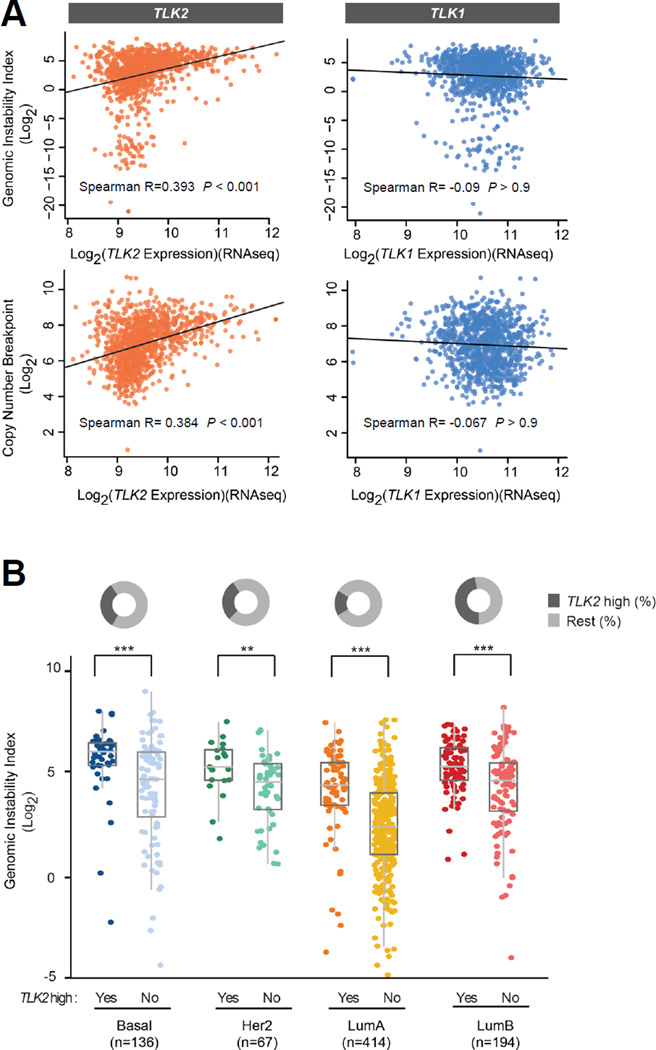
To examine if TLK2 overexpression correlates with CIN in breast tumors, we calibrated genome–wide genomic instability index (GII) for all TCGA breast tumor samples profiled by Affymetrix SNP 6.0 array. The GII score records the percentage of altered genome and is less affected by the noise of copy number datasets. As a result, we observed a significant positive correlation between TLK2 expression and GII (Spearman’s correlation: R = 0.393, P < 0.001), suggesting the role of TLK2 in the instability of the breast cancer genome (Fig. 1A; upper panel). Interestingly, this correlation with GII was not observed for TLK1 expression (Spearman’s R = −0.09, P > 0.9). In addition to GII, we also enumerated the genome-wide copy number breakpoints (CNB, also known as copy number transitions) for TCGA breast tumors as a quantitative measure of the rearrangement events leading to copy number aberrations (4, 16). The CNB approach will complement the GII approach that does not reflect the complex rearrangement events underlying these copy number alterations. As a result, this revealed a significant association of TLK2 expression with CNB index (Spearman’s R=0.384, P < 0.001), but not TLK1 expression (Spearman’s R = −0.067, P > 0.9) (Fig. 1A; lower panel). These data support the correlation of TLK2 upregulation with increased CIN in breast cancer and its distinct function from TLK1.
Figure 1. TLK2 overexpression correlates with chromosomal instability measured by copy number data.
(A) Correlation of the genomic instability index and copy number breakpoint index with TLK2 or TLK1 expression (RNAseq) in 1083 invasive breast cancers based on Spearman’s correlation statistics (copy number and RNAseq data are from TCGA). (B) Genomic Instability Index of TCGA breast tumors from different intrinsic breast cancer subtypes classified based on TLK2 expression. P-values were calculated based on t-test. **P<0.01, ***P<0.001. The samples categorized by PAM-50 subtypes are further classified as TLK2 overexpressing samples and the “rest” samples (see methods for the cut-off of TLK2 overexpression). All intrinsic subtypes show significantly higher genomic instability in samples with TLK2 overexpression.
To assess the association of TLK2 overexpression with CIN in different breast cancer subtypes, we compared GII index of breast tumors classified by intrinsic breast cancer subtypes in the presence or absence of TLK2 overexpression. Here the intrinsic subtyping is based on the 50-gene PAM50 predictor (17) (see methods). The GII indexes are significantly higher in TLK2-overexpressing luminal B, luminal A, and basal tumor samples as compared to TLK2 low samples (P < 0.001), and to a lesser degree in Her2 subtype samples (P < 0.01) (Fig. 1B). This suggests that the increased GII associated with TLK2 overexpression may not be attributed to the enrichment of TLK2 overexpression in the luminal B tumors known to harbor increased CIN (1). To access if TLK2 overexpression correlates with other known drivers of CIN in breast cancer such as AURKA and PLK1 (7, 18) (Fig. S1), we charted the status of TLK2, AURKA, and PLK1 overexpression together with chromosome instability scores (Fig. S2). The resulting heat map showed that TLK2 overexpression is independent of AURKA or PLK1 overexpression, suggesting TLK2 as an independent factor in promoting CIN in breast cancer (Fig. S2A). In addition, the luminal B breast tumors overexpressing all three kinases (TLK2, AURKA, and PLK1) show a higher genomic instability index compared to the tumors overexpressing only one or two kinases, and the luminal B tumors that are negative for all three kinases showed the lowest average GII level (Fig. S2B). These observations suggest the role of TLK2 overexpression in promoting CIN of breast cancers.
TLK2 overexpression impairs DNA-damage repair process
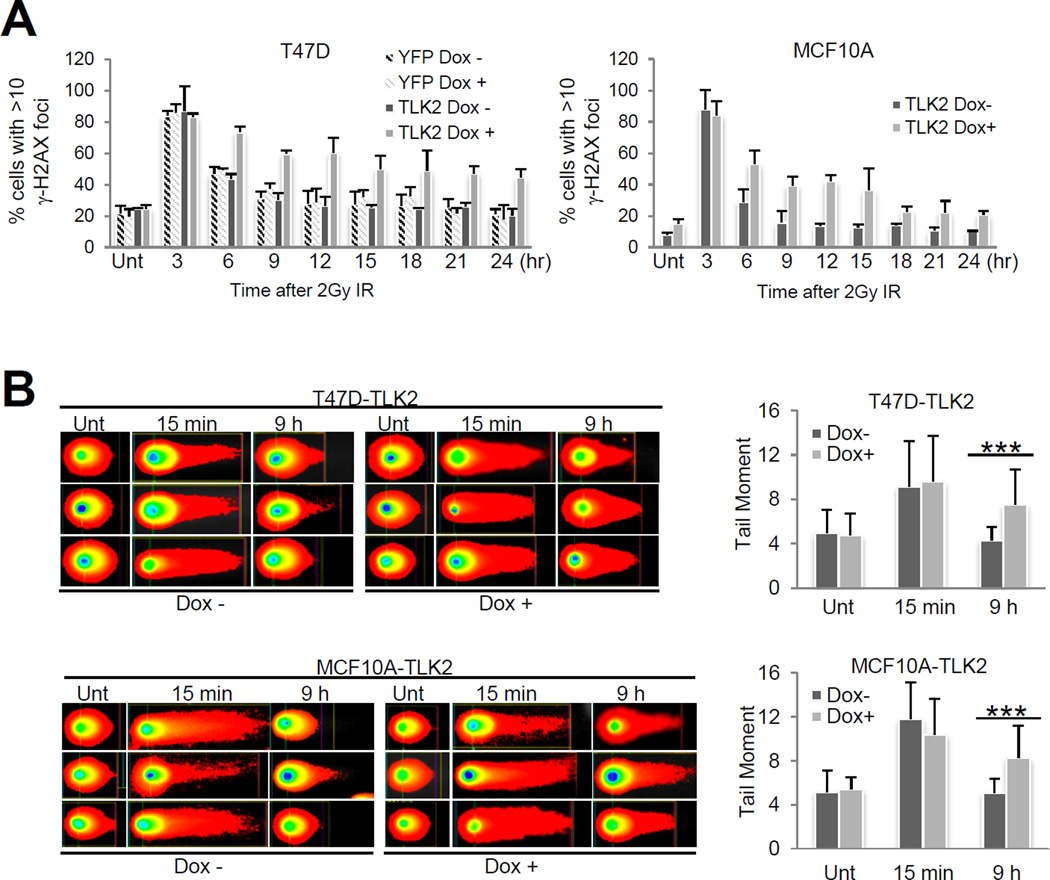
Since TLK kinase activity is quickly inhibited following IR-induced DNA damage response (DDR), we hypothesized that TLK2 amplification and overexpression may override the DNA damage response signaling, leading to impaired double-strand break (DSB) repair. We thus assessed the DSB repair process induced by γ-irradiation (IR) in T47D and MCF10A cells inducibly expressing TLK2 using γ-H2AX foci formation assay (γ-H2AX is a biomarker for DSBs) (19). Interestingly, induction of TLK2 overexpression in T47D or MCF10A cells treated with 2Gy IR prominently delayed the DSB repair process, leading to an increase of DSBs compared to the controls without TLK2 induction, which is sustained for a prolonged period of time (Fig. 2A; Fig. S3). This result was corroborated by the neutral comet assay directly visualizing the DSBs in the individual irradiated T47D cells and MCF10A overexpressing TLK2 (Fig. 2B). In addition, immunofluorescence staining suggests that TLK2 forms nuclear foci in irradiated MCF10A cells, which partially co-localize with γ-H2AX foci (Fig. S4). This implies the intimate association of TLK2 with DNA damage response. Moreover, TLK2-driven DSB repair defect seems to be independent of p53 status since TLK2-overexpressing MCF10A (p53 wild type) and T47D (p53 mutant) presented similar DDR alternations (Fig. 2; Fig. S3).
Figure 2. TLK2 overexpression undermines double-strand break repair.
(A) TLK2 overexpression in T47D cells (left) or MCF10A (right) cells delays the DSB repair process in response to IR. As a control, Yellow Fluorescent Protein (YFP) was expressed in T47D cells. TLK2 or YFP expression was induced in T47D cells or MCF10A cells by treating 100 ng/ml of Dox for 48hr. DSB foci induced by two Gray of IR were assessed by γ-H2AX staining. The charts show the quantification results counting the percentage of cells with more than ten γ-H2AX foci. The representative microscope images are shown in Fig. S3. Dox, doxycycline. DSB, DNA double strand breaks. (B) DSBs were visualized by neutral comet assays in T47D (upper panel) and MCF10A cells (lower panel) after IR with or without TLK2 induction. TLK2 was overexpressed in T47D or MCF10A cells by treating 100 ng/ml of Dox for 48hr. Eight Gy of IR was applied and cells were incubated for 0, 15 min, 6hr or 9hr at 37 degrees. Then, neutral comet assay was performed. Left panel shows the representative images and right panel shows the quantification results. IR, γ-irradiation. P-values were calculated based on t-test. ***P<0.001. DSB, DNA double strand breaks. Dox, doxycycline.
TLK2 overexpression leads to G2/M checkpoint defect
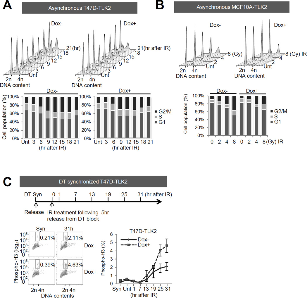
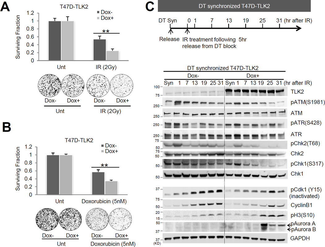
Next, we performed cell cycle analysis to determine if T47D and MCF10A cells overexpressing TLK2 have a defect in cell cycle arrest after DNA damage (Fig. 3A-B). The asynchronized T47D cells inducibly expressing TLK2 were irradiated with 2 Gy IR, and collected at different time points until 21 hours. The asynchronized MCF10A cells inducibly expressing TLK2 were treated with different doses of IR, and collected after 18 hrs. Cell cycle changes were assessed by flow cytometry measuring DNA content. In both models, a prominently delayed accumulation of G2-M phase cells was observed after IR when TLK2 was overexpressed, suggesting a possible defect of G2/M arrest. To verify the G2/M checkpoint defect, we synchronized the T47D cells at G1/S border by double thymidine (DT) block. At 5 hours after release from DT block, but before entering G2-M phase (Fig. 3C; Fig.S5), cells were irradiated, and mitotic entry was analyzed by phospho-H3 staining (a mitotic biomarker). This revealed a marked increase in mitotic cells (phosphor-H3 %) in T47D cells overexpressing TLK2 after irradiation, supporting a G2/M checkpoint defect (Fig. 3C). As G2/M checkpoint defect has been linked to increased sensitivity to irradiation and genotoxic agents, as seen in the cancer cells treated with Chk1 inhibitors (20), we postulate that TLK2 overexpression may have similar effect in breast cancer cells. We thus treated the T47D cells overexpressing TLK2 with irradiation or doxorubicin. Indeed, a modest but significant sensitizing effect was observed for both treatments with TLK2 overexpression, which supports our reasoning (Fig. 4A-B).
Figure 3. TLK2 overexpression impairs G2/M checkpoint induced by DNA damage.
IR-induced cell-cycle changes were assessed by flow cytometry measuring DNA content in T47D and MCF10A cells overexpressing TLK2. TLK2 expression was induced for 24h in T47D and MCF10A cells using 200 ng/ml Dox. (A) T47D cells were then irradiated with 2 Gy IR, and collected at indicated times. (B) MCF10A cells were treated with 2 Gy, 4 Gy, or 8 Gy of IR, and collected after 18 hr. Cell cycle changes were assessed by flow cytometry measuring DNA content. (C) Increased mitotic cell population (phospho-H3 positive) after IR in T47D cells overexpressing TLK2. T47D cells with or without TLK2 overexpression were synchronized at the G1/S border by DT block (10 mM); 5 hours after release, but before entering into G2-M phase (Fig. S5A), cells were treated with 8 Gy IR and then collected at indicated times. The percentage of phospho-H3 positive cells was quantified by flow cytometry. IR, γ-irradiation. Dox, doxycycline. Unt, untreated. Syn, cells synchronized at G1/S border by DT block. DT, double-thymidine.
Figure 4. TLK2 overexpression impairs DNA damage checkpoint signaling, and modestly increases the sensitivity of T47D luminal breast cancer cells to genotoxic agents.
A-B, TLK2 overexpression in T47D cells led to modest but significant increase of cancer cell sensitivity to irradiation and doxorubicin treatment. 100 ng/ml Dox was administered for 2 weeks to induce TLK2 overexpression in T47D cells, then either 2Gy of IR (A) or 5nM of doxorubicin (B) was administered for 2 weeks. Clonogenic assays were performed to measure cell viability after IR or doxorubicin treatment. P-values were calculated based on the t-test. **P<0.01. (C) Alternations of IR-induced checkpoint signaling after TLK2 overexpression in T47D cells. Western blot analysis was performed using the cell lysates obtained from the same experiment as in Fig. 3C. Dox, doxycycline. Unt, untreated. Syn, cells synchronized at G1-S border by DT block. DT, double-thymidine. IR, γ-irradiation.
TLK2 overexpression impairs cell cycle checkpoint signaling in response to DNA damage
Next, we went on to investigate how TLK2 overexpression leads to a G2/M checkpoint defect. The key step to initiate the mitotic processes is activation of the Cdk1/Cyclin B complex by dephosphorylation of Cdk1 on the inhibitory tyrosine pY15 residue (5), which is directly controlled by the G2/M checkpoint signaling (21). We thus performed Western blot analysis to detect alterations in IR-induced G2/M checkpoint signaling in T47D cells overexpressing TLK2 (Fig. 4C). Impressively, TLK2 overexpression markedly repressed the phosphorylation of Chk1 and Chk2 in response to IR. In addition, a decrease in pY15 Cdk1 (inactive form) and cyclin B1 level is observed with TLK2 overexpression followed by IR. Cyclin B1 starts to accumulate in G2 phase, whereas dephosphorylation of pY15 Cdk1 and activation of Cdk1 is specific to mitotic cells (5). As a vast majority of cells with 4n DNA content (G2-M population) attributes to G2 phase, the total Cyclin B1 level will be primarily affected by the G2 cell population (22). Thus the decrease of pY15 Cdk1 and Cyclin B1 implies an increase in the mitotic cell population and a decrease in the G2 population following TLK2 overexpression. Further, consistent with the G2/M checkpoint defect, TLK2 overexpression leads to increased phosphorylation and activation of mitotic kinases such as Aurora A, and increased phospho-H3 (Fig. 4C). Together, these data suggest that overexpression of TLK2 may override the DNA damage checkpoint signaling via repressing Chk1/2, leading to G2/M checkpoint defect, and delayed DSB repair. A schematic illustrating the mechanisms of G2/M checkpoint signaling impaired by TLK2 overexpression is shown in Fig. S6.
DISCUSSION
Our genome–wide genomic instability index (GII) and copy number breakpoint (CNB) analysis of TCGA breast tumor samples revealed that TLK2 amplification might be one of the genetic factors contributing to the outbreak of chromosome instability (CIN) in the luminal B breast tumors. Interestingly, a most recent phosphoproteomics study of TCGA breast cancers by The Clinical Proteomic Tumor Analysis Consortium (CPTAC) independently identified TLK2 as a highly phosphorylated kinases associated with genomic amplifications at this loci that preferentially present in luminal breast cancer (23). This further supports the importance of TLK2 amplification in luminal tumors. The present study will timely complement the CPTAC study revealing the role of TLK2 amplification in G2/M checkpoint defect and CIN. Our experimental data suggest that ectopic expression of TLK2 in the T47D luminal breast cancer cells or MCF10A benign breast epithelial leads to delayed DNA repair as evidenced by the γ-H2AX foci formation and neutral comet assays. Further cell cycle analysis after irradiation of T47D or MCF10A cells suggested a G2/M checkpoint defect associated with TLK2 overexpression, which is further verified via phospho-H3 staining (a mitotic biomarker). These data support the role of TLK2 in G2/M checkpoint defect and CIN. Presumably, such a G2/M checkpoint defect will allow unrepaired DSBs to enter into mitosis, leading to accumulation of DNA copy number aberrations as described previously (24). In addition, our mechanistic studies show that TLK2 overexpression in T47D cells potently represses the phosphorylation of Chk1 (S317) and Chk2 (T68), which may explain the impaired G2/M checkpoint signaling (Fig. 4C; Fig. S6). Finally, our result revealed that TLK2 overexpression in T47D cells modestly increase the sensitivity of breast cancer cells to DNA damaging agents such as γ-irradiation (IR) and doxorubicin (Fig. 4A-B), consistent with the G2/M checkpoint defect in TLK2 overexpressing cells. Future studies will be required to further examine the effect of TLK2 overexpression on breast cancer cell sensitivity to DNA damaging agents, and the dependence of such effect on P53 status. In addition, it will be interesting to further examine whether TLK2 overexpression induces chromosomal instability during tumor initiation process.
More important, our result revealed the distinct functions of TLK2 from TLK1 in DNA damage response. TLK1 has been considered as a guardian of genome integrity (12). As opposed to the CIN driven by TLK2 overexpression, upregulation of TLK1 led to enhanced DNA repair and increased genomic stability (25). In response to DNA damage, TLK1 localizes to DSBs and functions as a molecular chaperone to recruit the Rad9-Hus1-Rad1 (9-1-1) complex (25), which initiates an ATR-Chk1-mediated cell cycle checkpoint (26). In our result, overexpressed TLK2 in MCF10A was also recruited to DNA damage site after IR (Fig. S4), and TLK2 overexpression led to delayed DNA repair showing 50–100% higher number of cells containing more than ten γ-H2AX foci compare to control after IR (Fig. 2A). As TLK2 shares less homology with TLK1 in the non-STK domains (10), it may be possible that TLK2 may lack the DDR chaperone function possessed by TLK1 and thus competitively inhibit the TLK1 function as a DDR chaperone after DNA damage. Furthermore, a new study just published during our submission has reported TLK2 as a key regulator of checkpoint recovery from DNA-damage induced G2 arrest (27). Thus besides repressing IR-induced Chk1/2 activation, TLK2 overexpression may also contribute to the premature mitotic entry via its function in G2 checkpoint recovery. Future studies are needed to investigate the interaction of the two tousled-like kinases in DNA damage response (DDR), and pinpoint the precise mechanisms engaged by TLK2 to repress Chk1/2, and impair G2/M checkpoint.
Together, our data suggest that TLK2 amplification may contribute to the increased CIN of luminal breast cancer via impairing the G2/M DNA damage checkpoint. In addition to this observation, our previous study showed that TLK2 overexpression promotes more aggressive phenotypes in luminal breast cancers and correlates with poor prognosis regardless of endocrine therapy. Thus targeting TLK2 may present an attractive therapeutic strategy for the luminal breast tumors harboring TLK2 amplifications with enhanced aggressiveness and increased chromosomal instability.
Supplementary Material
Acknowledgments
The results published here are in part based upon data generated by TCGA (dbGaP accession: phs000178.v6.p6). The computational infrastructure was supported by BCM Dan L. Duncan Cancer Center Biostatistics and Informatics Shared Resource (supported by NCI P30 CA125123). This project was also supported by the Cytometry and Cell Sorting Core at BCM with funding from the NIH (P30 AI036211, P30 CA125123, and S10 RR024574). We thank Dr. Dean P. Edwards for providing cell lines from the ATCC ICBP45 cell line panel. Funding: This study was supported by Susan G. Komen foundation PDF12231561 (JA.K.), CDMRP grants W81XWH-12-1-0166 (X-S.W.), W81XWH-12-1-0167 (R.S.), W81XWH-13-1-0431 (J.V.), and NIH grants CA181368 (X-S.W), CA183976 (X-S.W), and P30-125123-06.
Footnotes
DISCLOSURE OF POTENTIAL CONFLICTS OF INTEREST:
The authors report no conflict of interest.
References
- 1.Smid M, Hoes M, Sieuwerts AM, Sleijfer S, Zhang Y, Wang Y, et al. Patterns and incidence of chromosomal instability and their prognostic relevance in breast cancer subtypes. Breast Cancer Res Treat. 2011;128:23–30. doi: 10.1007/s10549-010-1026-5. [DOI] [PubMed] [Google Scholar]
- 2.McGranahan N, Burrell RA, Endesfelder D, Novelli MR, Swanton C. Cancer chromosomal instability: therapeutic and diagnostic challenges. EMBO Rep. 2012;13:528–538. doi: 10.1038/embor.2012.61. [DOI] [PMC free article] [PubMed] [Google Scholar]
- 3.Burrell RA, McGranahan N, Bartek J, Swanton C. The causes and consequences of genetic heterogeneity in cancer evolution. Nature. 2013;501:338–345. doi: 10.1038/nature12625. [DOI] [PubMed] [Google Scholar]
- 4.Fridlyand J, Snijders AM, Ylstra B, Li H, Olshen A, Segraves R, et al. Breast tumor copy number aberration phenotypes and genomic instability. BMC Cancer. 2006;6:96. doi: 10.1186/1471-2407-6-96. [DOI] [PMC free article] [PubMed] [Google Scholar]
- 5.Norbury C, Blow J, Nurse P. Regulatory phosphorylation of the p34cdc2 protein kinase in vertebrates. Embo J. 1991;10:3321–3329. doi: 10.1002/j.1460-2075.1991.tb04896.x. [DOI] [PMC free article] [PubMed] [Google Scholar]
- 6.Bouwman P, Jonkers J. The effects of deregulated DNA damage signalling on cancer chemotherapy response and resistance. Nat Rev Cancer. 2012;12:587–598. doi: 10.1038/nrc3342. [DOI] [PubMed] [Google Scholar]
- 7.Li JJ, Li SA. Mitotic kinases: the key to duplication, segregation, and cytokinesis errors, chromosomal instability, and oncogenesis. Pharmacol Ther. 2006;111:974–984. doi: 10.1016/j.pharmthera.2006.02.006. [DOI] [PubMed] [Google Scholar]
- 8.Gjertsen BT, Schoffski P. Discovery and development of the Polo-like kinase inhibitor volasertib in cancer therapy. Leukemia. 2015;29:11–19. doi: 10.1038/leu.2014.222. [DOI] [PMC free article] [PubMed] [Google Scholar]
- 9.Kollareddy M, Zheleva D, Dzubak P, Brahmkshatriya PS, Lepsik M, Hajduch M. Aurora kinase inhibitors: progress towards the clinic. Invest New Drugs. 2012;30:2411–2432. doi: 10.1007/s10637-012-9798-6. [DOI] [PMC free article] [PubMed] [Google Scholar]
- 10.Sillje HH, Takahashi K, Tanaka K, Van Houwe G, Nigg EA. Mammalian homologues of the plant Tousled gene code for cell-cycle-regulated kinases with maximal activities linked to ongoing DNA replication. Embo J. 1999;18:5691–5702. doi: 10.1093/emboj/18.20.5691. [DOI] [PMC free article] [PubMed] [Google Scholar]
- 11.Groth A, Lukas J, Nigg EA, Sillje HH, Wernstedt C, Bartek J, et al. Human Tousled like kinases are targeted by an ATM- and Chk1-dependent DNA damage checkpoint. Embo J. 2003;22:1676–1687. doi: 10.1093/emboj/cdg151. [DOI] [PMC free article] [PubMed] [Google Scholar]
- 12.De Benedetti A. The Tousled-Like Kinases as Guardians of Genome Integrity. ISRN Mol Biol. 2012;2012:627596. doi: 10.5402/2012/627596. [DOI] [PMC free article] [PubMed] [Google Scholar]
- 13.Genovese G, Ergun A, Shukla SA, Campos B, Hanna J, Ghosh P, et al. microRNA regulatory network inference identifies miR-34a as a novel regulator of TGF-beta signaling in glioblastoma. Cancer Discov. 2012;2:736–749. doi: 10.1158/2159-8290.CD-12-0111. [DOI] [PMC free article] [PubMed] [Google Scholar]
- 14.Debnath J, Muthuswamy SK, Brugge JS. Morphogenesis and oncogenesis of MCF-10A mammary epithelial acini grown in three-dimensional basement membrane cultures. Methods. 2003;30:256–268. doi: 10.1016/s1046-2023(03)00032-x. [DOI] [PubMed] [Google Scholar]
- 15.Liang Y, Gao H, Lin SY, Peng G, Huang X, Zhang P, et al. BRIT1/MCPH1 is essential for mitotic and meiotic recombination DNA repair and maintaining genomic stability in mice. PLoS Genet. 2010;6:e1000826. doi: 10.1371/journal.pgen.1000826. [DOI] [PMC free article] [PubMed] [Google Scholar]
- 16.Korbel JO, Urban AE, Grubert F, Du J, Royce TE, Starr P, et al. Systematic prediction and validation of breakpoints associated with copy-number variants in the human genome. Proceedings of the National Academy of Sciences of the United States of America. 2007;104:10110–10115. doi: 10.1073/pnas.0703834104. [DOI] [PMC free article] [PubMed] [Google Scholar]
- 17.Parker JS, Mullins M, Cheang MC, Leung S, Voduc D, Vickery T, et al. Supervised risk predictor of breast cancer based on intrinsic subtypes. Journal of clinical oncology : official journal of the American Society of Clinical Oncology. 2009;27:1160–1167. doi: 10.1200/JCO.2008.18.1370. [DOI] [PMC free article] [PubMed] [Google Scholar]
- 18.Zhou H, Kuang J, Zhong L, Kuo WL, Gray JW, Sahin A, et al. Tumour amplified kinase STK15/BTAK induces centrosome amplification, aneuploidy and transformation. Nat Genet. 1998;20:189–193. doi: 10.1038/2496. [DOI] [PubMed] [Google Scholar]
- 19.Kuo LJ, Yang LX. Gamma-H2AX - a novel biomarker for DNA double-strand breaks. In Vivo. 2008;22:305–309. [PubMed] [Google Scholar]
- 20.Morgan MA, Parsels LA, Zhao L, Parsels JD, Davis MA, Hassan MC, et al. Mechanism of radiosensitization by the Chk1/2 inhibitor AZD7762 involves abrogation of the G2 checkpoint and inhibition of homologous recombinational DNA repair. Cancer Res. 2010;70:4972–4981. doi: 10.1158/0008-5472.CAN-09-3573. [DOI] [PMC free article] [PubMed] [Google Scholar]
- 21.Donzelli M, Draetta GF. Regulating mammalian checkpoints through Cdc25 inactivation. EMBO Rep. 2003;4:671–677. doi: 10.1038/sj.embor.embor887. [DOI] [PMC free article] [PubMed] [Google Scholar]
- 22.Hershko A. Mechanisms and regulation of the degradation of cyclin B. Philos Trans R Soc Lond B Biol Sci. 1999;354:1571–1575. doi: 10.1098/rstb.1999.0500. discussion 5-6. [DOI] [PMC free article] [PubMed] [Google Scholar]
- 23.Mertins P, Mani DR, Ruggles KV, Gillette MA, Clauser KR, Wang P, et al. Proteogenomics connects somatic mutations to signalling in breast cancer. Nature. 2016;534:55–62. doi: 10.1038/nature18003. [DOI] [PMC free article] [PubMed] [Google Scholar]
- 24.Xu X, Weaver Z, Linke SP, Li C, Gotay J, Wang XW, et al. Centrosome amplification and a defective G2-M cell cycle checkpoint induce genetic instability in BRCA1 exon 11 isoform-deficient cells. Mol Cell. 1999;3:389–395. doi: 10.1016/s1097-2765(00)80466-9. [DOI] [PubMed] [Google Scholar]
- 25.Sunavala-Dossabhoy G, De Benedetti A. Tousled homolog, TLK1, binds and phosphorylates Rad9; TLK1 acts as a molecular chaperone in DNA repair. DNA Repair (Amst) 2009;8:87–102. doi: 10.1016/j.dnarep.2008.09.005. [DOI] [PubMed] [Google Scholar]
- 26.Parrilla-Castellar ER, Arlander SJ, Karnitz L. Dial 9-1-1 for DNA damage: the Rad9-Hus1-Rad1 (9-1-1) clamp complex. DNA Repair (Amst) 2004;3:1009–1014. doi: 10.1016/j.dnarep.2004.03.032. [DOI] [PubMed] [Google Scholar]
- 27.Bruinsma W, van den Berg J, Aprelia M, Medema RH. Tousled-like kinase 2 regulates recovery from a DNA damage-induced G2 arrest. EMBO Rep. 2016 doi: 10.15252/embr.201540767. [DOI] [PMC free article] [PubMed] [Google Scholar]
Associated Data
This section collects any data citations, data availability statements, or supplementary materials included in this article.


